Tag Archive for: radical prostatectomy

Posts

Editorial: Somatic-autonomic neurorrhaphy for erectile function restoration after radical prostatectomy

The authors of the present study [1] are to be commended for their efforts in describing their 3-year experience with a novel bilateral end-to-side somatic-autonomic neurorrhaphy intended to restore erectile function at least 24 months after radical prostatectomy, after which spontaneous return of erectile function is unlikely [2]. Using the principles of brain plasticity and neurotization, the authors describe, for the first time, bilateral sural neurografting between the femoral nerve and both the corpus cavernosum and the dorsal nerve of the penis to achieve penile re-innervation. The employed side-to-end neurorrhaphy theoretically limits functional damage during axonal sprouting, reinforces sensory–motor communications to the cavernous nerves, and promotes glans penis sensitivity, although direct evidence of these physiological and clinical outcomes is yet to be demonstrated. Furthermore, the authors astutely capitalize on the potential advantages of using a femoral donor nerve, including its proximity to the proximal penis, its diameter, the sufficient axon count, the mixed composition of sensory and motor fibres, and its secretion of acetylcholine which is an essential neurotransmitter in the nitric oxide-mediated pathway leading to penile tumescence.

What is especially singular about this technique is its application months after radical prostatectomy following demonstrated post-surgical loss of erectile function. Previous studies have focused on unilateral or bilateral sural [3-6] or genitofemoral [7] neurografting of the cavernous nerves at the time of radical prostatectomy before post-surgical loss of erectile function could be substantiated. Such studies have had mixed results, attributable in part to the success of the nerve-sparing radical prostatectomy technique in the hands of experienced high-volume surgeons [3, 6], and post-surgical exposure of the cavernous nerves to androgen deprivation or radiation therapy in some patients [3, 5]. Whereas post-radical prostatectomy re-innervation of the cavernous nerves may not be feasible or efficacious secondary to post-surgical and post-radiation fibrosis, the described technique does not require abdominopelvic access, is associated with a quick recovery time and minimal complications, and does not preclude subsequent penile prosthesis implantation if necessary.

The results of the present study show significant improvement in general sexual satisfaction from baseline to 6 months post intervention corresponding to achievement of flaccid erection for all patients; significant improvement in erectile dysfunction from baseline to 12 months post intervention corresponding to achievement of semi-rigid or rigid erections in 8/10 patients; and significant improvement in satisfaction with sexual intercourse from baseline to 18 months post intervention corresponding to achievement of penetration for 6/10 patients. It should be noted, however, that administration of the Clinical Evolution of Erectile Function instrument at 36 months postoperatively may have introduced recall bias in patient-reported erectile function. As would be expected, no significant differences were noted in sexual desire or orgasm satisfaction during the study period. These results were achieved without evidence of atrophy, fibrosis, or significant differences in vascular flow of the bilateral corpora cavernosa; and with minimal complications over the study period.

The present study provides preliminary data regarding the safety, feasibility and efficacy of bilateral sural neurografting between the femoral nerve and both the corpus cavernosum and the dorsal nerve of the penis to restore post-radical prostatectomy erectile function in a limited pool of 10 men. If these preliminary results are substantiated with long-term follow-up in a significant number of patients, it should prompt multi-institutional collaborations to appropriately power comparative effectiveness analyses of post-radical prostatectomy neurorrhaphy, such as the described technique to a regulatory-approved ethical sham intervention, vacuum assist device therapy, urethral suppository therapy, or intracavernosal injection therapy. Ideally, patients should be matched or statistical analyses should be controlled for patient age, relevant comorbidities, prostate cancer stage, pre-radical prostatectomy erectile function, nerve-sparing radical prostatectomy technique, androgen deprivation therapy, radiation therapy, response to phosphodiesterase-5 inhibition, and time interval between radical prostatectomy and intervention. Such multi-institutional studies would benefit from: standardized protocols for preoperative evaluation, operative technique, peri-operative care and post-surgical sexual stimulation; repeated-measure analyses of both objective assessments of the quality and duration of penile tumescence, as well as patient-reported outcomes of erectile function and disease-specific quality of life using validated instruments; report of oncological outcomes; and long-term follow-up. We would encourage the authors to produce a technical video demonstrating their technique so that it may be attempted and validated by other programmes. Urological surgeons with expertise in oncology, sexual function, and reconstruction may collaborate and pool patient data to achieve high-powered quality studies of novel techniques, such as the one described in the present study, for post-radical prostatectomy erectile function restoration.

Jaime A. Cavalloand Ashutosh K. Tewari, Chairman
Milton and Carroll Petrie Department of Urology, Icahn School of Medicine at M ount Sinai, New York, NY, USA


How to Cite

Cavallo, J. A. and Tewari, A. K. (2017), Somatic-autonomic neurorrhaphy for erectile function restoration after radical prostatectomy. BJU International, 119: 816–818. doi: 10.1111/bju.13858

References


The 5th BJUI Social Media Awards

It’s hard to believe that we have been doing the BJUI Social Media Awards for five years now! I recall vividly our inaugural BJUI Social Media Awards in 2013, as the burgeoning social media community in urology gathered in the back of an Irish Bar in San Diego to celebrate all things social. At that time, many of us had only got to know each other through Twitter, and it was certainly fun going around the room putting faces with twitter handles for the first time. That spirit continues today as the “uro-twitterati” continues to grow, and the BJUI Awards, (or the “Cult” Awards as our Editor-in-Chief likes to call them), remains a fun annual focus for the social-active urology community to meet up in person.

As you may know, we alternate the Awards between the annual congresses of the American Urological Association (AUA) and of the European Association of Urology (EAU). Last year, we descended on Munich, Germany to join the 13,000 or so other delegates attending the EAU Annual Meeting and to enjoy all the wonderful Bavarian hospitality on offer. This year, we set sail for the #AUA17 Annual Congress in Boston, MA, along with over 16,000 delegates from 100 different countries. What a great few days in beautiful Boston and a most welcome return for the AUA to this historic city. Hopefully it will have a regular spot on the calendar, especially with the welcome dumping of Anaheim and Orlando as venues for the Annual Meeting.

Awards

On therefore to the Awards. These took place on Saturday 13th May 2017 in the City Bar of the Westin Waterfront Boston. Over 80 of the most prominent uro-twitterati from all over the world turned up to enjoy the hospitality of the BJUI and to hear who would be recognised in the 2017 BJUI Social Media Awards. We actually had to shut the doors when we reached capacity so apologies to those who couldn’t get in! Individuals and organisations were recognised across 12 categories including the top gong, The BJUI Social Media Award 2017, awarded to an individual, organization, innovation or initiative who has made an outstanding contribution to social media in urology in the preceding year. The 2013 Award was won by the outstanding Urology Match portal, followed in 2014 by Dr Stacy Loeb for her outstanding individual contributions, and in 2015 by the #UroJC twitter-based journal club. Last year’s award went to the #ilooklikeaurologist social media campaign which we continue to promote.

This year our Awards Committee consisted of members of the BJUI Editorial Board – Declan Murphy, Prokar Dasgupta, Matt Bultitude, Stacy Loeb, John Davis, as well as BJUI Managing Editor Scott Millar whose team in London (Max and Clare) drive the content across our social platforms. The Committee reviewed a huge range of materials and activity before reaching their final conclusions.

The full list of winners is as follows:

Most Read Blog@BJUI – “The optimal treatment of patients with localized prostate cancer: the debate rages on”. Dr Chris Wallis, Toronto, Canada

1-1

Most Commented Blog@BJUI – “It’s not about the machine, stupid”. Dr Declan Murphy, Melbourne, Australia

1-2

Most Social Paper – “Novel use of Twitter to disseminate and evaluate adherence to clinical guidelines by the European Association of Urology”. Accepted by Stacy Loeb on behalf of herself and her colleagues.

1-3

Best BJUI Tube Video – “Combined mpMRI Fusion and Systematic Biopsies Predict the Final Tumour Grading after Radical Prostatectomy”. Dr Angela Borkowetz, Dresden, Germany

AUA

Best Urology Conference for Social Media – #USANZ17 – The Annual Scientific Meeting of the Urological Association of Australia & New Zealand (USANZ) 2017. Accepted by Dr Peter Heathcote, Brisbane, Australia. President of USANZ.

1-5

Best Urology App – The EAU Guidelines App. Accepted by Dr Maria Ribal, Barcelona, Spain, on behalf of the EAU.

1-6

Innovation Award – BJUI Urology Ontology Hashtags keywords. Accepted by Dr Matthew Bultitude, London, UK, on behalf of the BJUI.

1-7

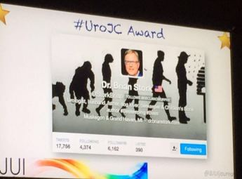
#UroJC Award – Dr Brian Stork, Michigan, USA. Accepted by Dr Henry Woo of Brian’s behalf.

UroJC
Most Social Trainee – Dr Chris Wallis, Toronto, Canada

1-9

Best Urology Journal for Social Media –Journal of Urology/Urology Practice. Accepted by Dr Angie Smith, Chapel Hill, USA, on behalf of the AUA Publications Committee.

1-10

Best Urology Organisation – Canadian Urological Association. Accepted by Dr Mike Leveridge, Vice-President of Communications for CUA.

1-11

The BJUI Social Media Award 2017 – The Urology Green List, accepted by Dr Henry Woo, Sydney, Australia.

1-12

All the Award winners (except Dr Brian Stork who had to get home to work), were present to collect their awards themselves. A wonderful spread of socially-active urology folk from all over the world, pictured here with BJUI Editor-in-Chief, Prokar Dasgupta.

1-13

A special thanks to our outstanding BJUI team at BJUI in London, Scott Millar, Max Cobb and Clare Dunne, who manage our social media and website activity as well as the day-to-day running of our busy journal.

See you all in Copenhagen for #EUA18 where we will present the 6th BJUI Social Media Awards ceremony!


Declan Murphy

Peter MacCallum Cancer Centre, Melbourne, Australia

Associate Editor, BJUI

@declangmurphy

TRoMbone is launched!

TRoMbone is launched! The UK feasibility randomised trial Testing radical prostatectomy in men with prostate cancer and oligoMetastases to the bone has opened at Oxford University Hospitals (PI Freddie Hamdy), University College London Hospitals (PI John Kelly) and Royal Surrey County Hospital (PI Chris Eden). Men <75 with newly-diagnosed prostate cancer and 1-3 skeletal lesions on any standard-of-care imaging (CT, bone scan, MRI, or PET) who are fit for surgery and deemed to be technically operable are eligible. Emerging but lower-quality data suggests a role for treatment of the primary tumour in men with oligo-metastatic prostate cancer and this UK study will investigate this question with level 1 evidence.

Participants will be randomly allocated to standard-of-care treatment (hormones +/- chemotherapy) versus standard-of-care treatment plus surgery to remove the prostate and draining lymph nodes (radical prostatectomy plus extended pelvic lymphadenectomy). A qualitative recruitment investigation to optimise accrual will be conducted by the University of Bristol (Caroline Wilson) and biologic samples will be collected, processed and stored in a repository at the Institute of Cancer Research (Gerhardt Attard).

We will assess technical feasibility, safety and complications of surgery in oligo-metastatic prostate cancer, and examine ways to improve recruitment in this pilot study. TRoMbone is managed by the Surgical Intervention Trials Unit at the University of Oxford and funded by the Prostate Cancer Foundation and The Urology Foundation.

We need to recruit 50 men over a 12-month period, and are seeking referrals from other centres to increase accrual. Centres that demonstrate ability to refer eligible patients will be able to take part in the main trial if we can demonstrate feasibility in this phase and get funding for the larger study.

So please look out for these patients and send them to me at UCLH, Freddie in Oxford, or Chris in Guildford. One of the three of us will do the surgery if they get randomised to it, but of course you’re welcome to come with the patients. If you have any queries please contact me, the study CI (P. Sooriakumaran (PS); [email protected]) or the study co-ordinator (Neelam Hassanali; [email protected]). You can start them on androgen deprivation and ‘stop the clock’ before you refer them to us. The extra burden of participating in this study is minimal. They will require one visit for consent, and one follow-up visit at 3 months after randomisation. The surgical group will also have two other visits for their surgery and catheter removal. The rest of the follow-up can be done back at your referring centre or with us, whatever you and the patient prefer. If it’s your standard policy to give them chemotherapy or metastasis-directed therapy with SBRT then you can still do that as part of the study.


With your help we can demonstrate that this study is feasible in the UK and we can lead the way in the surgical management of oligo-metastatic prostate cancer.


P. Sooriakumaran [social type=”facebook” opacity=”dark’ label=’PLACE_LINK_HERE[/social]

BMedSci(Hons) BMBS(Hons) PhD PGCMedLaw ADCClinInv FRCS(Urol) FEBU USLME

Consultant Urological Surgeon, UCL Hospitals NHS Foundation Trust & Honorary Clinical Senior Researcher, University of Oxford


March 2017 #urojc summary: Pelvic Lymph Node Dissection with Radical Prostatectomy – Is there enough evidence for and against?

The twitter-based international urology journal club @iurojc #urojc is back with a splash after a brief hiatus. For the March 2017 #urojc, a lively discussion takes the theme of pelvic node dissection (PLND) on radical prostatectomy (RP) reviewing a timely article by Nicola Fossati et al. The paper was made available open access courtesy of European Urology @EUplatinum.

A systematic review of the literature was performed including all comparative studies of both randomized and non randomized studies, with at least one experimental and one control arm. This summarised 66 studies including more than 250.000 patients with particular focus on different extents of pelvic lymphadenectomy as proposed by the European Association of Urology. Outcome measures studied included oncological features of biochemical recurrence, development of metastases, cancer-specific survival, and overall survival. Adverse events were covered under secondary outcomes, both intra- and postoperatively observed. Finally, quality of PLND was addressed in terms of total number of nodes and total number of positive nodes. Risk of bias was assessed for all studies judging on basis of specific confounders.

The journal club ran for 48 hours from Sunday 5th march. The central question addressed is balance of benefits and drawbacks of lymph node dissection. The corresponding author of the manuscript, Steven Joniau from the University Hospitals of Leuven, Belgium highlighted the role of lymph nodes in prostate cancer recurrence.

However despite this idea, the benefit of PLND is heavily scrutinized from the start. Long term data from a single centre  suggested limited benefit.


However PLND has since earlier times been employed as a diagnostic tool, where an optimal template (presacral in addition to extended LND) may be optimal for staging and removal of lymph nodes.

Despite the current state of evidence, PLND is frequently mentioned in the various guidelines available for prostate cancer. However the exact situations when to employ them is questioned by some participants.

The various therapeutic options for lymph node metastases also coloured the discussion.

The discussion further continued to the important issue of morbidity, and the associated question of performing an extended PLND (ePLND).

Despite the current state of evidence, PLND is frequently mentioned in the various guidelines available for prostate cancer. However the exact situations when to employ them is questioned by some participants.

The

The discussion further continued to the important issue of morbidity, and the associated question of performing an extended PLND (ePLND).

The increasing use of PSMA PET/CT provided other spread pattern data to be considered. And finally temporal changes in PSA testing is observed to affect the need for LND.


From the poll which ran during the discussion, about half responders would perform extended PLND for staging, while the rest were divided almost equally between therapeutic benefit and adherence to guideline recommendations.

Probably all participants of the discussion agrees for the need of a proper randomised study addressing role of PLND.

At the end of a busy 48 hours, the discussion had been joined by top experts in the field of prostate cancer, generated more than 200 tweets and reached more than 700 thousand impressions the world over.

Yodi Soebadi (@yodisoebadi) is an Indonesian urologist, trained at Universitas Airlangga, currently pursuing doctoral research at KU Leuven in Belgium.


Article of the Month: 68Ga-PSMA PET/CT for LN staging in PCa

Every Month the Editor-in-Chief selects an Article of the Month from the current issue of BJUI. The abstract is reproduced below and you can click on the button to read the full article, which is freely available to all readers for at least 30 days from the time of this post.

In addition to the article itself, there is an accompanying editorial written by a prominent member of the urological community. This blog is intended to provoke comment and discussion and we invite you to use the comment tools at the bottom of each post to join the conversation.

If you only have time to read one article this week, it should be this one.

Prospective evaluation of 68Gallium-prostate-specific membrane antigen positron emission tomography/computed tomography for preoperative lymph node staging in prostate cancer

Pim J. van Leeuwen*, Louise Emmett,§, Bao Ho, Warick Delprado, Francis Ting*Quoc Nguyen† and Phillip D. Stricker*


*St Vincents Prostate Cancer Centre, St Vincents Clinic, Australian Prostate Cancer Research Centre, New South Wales, The Garvan Institute of Medical Research/The Kinghorn Cancer Centre, Department of Diagnostic Imaging, St Vincents Public Hospital, §University of New South Wales, Sydney, and University of Notre Dame, Darlinghurst, NSW, Australia


Read the full article

Abstract

Objectives

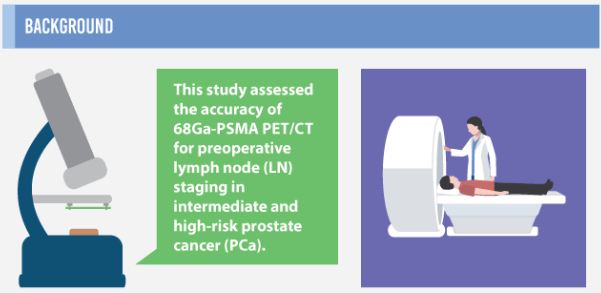
To assess the accuracy of 68Gallium-prostate-specific membrane antigen (68Ga-PSMA) positron emission tomography/computed tomography (PET/CT) for lymph node (LN) staging in intermediate- and high-risk prostate cancer (PCa).

Materials and Methods

From April to October 2015, 30 patients with intermediate- (n = 3) or high-risk (n = 27) PCa were prospectively enrolled. Patients underwent preoperative 68Ga-PSMA PET/CT. Both visual and semi-quantitative analyses were undertaken. Subsequently, all patients underwent radical prostatectomy (RP) with an extended pelvic lymph node dissection. The sensitivity, specificity, and positive (PPV) and negative predictive value (NPV) for LN status of 68Ga-PSMA were calculated using histopathology as reference.

aotmfeb2017-results

Results

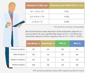
Eleven patients (37%) had lymph node metastases (LNMs); 26 LNMs were identified in the 11 patients. Patient analysis showed that 68Ga-PSMA PET/CT had a sensitivity of 64% for the detection of LNMs, its specificity was 95%, the PPV was 88%, and the NPV was 82%. In total, 180 LN fields were analysed. In the LN-region-based analysis, the sensitivity of 68Ga-PSMA PET/CT for detection of LNMs was 56%, the specificity was 98%, the PPV was 90% and the NPV was 94%. The mean size of missed LNMs was 2.7 mm. Receiver-operating characteristic curve analysis showed a high accuracy of maximum standardized uptake value (SUVmax) for the detection of LNMs, with an area under the curve of 0.915 (95% confidence interval 0.847–0.983); the optimum SUVmax was 2.0.

Conclusions

In patients with intermediate- to high-risk PCa, 68Ga-PSMA PET/CT had a high specificity and a moderate sensitivity for LNM detection. 68Ga-PSMA PET/CT had the potential to replace current imaging for LN staging of patients with PCa scheduled for RP.

Read more articles of the week

info-feb-2017

Click on image for full infographic


Editorial: Bringing clarity or confusion? The role of prostate-specific membrane antigen positron-emission/computed tomography for primary staging in prostate cancer

The use of 68Ga-labelled prostate-specific membrane antigen (PSMA) positron-emission tomography (PET)/CT for staging prostate cancer in Australia has reached almost plague-like proportions. Despite what must be admitted is little high-level evidence to guide us in the accuracy or appropriateness of this imaging technique for either primary staging or prostate cancer recurrence, hundreds of these scans are being performed every week around Australia, and in many cases we simply do not know what to do with the results. We performed the first such scan at our centre in Melbourne in August 2014, and were soon receiving 10 requests per day, with patients waiting up to 3 months to be scanned. Fast-forward 2 years, and there are now eight centres offering PSMA PET/CT in Melbourne, a city of 4.5 million people. Scans can be obtained within 24 h of referral and costs have dropped to €500. A similar situation exists in Germany where this imaging method was pioneered [1], and interest is also growing in Belgium, Italy, India and a number of other countries (the USA being a notable exception). But do we really understand the impact of the decision to perform PSMA/PET scanning, and do we have enough evidence to guide us on the most appropriate setting for its use?

The current interest in PSMA PET/CT has been triggered by the development of small molecule ligands which bind to the extracellular domain of the PSMA molecule, leading to increased sensitivity and specificity when compared with conventional imaging [2]. Previously, the use of PET imaging for prostate cancer detection was greatly limited by the relatively poor performance characteristics of choline-based PET/CT, and limited availability and high costs associated with this type of imaging. The introduction of 68Ga-labelled PSMA PET/CT has addressed many of these concerns, although high-quality evidence is still lacking to help guide its most appropriate utility. The best data exist for identification of prostate recurrence in patients with biochemical recurrence (BCR) after previous definitive therapy. In our recent systematic review and meta-analysis of this topic, we reported pooled data on 1309 men with BCR undergoing PSMA PET/CT [3]. When stratified by PSA level post-radical prostatectomy, positive scans are reported in 42, 58, 76 and 95% of patients with PSA levels of 0–0.2, 0.2–1, 1–2, and >2 ng/mL, respectively. Fewer data exist for the role of PSMA PET/CT in the primary staging setting.

In this interesting paper from some of our Australian colleagues, van Leeuwen et al. [4] report their experience of PSMA PET/CT in the primary staging setting, in particular to evaluate the performance of PSMA PET/CT to evaluate lymph node positivity in patients with intermediate- and high-risk disease, scheduled for radical prostatectomy. A total of 30 patients underwent preoperative PSMA PET/CT, of which 27 were stratified as high risk, and all subsequently underwent radical prostatectomy and pelvic lymph node dissection. In total, 11 patients (37%) had histologically proven lymph node metastases. On a per-patient basis, PSMA PET had a sensitivity of 64%, specificity of 95%, positive predictive value of 88%, and negative predictive value of 82%. The average size of positive lymph nodes not detected by PSMA PET/CT was 2.7 mm; therefore, in this population of patients with predominately high-risk prostate cancer, PSMA PET/CT had very high specificity and moderate sensitivity for lymph node metastasis detection.

In a larger experience from Munich, Maurer et al. [5] compared pathology findings of 130 patients with intermediate- and high-risk disease who underwent radical prostatectomy and pelvic lymph node dissection, with preoperative PSMA PET/CT or PET/MRI findings. They reported similar sensitivity, specificity and accuracy of 65.9, 98.9 and 88.5%, respectively. On receiver-operating characteristic analysis, PSMA-PET performed significantly better than conventional imaging alone on patient and template-based analyses (P = 0.002 and <0.001, respectively).

Just as there appears to be some clarity, however, in the role of PSMA PET/CT in patients with BCR, and in improving the detection of lymph node metastases preoperatively, there are many instances in which the high specificity of this scanning method leaves us in a decision-making quandary. As van Leeuwen et al. identified in their paper, and as we have frequently observed ourselves, PSMA PET/CT may identify prostate cancer in hitherto unidentified and unusual locations such as the mesorectum (Fig. 1). Disease may also be identified in quite distant locations despite relatively low PSA levels, thereby disrupting traditional management algorithms including the use of postoperative radiotherapy [6]. Should we alter patients’ management based on novel imaging, or should we assess the decision impact more formally in prospective studies? The answer should obviously be the latter, but the current plague of PSMA PET imaging means such decisions are already being taken in the absence of high-quality evidence.

image

Figure 1. 68Ga-labelled prostate-specific membrane antigen (PSMA) positron-emission tomography (PET)/CT in a 72-year-old man with biochemical recurrence after previous radical prostatectomy. His PSA level was 0.21 ng/mL and conventional staging including CT and bone scan showed no evidence of disease. PSMA PET/CT demonstrates intense avidity in an 11-mm mesorectal node near the recto-sigmoid junction on the left side. (a) CT demonstrates non-specific findings in area of subsequent avidity; (b) PSMA PET raw data demonstrating avidity in mesorectal node; (c) fused PSMA PET/CT image provides anatomical correlation; (d) coronal fused PET/CT image.

Nonetheless, PSMA PET imaging is here to stay, and will doubtless have a positive impact in improving decision-making in prostate cancer management as a result of the more accurate staging which it heralds. We must await more formal evaluation of the decision impact before defining the patient population who will benefit the most from this exciting imaging method.

Read the full article
Declan G. Murphy, Urologist*,, Michael Hofman, Nuclear Medicine Physician, Nathan Lawrentschuk, Urologist*,§ and Tobias Maurer, Urologist


*Division of Cance r Surgery, Peter MacCallum Cancer Centre, University of Melbourne, Epworth Prostate Centre, Epworth Hospital, Department of Cancer Imaging, Peter MacCallum Cancer Centre, Melbourne, §Department of Surgery, The Austin Hospital, University of Melbourne, Heidelberg, Vic.Australia and Department of Urology, Technische Universitat Munchen, Klinikum rechts der Isar, Munich, Germany


References


Infographic: 68Ga-PSMA PET/CT for LN staging in PCa

Infographic to accompany the February 2017 Article of the Month

68ga-psma-pet-ct-infographic

Read the full article

See more infographics

Article of the Week: Patient expectations of sexual function following RP

Every Week the Editor-in-Chief selects an Article of the Week from the current issue of BJUI. The abstract is reproduced below and you can click on the button to read the full article, which is freely available to all readers for at least 30 days from the time of this post.

In addition to the article itself, there is an accompanying editorial written by a prominent member of the urological community. This blog is intended to provoke comment and discussion and we invite you to use the comment tools at the bottom of each post to join the conversation.

If you only have time to read one article this week, it should be this one.

A survey of patient expectations regarding sexual function following radical prostatectomy

Serkan Deveci*,, Geoffrey T. Gotto*, Byron Alex*, Keith OBrien* and John P. Mulhall*


*Department of Urology, Memorial Sloan Kettering Cancer Center, New York, NY, USA, and Department of Urology, Medical School of Acibadem University, Istanbul, Turkey


Read the full article

Objective

To assess the understanding of patients, who had previously undergone radical prostatectomy (RP), about their postoperative sexual function, as clinical experience suggests that some RP patients have unrealistic expectations about their long-term sexual function.

Patients and Methods

Patients presenting within 3 months of their open RP or robot-assisted laparoscopic prostatectomy (RALP) were questioned about the sexual function information that they had received preoperatively. Patients were questioned about erectile function (EF), postoperative ejaculatory status, orgasm, and postoperative penile morphology changes. Statistical analyses were performed to assess for differences between patients who underwent open RP vs RALP.

octaotw4-results

Results

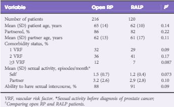
In all, 336 consecutive patients (from nine surgeons) with a mean (SD) age of 64 (11) years had the survey instrument administered (216 underwent open RP and 120 underwent RALP). There were no significant differences in patient age or comorbidity profiles between the two groups. Only 38% of men had an accurate recollection of their nerve-sparing status. The mean (SD) elapsed time after RP at the time of postoperative assessment was 3 (2) months. RALP patients expected a shorter EF recovery time (6 vs 12 months, P = 0.02), a higher likelihood of recovery back to baseline EF (75% vs 50%, P = 0.01), and a lower potential need for intracavernosal injection therapy (4% vs 20%, P = 0.01). Almost half of all patients were unaware that they were rendered anejaculatory by their surgery. None of the RALP patients and only 10% of open RP patients recalled being informed of the potential for penile length loss (P < 0.01) and none were aware of the association between RP and Peyronie’s disease.

Conclusions

Patients who have undergone RP have largely unrealistic expectations about their postoperative sexual function.

Read more articles of the week

Editorial: Managing expectations after radical prostatectomy; time to change

There have been numerous advances in the management of prostate cancer. Developments in imaging, surgical and radiotherapy technology, and pathological grading, have led to improvements in the diagnosis and management of this common malignancy. Such progress has translated to earlier diagnosis and improved disease-specific outcomes.

With improved outcomes comes increased attention on life after treatment. Survivorship in cancer has been an area of increasing focus. The aim; to live as healthy and as good a quality of life for as long as possible after diagnosis, by managing the consequences of the cancer and its treatment. In prostate cancer, the functional impact of surgery on quality of life can be considerable, especially given the falling age at first presentation. The consequences of treatment are often the reason patients’ select one form of therapy over another.

Both sexual function and urinary continence can be significantly affected after radical prostatectomy (RP). Despite numerous consequences of surgery on sexual function, the greatest focus in publications is erectile dysfunction (ED). The quoted incidence in published studies can vary from 20% to 90%, depending on whether the return of ‘normal’ erectile function is classed as the return of spontaneous erections, a ‘return to baseline function’, or functional recovery only with pharmacological assistance. This lack of agreed definition of ED after RP hampers progress by underestimating the impact of surgery and making an uneven playing field when comparing studies.

The tendency for surgeons and studies to focus solely on erectile function, when there are so many changes in sexual function after RP, does not give patients a realistic expectation of the impact of surgery on their life after treatment. Patients should be made aware 100% will experience some change in their sexual function after surgery. Patients should be aware of all the possible risks, including;

  1. ED: All will develop a degree of ED after any RP and should be aware that their risk is dependent on their baseline function, comorbidity, and nerve-spare status. They should be made aware of the protracted time course for recovery, even when the nerves are spared, the need for possible injection therapy, and the possible future dependence on some form of therapy to achieve functional erections in the long-term.
  2. Changes in ejaculation: All patients should be made aware of the loss of ejaculation as a permanent feature, and the impact this will have on their natural fertility. They should also be aware that some develop climacturia, and the possible risk of ejaculatory pain.
  3. Changes to penile size/shape: Patients should be aware of the possible reduction in penile length after RP, and the increased risk of developing Peyronie’s disease after RP, which can further impact their sexual function.

With high profile advances such as the rise of robot-assisted RP (RARP), much has been made of the improved view of local anatomy, and ability to manoeuvre within the confined space. The expectation that this translates to improved functional outcome existed way before any studies had been conducted to show any benefit. It is of no surprise, therefore, that patients’ expectations of outcomes after RP have been unrealistically raised by such technologies.

In this month’s BJUI, Deveci et al. [1] present their survey looking at patient expectations of sexual function after RP. In this study, patients who had undergone RP (open or robot-assisted) in the last 3 months were asked to recall the counselling they had received about possible changes in sexual function preoperatively. The comprehensive approach of this study examined patients’ expectations in all the different facets of sexual function, including erectile function, expected time to full recovery of erections, the possible need for intracavernosal injections (ICI), changes in ejaculation including intensity, pain, and climacturia, and awareness of penile length changes and risk of postoperative Peyronie’s disease.

Compared with those undergoing open RP, patient’s expectations after RARP were greater, with more expecting a shorter recovery time (6 vs 12 months, P = 0.02), a higher expectation of a recovery back to baseline erectile function (75% vs 50%, P = 0.01), and lower expected need for ICI (4% vs 20%, P = 0.01) [1]. This greater expectation of newer technology leads to greater regret, and a greater need to manage expectations preoperatively [1, 2].

In addition to the misconceptions on erectile function, ~50% were unaware of the risk of anejaculation, <10% were aware of changes to penile length, and none were aware of risks of developing Peyronie’s disease after RP.

While the Deveci et al. [1] study does have flaws, primarily the lack of ability to differentiate between what patients were exactly told before RP by the nine different operating surgeons, and what could be recalled after RP, it does highlight an important point. No matter what patients were told before surgery, within 3 months of surgery their recollection and understanding of its possible impact on sexual function was poor. Previous studies have highlighted this disparity between clinician’s recall of discussions on the consequences of surgery and patient recall. In one study, while 100% of clinicians felt they had adequately addressed patients concerns on ED, <30% of patients felt the issues had been adequately addressed [3].

Effective management of patients’ expectations of the possible consequences of RP preoperatively allows for better informed consent, a realistic expectation of outcome and time course for recovery, better compliance with postoperative treatments for ED, and less regret of the initial surgical approach.

Given the limited time in consultations, there is not enough time to address all the possible consequences of surgery in detail. When being diagnosed with cancer, often the last thing on the patients mind is sexual function a year down the line. The more important issue at first is coming to terms with the cancer diagnosis, and just making it through the surgery. In addition, one has to wonder if it is fitting for the oncological surgeon to discuss the functional consequences, possible outcomes and their management when other specialists will manage this in the future. A discussion of sexual consequences of surgery is very different coming from the robotic surgeon, rather than the andrologist who would see them after.

The most ideal approach would be for all patients to see an andrologist and continence specialist before RP or be seen in preoperative ‘survivorship’ seminars, based on discussing possible consequences and optimising functional recovery after treatment. Such seminars should be run by the teams involved in managing sexual function and continence postoperatively. Patients should be given a simple, one page sheet outlining the possible consequences of their intended treatment, be that radiotherapy or surgery, on sexual function and continence. In the same way that patients are given a key contact for their cancer care, they should have access to a key contact for their functional recovery. In addition to follow-up visits with the operating surgeon, focusing on the oncological outcome, a separate follow-up based on functional outcome with an andrologist and continence specialist would focus on functional recovery.

There has been a drive to develop high-volume cancer centres of excellence, with pooled resources to allow excellence in imaging, pathology, as well as surgical and non-surgical treatments. The most utopian approach would see these centres also having andrology and continence specialists focused on the management of all postoperative functional consequences, including the ability to undertake penile implant and artificial sphincter surgery as required.

The progress in developing such an infrastructure has been slow. Research on optimising functional recovery has not been as extensive as the focus on diagnosis and treatment in prostate cancer, which can dominate many urology journals and meetings. This imbalance needs to be addressed, to provide not only the best treatment for prostate cancer, but also the best management of the consequences of treatment, aimed at improving quality of life after surgery.

Read the full article
Majid Shabbir
Guys Hospital, London, UK


References


1 Deveci S, Gotto GT, Alex B, OBrien K, Mulhall JP. A survey of patient


2 Schroeck FR, Krupski TL, Sun L et al. Satisfaction and regret after open


3 Crawford ED, Bennett CL, Stone NN et al. Comparison of perspectives


© 2024 BJU International. All Rights Reserved.