Tag Archive for: TRUS

Posts

Article of the week: Ultrasound characteristics of regions identified as suspicious by MRI predict the likelihood of clinically significant cancer on MRI–ultrasound fusion‐targeted biopsy

Every week, the Editor-in-Chief selects an Article of the Week from the current issue of BJUI. The abstract is reproduced below and you can click on the button to read the full article, which is freely available to all readers for at least 30 days from the time of this post.

In addition to the article itself, there is an editorial written by a prominent member of the urological community, and a video made by the authors. These are intended to provoke comment and discussion and we invite you to use the comment tools at the bottom of each post to join the conversation. 

If you only have time to read one article this week, it should be this one.


The ultrasound characteristics of regions identified as suspicious by magnetic resonance imaging (MRI) predict the likelihood of clinically significant cancer on MRI–ultrasound fusion‐targeted biopsy

Benjamin Press*, Andrew B. Rosenkrantz, Richard Huang and Samir S. Taneja§ 
 
*Rutgers New Jersey Medical School, Newark, NJ, Department of Radiology, Department of Urology, and §Departments of Urology and Radiology, NYU Langone Health, New York, NY, USA
 

Abstract

Objective

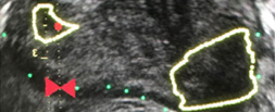
To determine whether the presence of an ultrasound hypoechoic region at the site of a region of interest (ROI) on magnetic resonance imaging (MRI) results in improved prostate cancer (PCa) detection and predicts clinically significant PCa on MRI–ultrasonography fusion‐targeted prostate biopsy (MRF‐TB).

Materials and Methods

Between July 2011 and June 2017, 1058 men who underwent MRF‐TB, with or without systematic biopsy, by a single surgeon were prospectively entered into an institutional review board‐approved database. Each MRI ROI was identified and scored for suspicion by a single radiologist, and was prospectively evaluated for presence of a hypoechoic region at the site by the surgeon and graded as 0, 1 or 2, representing none, a poorly demarcated ROI‐HyR, or a well demarcated ROI‐HyR, respectively. The interaction of MRI suspicion score (mSS) and ultrasonography grade (USG), and the prediction of cancer detection rate by USG, were evaluated through univariate and multivariate analysis.

Results

For 672 men, the overall and Gleason score (GS) ≥7 cancer detection rates were 61.2% and 39.6%, respectively. The cancer detection rates for USGs 0, 1 and 2 were 46.2%, 58.6% and 76.0% (P < 0.001) for any cancer, and 18.7%, 35.2% and 61.1% (P < 0.001) for GS ≥7 cancer, respectively. For MRF‐TB only, the GS ≥7 cancer detection rates for USG 0, 1 and 2 were 12.8%, 25.7% and 52.0%, respectively (P < 0.001). On univariate analysis, in men with mSS 2–4, USG was predictive of GS ≥7 cancer detection rate. Multivariable regression analysis showed that USG, prostate‐specific antigen density and mSS were predictive of GS ≥7 PCa on MRF‐TB.

Conclusions

Ultrasonography findings at the site of an MRI ROI independently predict the likelihood of GS ≥7 PCa, as men with a well‐demarcated ROI‐HyR at the time of MRF‐TB have a higher risk than men without.

Editorial: Is transrectal ultrasonography of the prostate obsolete in the MRI era?

Sampling of prostate tissue to confirm pathologically a clinical suspicion of cancer has undergone an exponential change. The random systematic prostate biopsy technique was the only method used for many decades, initially guided by the finger but, since 1989, performed with TRUS guidance. Now, within the space of only a few years, we have entered the era of performing prostate biopsies on the basis of high‐tech three‐dimensional multiparametric MRI images, including software that can track the exact course of the biopsy needle [1]. While new technical developments in general lead to better, more individually directed healthcare, there is always the risk of abandoning ‘old’ but well developed and extensively tested techniques too soon. In this issue of the BJUI, Press et al. [2] looked at the added value of the presence of an ‘old‐fashioned’ TRUS‐detected lesion in cancer‐suspicious regions on MRI to better predict the presence of clinically significant prostate cancer (csPCa) defined as Gleason score ≥7. In their study comprising 1058 men, it was shown that a well‐demarcated abnormal TRUS finding noted at the time of MRI‐TRUS fusion‐guided prostate biopsy coincides with an increased risk of csPCa detection, independent of MRI suspicion (Prostate Imaging Reporting and Data System [PI‐RADS] score).

Increasing PI‐RADS score is correlated with an increased percentage of csPCa after targeted biopsy, both at initial and repeat biopsy. In a review based on data from 8252 men, it was shown that there is a gradual increase in the detection of csPCa from PI‐RADS 3 to PI‐RADS 4 to PI‐RADS 5 index lesions. For example, at first biopsy, the overall rate of PCa detection and the percentage of csPCa were 39%, 62% and 92% and 54%, 63% and 76% for PI‐RADS 3, 4 and 5 lesions, respectively. This means that in men with PI‐RADS 3 lesions, representing approximately one‐third of men deemed eligible for further assessment, only 39% will be diagnosed with PCa and half of the PCa detected will be potentially indolent Gleason 6 PCa [3]. This makes this group of men extremely interesting for further risk stratification before biopsy. Multivariable risk stratification in which PSA density plays an important role has been shown to be of value in these men [4] but further refinement could potentially be made by including suspicious lesions identified at TRUS.

Apart from the added value of TRUS findings in terms of risk stratification, the performance of the MRI‐targeted biopsy itself could be improved by visual guidance of hypoechoic lesions. In the present study by Press et al [2], a hypoechoic TRUS lesion was present at or near the location of two‐thirds of cancer‐suspicious lesions on MRI. The authors more or less advise to direct the targeted biopsy cores not only to the MRI suspicious lesion, but also the TRUS suspicious lesion, both of which often do not fully overlay in a software‐assisted MRI‐TRUS fusion model. The extent to which this ‘correction for misregistration’ is already included during targeted biopsy in current clinical practice is unknown. Although feasible and seemingly important during software‐assisted fusion targeted biopsy, TRUS lesions in cancer‐suspicious MRI regions might be more frequently targeted during cognitive fusion‐targeted biopsy. Two recent studies underline the important message of the present study, and show that a considerable proportion of csPCa is missed in and around MRI‐suspicious lesions by targeted biopsies, as a result of sampling errors related to both misregistration and intra‐tumour heterogeneity [56]. As suggested by these studies, visual guidance by hypoechoic lesions and ‘focal saturation’ biopsy by additional (peri‐)lesional cores might improve the detection of csPCa.

In summary, ‘good old’ TRUS could be of value in those patients who are virtually always present in scenarios in which a grading system is being used, i.e. patients belonging to the so‐called grey zone. The challenge of risk stratification (i.e. personalized medicine) is to nibble at both sides of the grey zone by implementing new techniques or, more likely by implementing a combination of all available and relevant knowledge.

by Monique J. Roobol, Frank-Jan H. Drost and Arnout R. Alberts

References

  1. Verma, SChoyke, PLEberhardt, SC et al. The current state of MR imaging‐targeted biopsy techniques for detection of prostate cancer. Radiology 201728534356
  2. Press, BRosenkrantz, ABHuang, RTaneja, SSThe ultrasound characteristics of MRI suspicious regions predict the likelihood of clinically significant cancer on MRI‐ultrasound fusion targeted biopsy. BJUI 201912343946.
  3. Schoots, IGMRI in early prostate cancer detection: how to manage indeterminate or equivocal PI‐RADS 3 lesions? Transl Androl Urol 201877082
  4. Alberts, ARSchoots, IGBokhorst, LPLeenders, GJBangma, CHRoobol, MJRisk‐based patient selection for magnetic resonance imaging‐targeted prostate biopsy after negative transrectal ultrasound‐guided random biopsy avoids unnecessary magnetic resonance imaging scans. Eur Urol 201669112934
  5. Simmons, LAMKanthabalan, AArya, M et al. Accuracy of transperineal targeted prostate biopsies, visual estimation and image fusion in men needing repeat biopsy in the PICTURE trial. J Urol 2018200122734
  6. Leest, M, Cornel, EIsrael, B et al. Head‐to‐head comparison of transrectal ultrasound‐guided prostate biopsy versus multiparametric prostate resonance imaging with subsequent magnetic resonance‐guided biopsy in biopsy‐naive men with elevated prostate‐specific antigen: a large prospective multicenter clinical study. Eur Urol 2018; [Epub ahead of print]. https://doi.org/10.1016/j.eururo.2018.11.023.


Editorial: An end to the phenomenon of ‘upgrading’ in early prostate cancer?

The phenomenon of ‘upgrading’ in early prostate cancer is one of those unusual events that is both useful to us on the one hand and undesirable on the other; useful because the phenomenon gifts us a direct measure of the precision of our risk stratification methods for men recently diagnosed, and undesirable because the perfect pathway should, ideally, be free of any upgrading.

Upgrading occurs in a number of settings. We see it at play to some degree when an unreliable test is re-applied in the same subject. The REDUCE study [1] showed us that just under one fifth of men will convert from a status of ‘cancer-free’ to one of ‘cancer-present’ as a result of a second exposure to the same test; that is, TRUS-guided biopsy. We see it in full play when an unreliable test is followed by a more accurate test. Shaw et al. [2] have reminded us once again – as have a number of others – of our limited ability to risk-stratify patients with early prostate cancer. They reported a 50% upgrading when they compared the results of TRUS biopsy against the final pathology at radical prostatectomy. In other words, half the patients went on to their definitive therapy with an incorrect grade attribution [2].

It would be a great pity if, in the modern era, the only route available to patients who wanted to be sure of their risk status was to agree to surgical removal of the prostate. Surely, the value of accurate risk stratification is derived from using it to allocate appropriate and effective care. Risk stratification needs to be linked to or closely follow diagnosis if it is to be put to work for patients.

Nowhere is this need greater than in men whose treatment preference is tissue preservation. The study, in this issue of BJUI, by Pessoa et al. [3] adds to our knowledge on the subject and equips us with a strategy to mitigate some of the errors that are inherent to the standard diagnostic pathway.

In the present study, the authors evaluated the role of a single exposure to MRI (and the opportunity that resulted to undertake a targeted biopsy of an MRI-derived abnormality as well as systematic sampling) in 105 men who had been attributed a diagnosis of low-risk prostate cancer – and, as a result, were deemed to be suitable for active surveillance. The authors used prostate imaging reporting and data system (PIRADS) scoring to interpret and communicate MRI risk. In summary, men attributed a low PIRADS score (PIRADS 1–3) had a low probability of being re-classified to a higher risk. In contrast, men attributed PIRADS score 4 or 5 had a probability of 70–100% of being re-classified. The authors calculated a sensitivity of 93% for MRI to predict ‘re-classification’. This equates to a 93% sensitivity to predict the presence of clinically significant disease as re-classification occurred when there was a transition from low-risk to higher-risk disease.

These results concur with those of others who are working in this area [4] and are in line with current recommendations [5]. One observation that is worth highlighting – because it is a current controversy in the field – relates to the utility of the systematic (or semi-random) biopsies as a component of the confirmatory biopsy. Whilst targeted biopsy was superior to systematic biopsy at identifying clinically significant disease, omission of the systematic biopsies would have resulted in five significant cancers being overlooked. The less perfect the targeted biopsy, the greater the reliance on the systematic. In the present study, the lesion generation and the targeting may have been compromised by one or two issues. Using TRUS biopsy as the authors did (as opposed to transperineal biopsy) to access all areas of the prostate is always going to be a challenge. To do so without image registration makes it even harder. To use PIRADS – as opposed to a Likert scale – as a method of interpreting and communicating MRI outputs will, very likely, lead to an under-reporting of the smaller, high-grade lesions [6]. This is because PIRADS 2.0 is triggered by a volume threshold towards the upper end of the scale. Such lesions might be more prevalent in an apparently ‘low-risk’ population such as the one under scrutiny. If this is the case, they will not be identified as ‘targets’ by virtue of a high PIRADS score. As a consequence they cannot be identified by targeting but might be picked up by the random fall of the needles.

Mark Emberton
Division of Surgery and Interventional Science, University College London, London, UK

Read the full article

References

1 Andriole GL, Bostwick DG, Brawley OW et al. Rittmaster RS; REDUCE Study Group. Effect of dutasteride on the risk of prostate cancer. N Engl Med 2010; 362: 1192202


2 Shaw GL, Thomas BC, Dawson SN et al. Identication of pathologically insignicant prostate cancer is not accurate in unscreened men. Br Cancer 2014; 110: 240511


4 Nassiri N, Margolis DJ, Natarajan S et al. Targeted biopsy to detect Gleason score upgrading during active surveillance for men with low- vs. intermediate-risk prostate cancer. J Urol 2016; [Epub ahead of print]. doi: 10.1016/j.juro.2016.09.070.


5 Moore CM, Giganti F, Albertsen P et al. Reporting magnetic resonance imaging in men on active surveillance for prostate cancer: the PRECISE recommendations-a report of a European school of oncology task force. Eur Urol 2016; [Epub ahead of print]. doi: 10.1016/j.eururo.2016.06.011.


The times they are a-changin’

The other day, as the New York Times was getting excited about Nobel Laureate Bob Dylan new album ‘Triplicate’, I had the opportunity of remembering one of his classic songs. Let me explain. I turned up at the School of Surgery in central London for an academic committee meeting early that morning only to find that it had been cancelled. Due to a IT problem the email with this information never reached me! Rather than brave the London tube again, I decided to walk back to my hospital, which took me past my old hospital which sadly no longer exists.

middlesex-blog

The old hospital in question was The Middlesex Hospital in Mortimer Street, London (Fig.1). The original institution was built in 1745 at Windmill Street and moved in 1757 to Mortimer Street. I arrived there over 20 years ago to train at the Institute of Urology/St. Peter’s Hospital, a highly desirable post amongst surgical residents.

90a2fc7a-3d55-4a12-84b7-873eaa0f18e1

The Middlesex Hospital was closed in 2005 and sold to developers. It now houses swanky apartments and businesses around a beautiful Pearson Square, named after John Loughborough Pearson, who designed the Fitzrovia Chapel (Fig. 2) in 1890 inside the hospital. The Chapel survived the redevelopment as it is a protected building. So did one of the walls of the old hospital along Nassau Street which housed the radiotherapy building (Fig. 3). That facade has been preserved beautifully although there are no patients housed behind it anymore (Fig. 4).

dc533794-189e-4e5e-91b2-0f6cd0d1ea61

So why I am telling you all this? Nostalgia you may say. But in fact much more. The 3 mile walk that morning allowed me to reflect on my own contribution to science and that of two friends who although slightly ahead of me in the training program at The Middlesex Hospital are gentlemen that I greatly admire.

One is Mark Emberton, now Professor at UCL, who has, through the PROMIS study, established the use of MRI prior to prostate biopsies rather than random TRUS biopsies for patients with a raised PSA. The other is David Ralph, an acclaimed Andrologist, who has just published our Priapism Guidelines, a must read for everyone managing this emergency. There is no doubt that both have made significant contributions to British Urology and patient care in the last 20 years during which so many things have changed.

fb6ee749-c241-471c-8e34-6d979aa61a29

As for me, I headed to Queen Square from The Middlesex Hospital, where many years of basic research in a Medical Research Council (MRC) funded lab led to the description of the so called “Dasgupta technique” of injecting Botox into overactive bladders. I was pleasantly surprised to hear that it had made its way into a number of texts including Smith’s Textbook of Endourology.

There are however certain things that do not change much. Next to the Middlesex Hospital, on Cleveland Street was the legendary Ragam’s (Fig. 5), which many would regard as THE go to South Indian restaurant. The masala dosa (pancake with spicy potatoes and hot lentil soup) used to cost £3.95 in 1994; 20 years later the price has gone up by only £2 to £5.95 (Fig. 6), while the quality remains as outstanding as ever.

d14e8835-15c1-4b5d-844e-db6078944ba0

458caf67-1787-450e-bd45-c51f442a23e8

Prokar Dasgupta @prokarurol
Editor-in-Chief, BJUI 


Article of the Week: TRUS-Guided RB PCa Detection – Reasons for Targeted Biopsy Failure

Every Week the Editor-in-Chief selects an Article of the Week from the current issue of BJUI. The abstract is reproduced below and you can click on the button to read the full article, which is freely available to all readers for at least 30 days from the time of this post.

In addition to the article itself, there is an accompanying editorial written by a prominent member of the urological community. This blog is intended to provoke comment and discussion and we invite you to use the comment tools at the bottom of each post to join the conversation.

Finally, the third post under the Article of the Week heading on the homepage will consist of additional material or media. This week we feature a video from Hannes Cash and Patrick Asbach, discussing their paper.

If you only have time to read one article this week, it should be this one.

Prostate cancer detection on transrectal ultrasonography-guided random biopsy despite negative real-time magnetic resonance imaging/ultrasonography fusion-guided targeted biopsy: reasons for targeted biopsy failure

Hannes Cash*, Karsten Gunzel*, Andreas Maxeiner*, Carsten Stephan*, Thomas Fischer, Tahir Durmus, Kurt Miller*, Patrick Asbach, Matthias Haas† and Carsten Kempkensteffen*


*Department of Urology, and Department of Radiology, ChariteUniversity of Medicine Berlin, Berlin, Germany M. H. and C.K. contributed equally to the study.


Read the full article

Objective

To examine the value of additional transrectal ultrasonography (TRUS)-guided random biopsy (RB) in patients with negative magnetic resonance imaging (MRI)/ultrasonography (US) fusion-guided targeted biopsy (TB) and to identify possible reasons for TB failure.

Patients and Methods

We conducted a subgroup analysis of 61 men with prostate cancer (PCa) detected by 10-core RB but with a negative TB, from a cohort of 408 men with suspicious multiparametric magnetic resonance imaging (mpMRI) between January 2012 and January 2015. A consensus re-reading of mpMRI results (using Prostate Imaging Reporting and Data System [PI-RADS] versions 1 and 2) for each suspicious lesion was performed, with the image reader blinded to the biopsy results, followed by an unblinded anatomical correlation of the lesion on mpMRI to the biopsy result. The potential reasons for TB failure were estimated for each lesion. We defined clinically significant PCa according to the Epstein criteria and stratified patients into risk groups according to the European Association of Urology guidelines.

JulAOTW3Results

Results

Our analysis showed that RB detected significant PCa in 64% of patients (39/61) and intermediate-/high-risk PCa in 57% of patients (35/61). The initial mpMRI reading identified 90 suspicious lesions in the cohort. Blinded consensus re-reading of the mpMRI led to PI-RADS score downgrading of 45 lesions (50%) and upgrading of 13 lesions (14%); thus, negative TB could be explained by falsely high initial PI-RADS scores for 32 lesions (34%) and sampling of the target lesion by RB in the corresponding anatomical site for 36 out of 90 lesions (40%) in 35 of 61 patients (57%). Sampling of the target lesion by RB was most likely for lesions with PI-RADS scores of 4/5 and Gleason scores (GS) of ≥7. A total of 70 PCa lesions (67% with GS 6) in 44 patients (72%) were sampled from prostatic sites with no abnormalities on mpMRI.

Conclusion

In cases of TB failure, RB still detected a high rate of significant PCa. The main reason for a negative TB was a TB error, compensated for by positive sampling of the target lesion by the additional RB, and the second reason for TB failure was a falsely high initial PI-RADS score. The challenges that arise for both MRI diagnostics and prostate lesion sampling are evident in our data and support the integration of RB into the TB workflow.

Read more articles of the week

Editorial: MRI-Fusion Biopsy – Behind the Scenes

MRI information of the prostate is increasingly used for improving the diagnostic yield of prostate biopsies [1]. However, increasing complexity of a procedure makes it prone to errors at multiple technical and human levels. Incorporating MRI information and ultrasonography (US) images for MRI-fusion biopsies is a technically challenging task. It involves various steps such as the acquisition and fusion of MRI and US images, the needle guidance during biopsy, and the diligence of the pathological evaluation of biopsy specimens. These different steps and interfaces between different medical professions influence the diagnostic performance of MRI-fusion biopsies.

For example, in daily clinical practice, MRIs from different institutions still harbour a great variance of sequences and reporting, despite the European Society of Urogenital Urology (ESUR) recently introducing acquisition and imaging protocols and a new and advanced version of the Prostate Imaging Reporting and Data System (PIRADS) version 2.0 [2]. The usefulness of such reporting schemes is evidenced by a moderate-to-good interobserver agreement between uro-radiologists for tumour lesion interpretation and corresponding κ values ranging from 0.55 to 0.80 [3]. Important pitfalls of image interpretation are benign lesions such as prostatitis, BPH and fibrosis, which might score similarly to prostate cancer lesions. This problem is further aggravated by a high proportion of patients that receive their first multiparametric MRI (mpMRI) of the prostate in the repeat-biopsy setting with a high burden of post-biopsy artefacts (haemorrhage, capsular irregularity) and lower overall cancer detection rate. Also, during MRI-fusion biopsy patient movement, prostate deformation by the US probe, and mismatch of image planes can lead to a biopsy error exceeding 4 mm. Moreover, targeting error might be aggravated by MRI underestimation of the tumour volume compared with final pathology [4]. After various authors reported the advantages and accuracy of MRI/US-fusion biopsy approaches, Cash et al. [5] address potential reasons for targeted biopsy failure to detect prostate cancer compared with random biopsy. Within their analyses the authors address potential limitations and technical considerations. Based on different technical biopsy strategies (with the patient placed within the MRI scanner (‘in-bore’) vs outside) and different technical approaches, these considerations are very important.

In contrast to cognitive fusion, most MRI/US platforms allow needle tracking by archiving the needle orientation, either by an electromagnetic, image-based or stepper-based mechanism [1]. However, lesion targeting by needle guidance is highly dependent on the dimensions of the primary lesion, numbers of relevant lesions, localisation, and overall prostate volume, making MRI-US fusion and cognitive fusion more error prone (i.e. aiming off the mark with the needle) than in-bore biopsies. Moreover, different technical fusion approaches provide different degrees of manual/automated adjustment tools, with for example either rigid or elastic image transformation to facilitate MRI/US image alignment.

In their analyses, Cash et al. [5] found that 34% of negative targeted biopsies could be explained by initially too high estimated PIRADS scores that were downgraded at re-reading. Interestingly, the remaining lesions were without an mpMRI correlate but within this group 92.9% showed a primary Gleason 3 pattern in biopsy pathology, suggesting a high degree of invisibility on mpMRI. Subanalyses did not show an association of targeted biopsy failures in the ventral location. Therefore, the study by Cash et al. [5] is an important precursor for further analyses to address other underlying reasons for targeted biopsy failure. Moreover, it reveals the need for a tight collaboration of radiologists, urologists, and pathologists as interdisciplinary partners involved in MRI-fusion biopsy. Consequently, the optimal diagnostic performance of MRI-fusion biopsies can only be achieved through standardised MRI performance, reading and reporting of MRI findings, as well as final correlation of MRI findings with histopathological work up.

Read the full article

Lars Budaus and Sami-Ramzi Leyh-Bannurah
Martini-Clinic University Hospital Hamburg-Eppendorf, Hamburg, Germany


References


Video: TRUS-Guided RB Prostate Cancer Detection – Reasons for Targeted Biopsy Failure

Prostate cancer detection on transrectal ultrasonography-guided random biopsy despite negative real-time magnetic resonance imaging/ultrasonography fusion-guided targeted biopsy: reasons for targeted biopsy failure

Hannes Cash*, Karsten Gunzel*, Andreas Maxeiner*, Carsten Stephan*, Thomas Fischer, Tahir Durmus, Kurt Miller*, Patrick Asbach, Matthias Haas† and Carsten Kempkensteffen*


*Department of Urology, and Department of Radiology, ChariteUniversity of Medicine Berlin, Berlin, Germany M. H. and C.K. contributed equally to the study.


Read the full article

Objective

To examine the value of additional transrectal ultrasonography (TRUS)-guided random biopsy (RB) in patients with negative magnetic resonance imaging (MRI)/ultrasonography (US) fusion-guided targeted biopsy (TB) and to identify possible reasons for TB failure.

Patients and Methods

We conducted a subgroup analysis of 61 men with prostate cancer (PCa) detected by 10-core RB but with a negative TB, from a cohort of 408 men with suspicious multiparametric magnetic resonance imaging (mpMRI) between January 2012 and January 2015. A consensus re-reading of mpMRI results (using Prostate Imaging Reporting and Data System [PI-RADS] versions 1 and 2) for each suspicious lesion was performed, with the image reader blinded to the biopsy results, followed by an unblinded anatomical correlation of the lesion on mpMRI to the biopsy result. The potential reasons for TB failure were estimated for each lesion. We defined clinically significant PCa according to the Epstein criteria and stratified patients into risk groups according to the European Association of Urology guidelines.

JulAOTW3Results

Results

Our analysis showed that RB detected significant PCa in 64% of patients (39/61) and intermediate-/high-risk PCa in 57% of patients (35/61). The initial mpMRI reading identified 90 suspicious lesions in the cohort. Blinded consensus re-reading of the mpMRI led to PI-RADS score downgrading of 45 lesions (50%) and upgrading of 13 lesions (14%); thus, negative TB could be explained by falsely high initial PI-RADS scores for 32 lesions (34%) and sampling of the target lesion by RB in the corresponding anatomical site for 36 out of 90 lesions (40%) in 35 of 61 patients (57%). Sampling of the target lesion by RB was most likely for lesions with PI-RADS scores of 4/5 and Gleason scores (GS) of ≥7. A total of 70 PCa lesions (67% with GS 6) in 44 patients (72%) were sampled from prostatic sites with no abnormalities on mpMRI.

Conclusion

In cases of TB failure, RB still detected a high rate of significant PCa. The main reason for a negative TB was a TB error, compensated for by positive sampling of the target lesion by the additional RB, and the second reason for TB failure was a falsely high initial PI-RADS score. The challenges that arise for both MRI diagnostics and prostate lesion sampling are evident in our data and support the integration of RB into the TB workflow.

Read more articles of the week

© 2024 BJU International. All Rights Reserved.