Tag Archive for: urinary incontinence

Posts

Editorial: Penile vibratory stimulation (PVS) a novel approach for penile rehabilitation post nerve sparing radical prostatectomy

The reported incidence of erectile dysfunction (ED) after nerve-sparing radical prostatectomy (NS-RP) varies in the literature from 30 to 80% [1]. This can be explained by the state of neuropraxia which affects the cavernosal nerves, even if the nerves are anatomically intact. During this period there is a lack of nocturnal tumescence which leads to tissue hypoxia and ischaemic damage to the cavernosal smooth muscles leading to smooth muscle necrosis and fibrosis, which in turn causes veno-occlusive dysfunction (VOD). A study by Mulhall et al. [2] showed that, at 12 months after NS-RP, 50% of patients will have VOD and ED. The role of penile rehabilitation, therefore, is to maintain adequate tissue oxygenation until the cavernosal nerves recover with the return of the spontaneous nocturnal tumescence; thus, penile rehabilitation should not be confused with ED treatment. If you see yourself as religious, addiction may make you feel guilty or get you to feel isolated among your friends at your religious organization. A spiritual Christian rehab center in Orlando may be the right choice for you. Not only do you get to meet like-minded people to share your experiences in your journey to sobriety, but the process may also help you to rediscover your faith in God. Legacy Healing Center Tampa offer programs that make spiritual guidance an important part of every type of addiction treatment. Orange County law enforcement has taken steps to make sure the drugs are not as easily available as they once were. This has helped manage Orlando’s drug problem and kept it from turning worse. As important as prevention is to saving lives, however, to the hundreds who are already addicted, rehab is what helps. If you are religious or spiritual, faith-based drug rehab can be the answer to the challenges that you face. It’s important to remember that faith-based rehab only works well for those who are deeply spiritual or religious. Trying faith-based rehab when you are ambivalent about religion can work against you. You may find that you aren’t able to accept what you’re asked to practice, and you may find yourself rebelling. It’s important to choose a treatment approach that you can go along with in good conscience.

Several lines of treatment, including phosphodiesterase 5 inhibitors, intracavernous injection of alprostadil and vacuum pump therapy, have been used in penile rehabilitation but an agreed rehabilitation programme in terms of agents used, timing and duration of therapy does not yet exist [1].

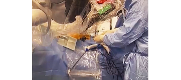
The present study by Fode et al. [3] reports a novel approach to penile rehabilitation using penile vibratory stimulation (PVS). The study looked into the effect of PVS on postoperative erection and continence. The Ferticare® vibrator (Fig. 1) was used at an amplitude of 2 mm and a vibration frequency of 100 Hz and applied to the frenulum once daily, with a sequence consisting of 10 s of stimulation followed by a 10-s rest and repeated 10 times.

The results showed a trend towards better erection in the PVS group (n = 30) compared with the control group (n = 38) as evidenced by the higher International Index of Erectile Function (IIEF) score, but the difference was not significant (P = 0.09). After 1 year, 16 patients (53%) in the PVS group had an IIEF score ≥18 compared with 12 (32%) patients in the control group (P = 0.07). The results did not show any effect of treatment on continence; at 12 months, 90% of the PVS group achieved continence compared with 94.7% of the control group (P = 0.46), although the PVS group had a significantly higher preoperative LUTS score which may explain the results.

The theory postulated is that application of PVS activates the parasympathetic erectile spinal centre (S2–S4), which in turn leads to activation of the cavernosal nerves, enhancing the healing process, and recovery from neuropraxia and restoration of spontaneous erections. Also this would lead to stimulation of the somatic S2–S4 spinal centre, which controls the pelvic floor muscles via the pudendal nerve, leading to the recovery of continence. Although this has been shown in patients with spinal cord injury as the authors mentioned; this may not be the case in post NS-RP with the nerves in a state of neurapraxia, whereas in patients with spinal cord injury the nerves are intact. It would have been of great value to conduct neurophysiological tests on these patients to demonstrate that, despite the cavernosal nerves being in a state of neurapraxia, nerve activity in response to PVS was actually present.

The rehabilitation protocol used in the present study started early but only continued for 6 weeks postoperatively. Studies have shown that the potential recovery time of erectile function after NS-RP is 6–36 months, with the majority recovering within 12–24 months [1,4]. The results might have shown statistical significance in favour of PVS, had treatment continued for a longer period. Starting PVS treatment in the early postoperative period may not be suitable in all patients; in this study six out of 36 patients (16.6%) were non-compliant with the protocol; four had prolonged catheterization and two experienced pain. Furthermore, neurophysiological testing is required to show that in the early postoperative period the cavernosal nerves are actually intact and therefore respond to PVS.

Although the results of the present study did not reach significance, they are encouraging, as there was a trend in favour of treatment with regard to erectile function. Further studies involving larger numbers of patients are warranted to investigate this new line of rehabilitation.

Amr Abdel Raheem* and David Ralph
*Andrology Department, Cairo University Hospital, Cairo, Egypt, and St. Peter’s Andrology Centre, Institute of Urology, London, UK

References

  1. Mulhall JP, Bivalacqua TJ, Becher EF. Standard operating procedure for the preservation of erectile function outcomes after radical prostatectomy. J Sex Med 2013; 10: 195–203
  2. Mulhall JP, Slovick R, Hotaling J et al. Erectile dysfunction after radical prostatectomy: hemodynamic profiles and their correlation with the recovery of erectile function. J Urol 2002; 167: 1371–5
  3. Fode M, Borre M, Ohl D, Lichtbach J, Sønksen J. Penile vibratory stimulation in the recovery of urinary continence and erectile function after nerve-sparing radical prostatectomy: a randomized, controlled trial. BJU Int 2014; 114: 111–7
  4. Rabbani F, Schiff J, Piecuch M et al. Time course of recovery of erectile function after radical retropubic prostatectomy: does anyone recover after 2 years? J Sex Med 2010; 7: 3984–90

Video: Penile vibratory stimulation after radical prostatectomy

Penile vibratory stimulation in the recovery of urinary continence and erectile function after nerve-sparing radical prostatectomy: a randomized, controlled trial

Mikkel Fode*, Michael Borre, Dana A. Ohl, Jonas Lichtbach§ and Jens Sønksen*

*Department of Urology, Herlev University Hospital, Herlev, Department of Urology, Aarhus University Hospital, Aarhus, Denmark, Department of Urology, University of Michigan, Ann Arbor, MI, USA, and §Department of Physiotherapy, Herlev University Hospital, Herlev, Denmark

OBJECTIVE

• To examine the effect of penile vibratory stimulation (PVS) in the preservation and restoration of erectile function and urinary continence in conjunction with nerve-sparing radical prostatectomy (RP).

PATIENTS AND METHODS

• The present study was conducted between July 2010 and March 2013 as a randomized prospective trial at two university hospitals. Eligible participants were continent men with an International Index of Erectile Function-5 (IIEF-5) score of at least 18, scheduled to undergo nerve-sparing RP.

• Patients were randomized to a PVS group or a control group. Patients in the PVS group were instructed in using a PVS device (FERTI CARE® vibrator).

• Stimulation was performed at the frenulum once daily by the patients in their own homes for at least 1 week before surgery. After catheter removal, daily PVS was re-initiated for a period of 6 weeks.

• Participants were evaluated at 3, 6 and 12 months after surgery with the IIEF-5 questionnaire and questions regarding urinary bother. Patients using up to one pad daily for security reasons only were considered continent. The study was registered at https://clinicaltrials.gov/ (NCT01067261).

RESULTS

• Data from 68 patients were available for analyses (30 patients randomized to PVS and 38 patients randomized to the control group).

• The IIEF-5 score was highest in the PVS group at all time points after surgery with a median score of 18 vs 7.5 in the control group at 12 months (P = 0.09), but the difference only reached borderline significance.

• At 12 months, 16/30 (53%) patients in the PVS group had reached an IIEF-5 score of at least 18, while this was the case for 12/38 (32%) patients in the control group (P = 0.07).

• There were no significant differences in the proportions of continent patients between groups at 3, 6 or 12 months. At 12 months 90% of the PVS patients were continent, while 94.7% of the control patients were continent (P = 0.46).

CONCLUSION

• The present study did not document a significant effect of PVS. However, the method proved to be acceptable for most patients and there was a trend towards better erectile function with PVS. More studies are needed to explore this possible effect further.


Article of the month: The race to urinary continence after RP

Every week the Editor-in-Chief selects the Article of the Week from the current issue of BJUI. The abstract is reproduced below and you can click on the button to read the full article, which is freely available to all readers for at least 30 days from the time of this post.

In addition to the article itself, there is an accompanying editorial written by prominent members of the urological community. This blog is intended to provoke comment and discussion and we invite you to use the comment tools at the bottom of each post to join the conversation.

Finally, the third post under the Article of the Week heading on the homepage will consist of additional material or media. This week we feature a video of Inge Geraerts discussing functional outcomes after ORP and RARP.

If you only have time to read one article this week, it should be this one.

Prospective evaluation of urinary incontinence, voiding symptoms and quality of life after open and robot-assisted radical prostatectomy

Inge Geraerts*, Hendrik Van Poppel†, Nele Devoogdt*‡, Ben Van Cleynenbreugel†, Steven Joniau† and Marijke Van Kampen*‡

Departments of *Rehabilitation Science, †Urology and ‡Physical Medicine and Rehabilitation, UZ Leuven, Leuven, Belgium

Read the full article
OBJECTIVE

• To compare functional outcomes, i.e. urinary incontinence (UI), voiding symptoms and quality of life, after open (ORP) and robot-assisted radical prostatectomy (RARP).

PATIENTS AND METHODS

• Between September 2009 and July 2011, 180 consecutive patients underwent radical prostatectomy; of these, 116 underwent ORP and 64 underwent RARP. We prospectively assessed the functional outcomes of each group during the first year of follow-up.

• We measured UI on the 3 days before surgery (24-h pad test) and daily after surgery until total continence, defined as 3 consecutive days of 0 g urine leak, was achieved. Additionally, all patients were assessed before surgery and at 1, 3, 6 and 12 months after surgery using the International Prostate Symptom Score (IPSS) and the King’s Health Questionnaire (KHQ).

• All patients received pelvic floor muscle training until continence was achieved.

• Kaplan–Meier analyses and Cox regression with correction for covariates were used to compare time to continence. A Mann–Whitney U-test was used to assess IPSS and KHQ.

RESULTS

• Patients in the RARP group had a significantly lower D’Amico risk group allocation and underwent more nerve-sparing surgery. Other characteristics were similar.

• Patients in the RARP group regained continence sooner than those in the ORP group (P = 0.007). In the RARP group, the median time to continence (16 vs 46 days, P = 0.026) was significantly shorter and the median amount of first day UI (44 vs 186 g, P < 0.01) was significantly smaller than in the ORP group. After correction for all covariates, the difference remained significant (P = 0.036, hazard ratio [HR] 1.522 (1.027–2.255). In addition, younger men, men with positive surgical margins and men without preoperative incontinence achieved continence sooner.

• A comparison of time to continence between groups with a sufficient number of patients (intermediate risk and/or bilateral nerve-sparing) still showed a faster return of continence after RARP, but the effect decreased in size and was nonsignificant (HR>1.2, P> 0.05).

• Only six patients (two in the RARP and four in the ORP group) still had UI after 1 year.• Patients in the RARP group had significantly better IPSS scores at 1 (P = 0.013) and 3 (P = 0.038) months, and scored better in almost all KHQ aspects.

CONCLUSION

• In this prospective trial, patients treated with RARP tended to regain urinary continence sooner than patients treated with ORP, but in subgroup analyses statistical significance disappeared and effect size decreased dramatically, indicating that the results must be interpreted with caution.


Read Previous Articles of the Week


Editorial: Regaining continence after radical prostatectomy: RARP vs. ORP

Functional outcomes represent relevant criteria to evaluate the success of radical prostatectomy (RP) in the treatment of localised and locally advanced prostate cancer. Indeed, while the primary goal of RP remains the complete extirpation of the primary tumour, patients’ satisfaction can be negatively affected by urinary incontinence and/or erectile dysfunction after RP.

In this issue of BJUI, Geraerts et al. [1] evaluated urinary continence recovery and voiding symptoms in a well-conducted, single-centre, prospective non-randomised study comparing two contemporary series of patients who underwent either open retropubic RP (RRP) or robot-assisted RP (RARP) for clinically localised or locally advanced prostate cancer. Patients were assigned to each group according to their or their surgeon’s preference. High-risk prostate cancers were preferably treated with an open access to offer a more accurate extended lymphadenectomy. The study showed that the urinary continence recovery rate was significantly shorter in the RARP group than in the RRP group (16 vs 46 days; P = 0.008). Interestingly, the RA approach remained an independent predictor of time to urinary continence recovery on multivariable Cox regression analysis (P = 0.03; hazard ratio [HR] 1.52, 95% CI 1.03–2.26). Therefore, this study confirmed previously published results. In 2003, Tewari et al. [2] reported a shorter time to recovery of urinary continence in patients who underwent RARP (44 days) than those who received RRP (160 days). In 2008, Kim et al. [3] reported a median time to continence in RARP patients of 1.6 months, significantly lower than the 4.3 months in the RRP patients. Interestingly, Geraerts et al. [1] identified other independent predictors of time to continence, such as patient’s age >65 years (P = 0.02; HR 0.67, 95% CI 0.45–0.96) and the preoperative continence status (P = 0.004; HR 1.69, 95% CI 1.18–2.43). In all, 28% of patients who received RRP and 34% of those who underwent RARP were preoperatively defined as incontinent using a symptom-specific questionnaire [1]. These patients classifiable as ‘Cx’ according to the Survival, Continence and Potency (SCP) classification [4] represent a confounding population in the Geraerts et al. study who should be evaluated separately.

An interesting question is whether the reported difference in time to continence in favour of RARP is also significant from the clinical perspective. Urinary continence in patients who underwent RARP recovered 1 month early than those treated with traditional RRP. The King’s Health questionnaire seems to confirm a positive effect of this outcome on the patient’s quality of life (QoL). Indeed, there were better results in the RARP compared with RRP group at 1 and 3 months after RP. Moreover, at 12 months after RP, patients who underwent RRP were more physically limited (P = 0.01) and took more precautions to avoid urine loss (P = 0.01) than those who received a RARP [1]. These data seem to be in conflict with the reported overlapping 12-month urinary continence rates (96% in RRP and 97% in RARP group). Moreover, looking at the 12-month urinary continence rate, the Geraerts et al. study does not confirm the results of a recent cumulative analysis of available comparative studies showing a better 12-month urinary continence rate after RARP compared with RRP (odds ratio 1.53; P = 0.03) [1].

Interestingly, the 12-month urinary continence rate reported after RRP by Geraerts et al. is significantly higher (96%) than the values reported in the comparative studies included in the meta-analysis (88.7%) and in the most important and recent RRP non-comparative series (60–93%) [5]. This aspect appears to confirm the important role of surgeon experience. Indeed, in this Belgium series most of the open procedures were performed by an expert surgeon with experience of >3000 RRPs, and thus able to reach excellent functional outcomes for urinary continence recovery. In favour of robotic surgeons, we could consider that they were able to reach overlapping results after <200 cases.

In conclusion, the study published by Geraerts et al. [1] showed that modern RP in expert hands is able to achieve excellent results for urinary continence recovery regardless of the approach. However, pure and RA laparoscopy has pushed open surgeons to improve technical and postoperative aspects to achieve comparable outcomes. RARP can offer some advantages over traditional RRP, above all for the time to reach urinary continence. This advantage seems to have generated a better QoL profile in patients who underwent RARP at 12 months after RP.

However, the choice between the two techniques must be taken according to all the most relevant parameters including perioperative, functional (continence and potency) and oncological outcomes. Therefore, we strongly support the publication of clinical series or comparative studies reporting results according to the ‘trifecta’, ‘pentafecta’ or SCP systems [6].

Vincenzo Ficarra°, Alessandro Iannettiand Alexandre Mottrie
OLV Vattikuti Robotic Surgery Institute, Aalst, Belgium, °Department of Experimental and Clinical Medical Sciences – Urology Unit – School of Medicine, University of Udine, 
and *Department of Surgical, Oncologic and Gastrointestinal Sciences, Padua, Italy

Read the full article

References

  1. Geraerts I, Van Poppel H, Devoogdt N, Van Cleynenbreugel B, Joniau S, Van Kampen M. Prospective evaluation of urinary incontinence, voiding symptoms and quality of life after open and robot-assisted radical prostatectomyBJU Int 2013; 112:936–943
  2. Tewari A, Srivasatava A, Menon M, Members of the VIP Team. A prospective comparison of radical retropubic and robot-assisted prostatectomy: experience in one institutionBJU Int 2003; 92: 205–210
  3. Kim SC, Song C, Kim W et al. Factors determining functional outcomes after radical prostatectomy: robot-assisted versus retropubicEur Urol 2011; 60: 413–419
  4. Ficarra V, Sooriakumaran P, Novara G et al. Systematic review of methods for reporting combined outcomes after radical prostatectomy and proposal of a novel system: the survival, continence, and potency (SCP) classificationEur Urol 2012; 61:541–548
  5. Ficarra V, Novara G, Rosen RC et al. Systematic review and meta-analysis of studies reporting urinary continence recovery after robot-assisted radical prostatectomyEur Urol 2012; 62: 405–417
  6. Ficarra V, Borghesi M, Suardi N et al. Long-term evaluation of survival, continence and potency (SCP) outcomes after robot-assisted radical prostatectomy (RARP)BJU Int 2013; 112: 338–345

Video: Functional outcomes after ORP and RARP

Prospective evaluation of urinary incontinence, voiding symptoms and quality of life after open and robot-assisted radical prostatectomy

Inge Geraerts*, Hendrik Van Poppel, Nele Devoogdt*, Ben Van Cleynenbreugel, Steven Joniau and Marijke Van Kampen*

Departments of *Rehabilitation Science, Urology and Physical Medicine and Rehabilitation, UZ Leuven, Leuven, Belgium

Read the full article
OBJECTIVE

• To compare functional outcomes, i.e. urinary incontinence (UI), voiding symptoms and quality of life, after open (ORP) and robot-assisted radical prostatectomy (RARP).

PATIENTS AND METHODS

• Between September 2009 and July 2011, 180 consecutive patients underwent radical prostatectomy; of these, 116 underwent ORP and 64 underwent RARP. We prospectively assessed the functional outcomes of each group during the first year of follow-up.

• We measured UI on the 3 days before surgery (24-h pad test) and daily after surgery until total continence, defined as 3 consecutive days of 0 g urine leak, was achieved. Additionally, all patients were assessed before surgery and at 1, 3, 6 and 12 months after surgery using the International Prostate Symptom Score (IPSS) and the King’s Health Questionnaire (KHQ).

• All patients received pelvic floor muscle training until continence was achieved.

• Kaplan–Meier analyses and Cox regression with correction for covariates were used to compare time to continence. A Mann–Whitney U-test was used to assess IPSS and KHQ.

RESULTS

• Patients in the RARP group had a significantly lower D’Amico risk group allocation and underwent more nerve-sparing surgery. Other characteristics were similar.

• Patients in the RARP group regained continence sooner than those in the ORP group (P = 0.007). In the RARP group, the median time to continence (16 vs 46 days, P = 0.026) was significantly shorter and the median amount of first day UI (44 vs 186 g, P < 0.01) was significantly smaller than in the ORP group. After correction for all covariates, the difference remained significant (P = 0.036, hazard ratio [HR] 1.522 (1.027–2.255). In addition, younger men, men with positive surgical margins and men without preoperative incontinence achieved continence sooner.

• A comparison of time to continence between groups with a sufficient number of patients (intermediate risk and/or bilateral nerve-sparing) still showed a faster return of continence after RARP, but the effect decreased in size and was nonsignificant (HR>1.2, P> 0.05).

• Only six patients (two in the RARP and four in the ORP group) still had UI after 1 year.• Patients in the RARP group had significantly better IPSS scores at 1 (P = 0.013) and 3 (P = 0.038) months, and scored better in almost all KHQ aspects.

CONCLUSION

• In this prospective trial, patients treated with RARP tended to regain urinary continence sooner than patients treated with ORP, but in subgroup analyses statistical significance disappeared and effect size decreased dramatically, indicating that the results must be interpreted with caution.

Step-by-Step: Robot-assisted AUS insertion


Robot-assisted laparoscopic artificial urinary sphincter insertion in men with neurogenic stress urinary incontinence

David R. Yates, Véronique Phé, Morgan Rouprêt, Christophe Vaessen, Jérôme Parra, Pierre Mozer and Emmanuel Chartier-Kastler

Academic department of Urology, Pitié-Salpétrière Hospital, Assistance Publique-Hôpitaux de Paris (AP-HP), Faculté de Médecine Pierre et Marie Curie, University Paris 6, Paris, France

The first two authors contributed equally to this article.

Read the full article
OBJECTIVES

• To describe for the first time the technique of robot-assisted artificial urinary sphincter (R-AUS) insertion in male patients with neurogenic incontinence.

MATERIALS AND METHODS

• From January 2011 to the present date, six patients with spinal cord injury have undergone R-AUS insertion at our academic institution and we have prospectively collected data on pre-, peri- and early postoperative outcomes.

• A transperitoneal five-port approach was used using a three-arm standard da Vinci® robot (Intuitive Surgical, Sunnyvale, CA, USA) in a 30° reverse Trendelenburg position.

• The artificial urinary sphincter (AUS) cuff was placed circumferentially around the bladder neck, the reservoir was left intra-abdominally in a lateral vesicular space and the pump was placed in a classic scrotal position.

RESULTS

• All six patients had successful robotic implantation of the AUS.

• The median patient age was 51.5 years, the median (range) operating time was 195 (175–250) min with no significant blood loss or intra-operative complications. The median (range) length of hospital stay was 4 (4–6) days.

• At a median (interquartile range) follow-up of 13 (6–21) months, all six patients had a functioning device with complete continence.

• To date, we have observed no incidence of early erosion, device infection or device malfunction.

CONCLUSIONS

• Allowing for the preliminary nature of our data, R-AUS insertion appears safe and technically feasible.

• Larger studies with long-term follow-up and comparison with open AUS insertion are necessary before definitive statements can be made for R-AUS in respect of complications and functional outcomes.

Article of the week: Implanting periurethral myofibres to treat incontinence

Every week the Editor-in-Chief selects the Article of the Week from the current issue of BJUI. The abstract is reproduced below and you can click on the button to read the full article, which is freely available to all readers for at least 30 days from the time of this post.

In addition to the article itself, there is an accompanying editorial written by a prominent member of the urological community. This blog is intended to provoke comment and discussion and we invite you to use the comment tools at the bottom of each post to join the conversation.

Finally, the third post under the Article of the Week heading on the homepage will consist of additional material or media. This week we feature a video of Prof. René Yiou discussing his article.

If you only have time to read one article this week, it should be this one.


Periurethral skeletal myofibre implantation in patients with urinary incontinence and intrinsic sphincter deficiency: a phase I clinical trial

René Yiou1, Jean-Yves Hogrel8, Catherine-Marie Loche2, François-Jerome Authier3, Philippe Lecorvoisier7, Pauline Jouany4, Françoise Roudot-Thoraval5 and Jean-Pascal Lefaucheur6

1Hôpital Henri Mondor, Service d’Urologie and CRCDC, 2Hôpital Albert Chenevier, Service de Médecine Physique et de Réadaptation, 3Hôpital Henri Mondor, Département de Pathologie, 4Hôpital Henri Mondor, Unité de Recherche Clinique (URC), Pôle Recherche Clinique et Santé Publique, 5Hôpital Henri Mondor, Département de Santé Publique, Pôle Recherche Clinique et Santé Publique, UPEC, 6Hôpital Henri Mondor, Service de Physiologie – Explorations Fonctionnelles, Université Paris-Est, Faculté de Médecine, APHP, 7Hôpital Henri Mondor, INSERM, Créteil, and 8Université Paris 6, UMR S974, INSERM U974, CNRS UMR 7215, GH Pitié-Salpêtrière, Institut de Myologie, Paris, France

This open-label nonrandomized phase I clinical trial was registered on clinicalTrials.gov (#NCT00472069)

Read the full article
OBJECTIVES

• To assess the safety of periurethral myofibre implantation in patients with urinary incontinence due to intrinsic sphincter deficiency (ISD).

• To assess the resulting myogenic process and effects on urinary continence.

PATIENTS AND METHODS

• An open-label non-randomised phase I clinical trial was conducted in five men and five women with ISD (mean age, 62.5 years).

• A free muscle strip from the patient’s gracilis muscle was implanted around the urethra as a means to deliver locally myofibres and muscle precursor cells (MPCs).

• Patients were assessed for collection formation and incomplete bladder emptying.

• The maximum urethral closure pressure (MUCP) and concomitant periurethral electromyographic (EMG) activity were recorded before surgery and 1 and 3 months after surgery. Continence was assessed using the 24-h pad test and self-completed questionnaires, for 12 months.

RESULTS

• There were no serious side-effects.

• Continence improved significantly during the 12-month follow-up in four of the five women, including two who recovered normal continence. In the women, MUCP increased two-fold and de novo EMG periurethral activity was recorded. In the men, MUCP and EMG recordings showed similar improvements but the effect on continence was moderate.

• The few patients enrolled could affect these results.

CONCLUSIONS

• This is the first report of a one-step procedure for transferring autologous MPCs via myofibre implantation in patients with ISD.

• EMG and urodynamic assessments showed improvement of periurethral muscle activity.

• Further work is needed to confirm and improve the therapeutic efficiency of this procedure.


Read Previous Articles of the Week


Editorial: A step toward simplicity

Although effective, current treatments of stress urinary incontinence (SUI) can be viewed as palliatives, as they do not regenerate the normal function of the urethral sphincter. Cell therapy pretends to do so.

Mesenchymal-derived stem cells, isolated from bone marrow, striated muscle or fat have been investigated. Transurethral injection of striated muscle-derived stem cells (MDSCs) has been the most studied technique in human. Although results were encouraging, the need for pre-implantation cell expansion to provide a sufficient amount of cells has two major drawbacks. First, the need for a complex long and expensive process of cell biology to treat a patient precludes diffusion of such a technique. Secondly, cell culture and manipulation are known to affect survival and myogenic potential of MDSCs.

After they first demonstrated in pigs, that an autologous graft of a striated muscle piece around the urethra resulted in degeneration of mature myocytes and an activation of satellite cells, which differentiated in well-oriented myotubes with synapses and contractility, for the first time, Yiou et al. report in BJUI clinical data on striated muscle cells used as a source of stem cells to repair the urethral sphincter without cell expansion in a ‘one-time’ procedure.

The step is remarkable and is a step toward simplicity. It has to be kept in mind that SUI pathophysiology does not only suppose a damaged muscle. Denervation is another mechanism as suggested by the observation of longer activation latency in the urethral sphincter before stress, cough or sneeze in incontinent female patients without neurological disease. Urethral fibrosis is implicated in post-prostatectomy SUI or after urethral complication of a first anti-incontinence procedure.

There is still need for an extensive and subtler understanding of the mechanisms leading to different forms of SUI. Cell therapy will find its specific indications among these different forms of SUI. Combined cell therapy approaches (muscle based, fat based and other) may be the way to regenerate the whole function of the urethral sphincter.


Gilles Karsenty*
*Aix-Marseille University, and Department of Urology and Kidney Transplantation, La Conception Hospital, Marseille, France

Read the full article

Video: Treating intrinsic sphincter deficiency with myofibre implantation


Periurethral skeletal myofibre implantation in patients with urinary incontinence and intrinsic sphincter deficiency: a phase I clinical trial

René Yiou1, Jean-Yves Hogrel8, Catherine-Marie Loche2, François-Jerome Authier3, Philippe Lecorvoisier7, Pauline Jouany4, Françoise Roudot-Thoraval5 and Jean-Pascal Lefaucheur6

1Hôpital Henri Mondor, Service d’Urologie and CRCDC, 2Hôpital Albert Chenevier, Service de Médecine Physique et de Réadaptation, 3Hôpital Henri Mondor, Département de Pathologie, 4Hôpital Henri Mondor, Unité de Recherche Clinique (URC), Pôle Recherche Clinique et Santé Publique, 5Hôpital Henri Mondor, Département de Santé Publique, Pôle Recherche Clinique et Santé Publique, UPEC, 6Hôpital Henri Mondor, Service de Physiologie – Explorations Fonctionnelles, Université Paris-Est, Faculté de Médecine, APHP, 7Hôpital Henri Mondor, INSERM, Créteil, and 8Université Paris 6, UMR S974, INSERM U974, CNRS UMR 7215, GH Pitié-Salpêtrière, Institut de Myologie, Paris, France

This open-label nonrandomized phase I clinical trial was registered on clinicalTrials.gov (#NCT00472069)

Read the full article
OBJECTIVES

• To assess the safety of periurethral myofibre implantation in patients with urinary incontinence due to intrinsic sphincter deficiency (ISD).

• To assess the resulting myogenic process and effects on urinary continence.

PATIENTS AND METHODS

• An open-label non-randomised phase I clinical trial was conducted in five men and five women with ISD (mean age, 62.5 years).

• A free muscle strip from the patient’s gracilis muscle was implanted around the urethra as a means to deliver locally myofibres and muscle precursor cells (MPCs).

• Patients were assessed for collection formation and incomplete bladder emptying.

• The maximum urethral closure pressure (MUCP) and concomitant periurethral electromyographic (EMG) activity were recorded before surgery and 1 and 3 months after surgery. Continence was assessed using the 24-h pad test and self-completed questionnaires, for 12 months.

RESULTS

• There were no serious side-effects.

• Continence improved significantly during the 12-month follow-up in four of the five women, including two who recovered normal continence. In the women, MUCP increased two-fold and de novo EMG periurethral activity was recorded. In the men, MUCP and EMG recordings showed similar improvements but the effect on continence was moderate.

• The few patients enrolled could affect these results.

CONCLUSIONS

• This is the first report of a one-step procedure for transferring autologous MPCs via myofibre implantation in patients with ISD.

• EMG and urodynamic assessments showed improvement of periurethral muscle activity.

• Further work is needed to confirm and improve the therapeutic efficiency of this procedure.


Classical Hodgkin’s lymphoma presenting as vesicovaginal fistula

This is a previously unreported presentation of Hodgkin’s lymphoma within the context of what is itself a rare complication of neglected pessary use. 

Authors:

Megan Murdocha;Paul Hiltonb
a) Clinical Research Associate, Newcastle University and Specialist Registrar in Obstetrics & Gynaecology, Northern Deanery
b) Consultant Gynaecologist & Urogynaecologist, Newcastle upon Tyne Hospitals NHS Foundation Trust

Corresponding Author: Paul Hilton, Consultant Gynaecologist & Urogynaecologist, Directorate of Women’s Services, Level 5, Leazes Wing Royal Victoria Infirmary, Newcastle upon Tyne , NE1 4LP, UK.   Email: [email protected]


Introduction


We report the case of a 79-year-old female who presented with acute onset urinary incontinence following the use of a shelf pessary in the management of her vaginal prolapse.  Examination revealed a large vesicovaginal fistula of unusual appearance; subsequent biopsy of the adjacent vaginal tissue confirmed classical Hodgkin’s lymphoma of mixed cellularity type.  This is a previously unreported presentation of Hodgkin’s lymphoma within the context of what is itself a rare complication of neglected pessary use.


Case report


The patient underwent abdominal hysterectomy in her forties and required a subsequent vaginal procedure for prolapse symptoms in her early seventies; as far as can be ascertained this was a conventional prolapse repair, without incorporation of an non-absorbable ‘mesh’ materials.  She developed recurrent prolapse shortly after surgery and this was managed with a shelf pessary.  Her symptoms were so well controlled that she did not attend for further follow-up, and the pessary was left in situ for 7 years without review.
Her past history included osteoporosis and hypertension; her medication at presentation was alendronate (once weekly), calcium carbonate (calcichew)  (once daily) and amlodopine (5mg daily).  She had left hip and left shoulder replacements.
She presented with sudden onset of continuous urinary incontinence having not had any urinary symptoms previously.  There were no reported bowel symptoms.  On examination under anaesthesia the shelf pessary was embedded in the anterior vaginal wall; after removal a 4x5cm vesicovaginal fistula was found extending from the vaginal vault to the level of the mid trigone.  There was third degree prolapse of the vaginal vault.  An intravenous urogram showed no evidence of ureteric obstruction.  An indwelling urethral catheter was inserted and left on free drainage for one month to encourage spontaneous closure.  This made no difference to her symptoms and she was referred to a tertiary urogynaecology unit specialising in fistula repair.  Examination under anaesthesia was repeated in order to plan the route and method of repair.  This revealed a hard necrotic area at the posterior edge of the fistula.  There was no extension into the rectum. (Figure 1a, b & c)


Figure 1a.  Initial view on examination showing prolapse of the bladder wall through the fistula.


Hayden 1a


1b. Posterior edge of fistula with Allis tissue forceps on the vaginal wall around the fistula edges, and a Landon blade retracting the prolapsed bladder wall.


Hayden 1b
1c. Close-up view as in figure 1b.
Hayden 1c


Provisional histology suggested changes secondary to chronic inflammation (consistent with her long-term pessary use), but also raised the possibility of lymphoma.  After discussion it was felt appropriate to proceed with surgery as planned, and vaginal vesicovaginal fistula repair combined with repair of the prolapse was undertaken by colpocleisis incorporating a Martius bulbocavernosus muscle & fat graft from the left labium majus; the ureters were stented during the procedure, and all residual visibly abnormal tissue in the area of the fistula was excised.


Figure 2a.  Cystoscopic appearance of fistula.


Hayden 2


Subsequent histology of the excised tissues was felt to represent an Epstein-Barr virus (EBV) -positive lymphoproliferative disease (LPD), consistent with either ‘LPD in the context of chronic inflammation in an elderly person’ or with a mixed cellularity type of classical Hodgkin’s lymphoma (MCCHL).  The clinical, radiological and histological findings were discussed at two independent specialist haematology/lymphoma MDT groups and the final agreed diagnosis, based on the finding of Reed-Sternberg cells, was of MCCHL.
After discharge from the urogynaecology unit, she was referred to her local haematologist for further imaging and clinical review.  CT images were difficult to interpret in view of the recent surgery, prior hysterectomy and a left total hip replacement.  MRI scan of the pelvis showed a small thickened area of tissue between the rectum and the left side of the bladder, i.e. in the area of the recent surgery, and above the level of the Martius fat graft (see figure 3 a & b).


Figure 3.  MRI images at 3 months postoperative: 3a showing thickened area to left of bladder base (arrowed).


Hayden 3a


3b. showing the Martius labial fat graft passing from left labium majus into the lower vagina (arrowed). 


Hayden 3b


Whilst it was not possible to establish with certainty whether this represented postoperative fibrosis or residual disease, it was felt appropriate to consider this as stage 1AE classical Hodgkin’s lymphoma.  Conventional treatment would have included ABVD chemotherapy (adiamycin, bleomycin, vincristine, dacarbazine) plus involved-field radiotherapy.  However, in a patient of almost 80 years of age, with at worst minimal residual disease, and in the absence of evidence of nodal or distant disease, it was decided to treat with radiotherapy alone.  The patient received external beam radiotherapy to the upper vagina in a dose of 30Gy in 15 fractions.  At subsequent review she was free from symptoms, signs or MRI evidence of disease progression; she remains under 6-monthly surveillance.  Urogynaecology review confirmed a successful fistula and prolapse repair and she has been discharged from follow up.


Discussion


In the UK, fistulae of the female genital tract are uncommon; a recent review of Hospital Episode Statistics suggest that around 120 urogenital fistulae are treated surgically each year.1  The use of mechanical devices (vaginal pessaries) in the management of pelvic organ prolapse (POP) is common.  Generally they are used for women who have not completed their family or those who do not wish to proceed to surgery, or are considered unsuitable for surgery.  Fistula as a result of pessary use has been reported, but is unusual.  A review in 2008 identified 39 major complications associated with pessary use, including eight vesicovaginal fistulas and four rectovaginal fistulas.2  Since the number of women treated by pessary for prolapse is unknown, the ‘fistula risk’ associated with this form of treatment cannot be determined.  In a recent case series of urogenital fistula in the UK, 11/348 (3.2%) were associated with the use of pessaries for the conservative management of POP.3
In 2008 there were 1730 new cases of Hodgkin’s lymphoma in the UK.  The incidence in females is 2.4 per 100,000; in women two peaks occur at the ages of 20-24 and 70-74 years.4  Extra-nodal lymphoma (with the primary disease other than in lymph nodes) may present at any site, and rare cases in the female genital tract have been reported.5, 6  It is said that 25% of malignant lymphomas arise at extra-nodal sites and in only 1% of women with extra-nodal tumours is the genital tract involved.5  This would suggest an incidence of primary genital tract Hodgkin’s of less than 1 per 100,000,000/ year .  The World Health Organisation classification divides Hodgkin’s lymphoma into several pathologic subtypes.7  In 15% the picture is described as classical Hodgkin’s lymphoma of mixed cellularity subtype, as in this case.4  The aetiology of Hodgkin’s lymphoma is unknown but studies have suggested a viral link; 50% of classical Hodgkin’s lymphomas overall, and up to 70% of MCCHL are EBV-positive.8  There is a known association between chronic inflammation and cancer and it is suggested that chronic inflammation triggered by EBV has a role in the aetiology of Hodgkin’s lymphoma.9  In the case presented, the possibility that this arose because of the presence of inflammation related to the long neglected vaginal pessary must be considered.
Conventionally it is recommended that vaginal pessaries are removed and the vagina examined for erosions on a regular basis.  This is often undertaken every 4-6 months although the optimum frequency has not been established and there is no consensus on the interval between pessary changes.10 The authors of the most recent Cochrane review of mechanical devices in the management of pelvic organ prolapse concluded that randomised controlled trials are needed to inform the best ways to manage long-term pessary use.10


Conclusion


A case of classical Hodgkin’s lymphoma presenting as a vesicovaginal fistula in a neglected vaginal pessary user is presented.  Although this is an extremely rare complication of pessary use, it emphasises the importance of regular review, including pessary removal, bimanual examination, and speculum visualisation of the vaginal walls.  With an increasing move to pessary management in primary care by General Practitioners and Practice Nurses, the development and implementation of an appropriate educational package is important.


References


1. Cromwell D, Hilton P. Patterns of care and outcomes of surgical treatment for urogenital fistula among English NHS hospitals between 2000 and 2009. 2012:(submitted).
2. Arias BE, Ridgeway B, Barber MD. Complications of neglected vaginal pessaries: case presentation and literature review. Int Urogynecol J & Pelvic Floor Dysfunct. 2008;19(8):1173-738.
3. Hilton P. Urogenital fistula in the UK – a personal case series managed over 25 years. BJU Int. 2011:(early view).
4. CancerHelp UK. Hodgkin’s Lymphoma.  2012; Available from: https://cancerhelp.cancerresearchuk.org/type/hodgkins-lymphoma/
5. Cheong IJ, Kim SH, Park CM. Primary Uterine Lymphoma: A Case Report. Korean Journal of Radiology. 2000;1(4):223-5.
6. Raggio ML, Bostrom SG, Harden EA. Hodgkin’s lymphoma of the uterus presenting as a refractory pelvic inflammatory disease. A case report. J Repro Med. 1988;33(10):827-30.
7. Swerdlow S. WHO classification of tumours of haematopoietic and lymphoid tissues. 4th ed. Lyon: International Agency for Research on Cancer, World Health Organization. ; 2008.
8. Thomas RK, Re D, Zander T, Wolf J, Diehl V. Epidemiology and etiology of Hodgkin’s lymphoma. Annals of Oncology. 2002;13(suppl. 4):147-52.
9. Khan GG. Epstein-Barr virus, cytokines, and inflammation: A cocktail for the pathogenesis of Hodgkin’s lymphoma? Experimental Hematology. 2006;34(4):399-406.
10. Adams EJ, Thomson AJM, Maher C, Hagen S. Mechanical devices for pelvic organ prolapse in women. Cochrane Database Syst Rev. 2004;Issue 2:Art. No.: CD004010.


Date added to bjui.org: 07/06/2012
DOI: 10.1002/BJUIw-2012-017-web


© 2024 BJU International. All Rights Reserved.