Tag Archive for: urothelial carcinoma

Posts

Article of the week: Impact of blood transfusion during radical cystectomy

Every week the Editor-in-Chief selects the Article of the Week from the current issue of BJUI. The abstract is reproduced below and you can click on the button to read the full article, which is freely available to all readers for at least 30 days from the time of this post.

In addition to the article itself, there is an accompanying editorial written by a prominent member of the urological community. This blog is intended to provoke comment and discussion and we invite you to use the comment tools at the bottom of each post to join the conversation.

Finally, the third post under the Article of the Week heading on the homepage will consist of additional material or media. This week we feature a video from Dr. Kluth discussing his paper.

If you only have time to read one article this week, it should be this one.

Impact of peri-operative blood transfusion on the outcomes of patients undergoing radical cystectomy for urothelial carcinoma of the bladder

Luis A. Kluth1,3, Evanguelos Xylinas1,4, Malte Rieken1,5, Maya El Ghouayel1, Maxine Sun1, Pierre I. Karakiewicz6, Yair Lotan7, Felix K.-H. Chun3, Stephen A. Boorjian8, Richard K. Lee1, Alberto Briganti9 , Morgan Rouprêt10, Margit Fisch3, Douglas S. Scherr1 and Shahrokh F. Shariat1,2,11

1Department of Urology and 2Division of Medical Oncology, Weill Cornell Medical College, New York Presbyterian Hospital, New York, NY, USA, 3Department of Urology, University Medical Centre Hamburg-Eppendorf, Hamburg, Germany, 4Department of Urology, Cochin Hospital, Assistance Publique-Hopitaux de Paris, Paris Descartes University, Paris, France, 5Department of Urology, University Hospital of Basel, Basel, Switzerland, 6Department of Urology, University of Montreal, Montreal, QC, Canada, 7Department of Urology, University of Texas Southwestern Medical Center, Dallas, TX, USA, 8Department of Urology, Mayo Medical School and Mayo Clinic, Rochester, MN, USA, 9Department of Urology, Vita-Salute University, Milan, Italy, 10Department of Urology of la Pitié-Salpétrière, Assistance Publique-Hôpitaux de Paris, University Paris VI, Faculté de Médicine Pierre et Marie Curie, Paris, France, and 11Department of Urology, Medical University of Vienna, Vienna, Austria

L.A.K. and E.X. contributed equally to this work

OBJECTIVE

• To determine the association between peri-operative blood transfusion (PBT) and oncological outcomes in a large multi-institutional cohort of patients undergoing radical cystectomy (RC) for urothelial carcinoma of the bladder (UCB).

PATIENTS AND METHODS

• We conducted a retrospective analysis of 2895 patients treated with RC for UCB.

• Univariable and multivariable Cox regression models were used to analyse the effect of PBT administration on disease recurrence, cancer-specific mortality, and any-cause mortality.

RESULTS

• Patients’ median (interquartile range [IQR]) age was 67 (60, 73) years and the median (IQR) follow-up was 36.1 (15, 84) months.

• Patients who received PBT were more likely to have advanced disease (P < 0.001), high grade tumours (P = 0.047) and nodal metastasis (P = 0.004).

• PBT was associated with a higher risk of disease recurrence (P = 0.003), cancer-specific mortality (P = 0.017), and any-cause mortality (P = 0.010) in univariable, but not multivariable, analyses (P > 0.05).

• In multivariable analyses, pathological tumour stage, pathological nodal stage, soft tissue surgical margin, lymphovascular invasion and administration of adjuvant chemotherapy were independent predictors of disease recurrence, cancer-specific mortality and any-cause mortality (all P values <0.002).

CONCLUSIONS

• Patients with UCB who underwent RC and received PBT had a greater risk of disease recurrence, cancer-specific mortality and any-cause mortality in univariable, but not multivariable, analysis.

• Although the greater need for PBT with more advanced disease is probably caused by a number of factors, including surgical and cancer-related factors, the present analysis showed that the disease characteristics rather than need for PBT led to worse outcomes.


Editorial: Radical cystectomy: how do blood transfusions affect oncological outcomes?

Kluth et al. [1] have conducted a large retrospective study from several institutions in North America and Europe to assess the impact of blood transfusion on oncological outcomes after radical cystectomy (RC) for bladder cancer. The hypothesis for a negative impact of transfusion on oncological outcomes stems from the observation that renal allograft survival is prolonged after pre-transplant blood transfusions because of its immuno-modulatory effects [2]. This finding prompted Gantt [3] to express concern about the possible adverse effects of transfusions in patients being treated for cancer. Since then, there have been numerous publications addressing this issue in various surgical journals including those of urology with conflicting messages.

Sadeghi et al. [4] queried the Columbia University Urologic Oncology Database. This included 638 patients undergoing RC between 1989 and 2010. Of these, 209 (32.8%) received perioperative blood transfusions. On univariate analysis, the number of units transfused was inversely related to overall and cancer-specific survival. However, on multivariate analysis, it did not prove to be an independent predictor of cancer-specific survival.

As the authors highlighted in this paper, Linder et al. [5] reported a large series of patients from the Mayo Clinic, which included 2060 patients undergoing RC over 25 years. Of this large cohort, 1279 (62%) received perioperative blood transfusion with adverse outcomes, not only in terms of overall and cancer-specific mortality, but also postoperative tumour recurrence.

RC is one of the most major surgical procedures performed in urological surgery. The vast majority of patients with bladder cancer requiring RC are in their mid-sixties, overweight and have several co-morbidities. Some of these patients present late and are anaemic at presentation.

Blood loss during open RC varies depending upon surgeons’ experience, patients’ body mass index, disease stage and availability of modern equipment, e.g. LigaSure™ or stapling devices. Blood transfusion may be required because of pre-existing anaemia or excessive blood loss during surgery. Variations exist in thresholds of anaesthesiologists and the surgeons for transfusions. All of these factors account for variation in reported frequency of transfusion rates for this operation and this is well reflected in many large series of RC.

As there are many confounding factors that may influence overall and cancer-specific survival in patients undergoing RC including stage of the disease, histological nature of the tumour, lymph node status and competing co-morbidities, it is very challenging to control for these factors in retrospective series. Hence, prospective well-controlled multicentre studies are the only way forward to answer this question.

While we await robust evidence on the influence of perioperative transfusion on oncological outcomes, several potential options could be explored to avoid homologous blood transfusion. These include preoperative optimisation of haemoglobin levels through iron infusions, administration of erythropoietin where appropriate, and preoperative autologous-banking. Intraoperatively meticulous surgical technique, use of modern devices, e.g. LigaSure/stapler and Cell Savers, could be used to avoid homologous blood transfusion.

Fortunately, these studies aimed at raising awareness of potential risks of transfusions are appearing in the urological literature at a time when urologists are moving away from open to minimally invasive oncological surgery with a steady decline in the need for perioperative blood transfusion. This is one of the important steps in the right direction and will have a major impact on the need for blood transfusion in foreseeable future.

Muhammed S. Khan
Department of Urology, Guy’s Hospital and King’s College London School of Medicine, London, UK


References

  1. Kluth LA, Xylinas E, Rieken M et al. Impact of perioperative blood transfusion on the outcome of patients undergoing radical cystectomy for urothelial carcinoma of the bladderBJU Int 2014; 113: 393–398
  2. Opelz G, Sengar DP, Mickey MR, Terasaki PI. Effect of blood transfusions on subsequent kidney transplantsTransplant Proc 1973; 5: 253–259
  3. Gantt CL. Red blood cells for cancer patientsLancet 1981; 2: 363
  4. Sadeghi N, Badalato GM, Hruby G, Kates M, McKiernan JM. The impact of perioperative blood transfusion on survival following radical cystectomy for urothelial carcinomaCan J Urol 2012; 19: 6443–6449
  5. Linder BJ, Frank I, Cheville JC et al. The impact of perioperative blood transfusion on cancer recurrence and survival following radical cystectomyEur Urol 2013; 63: 839–845

Video: Peri-operative blood transfusion: outcomes in patients with bladder cancer

Impact of peri-operative blood transfusion on the outcomes of patients undergoing radical cystectomy for urothelial carcinoma of the bladder

Luis A. Kluth1,3, Evanguelos Xylinas1,4, Malte Rieken1,5, Maya El Ghouayel1, Maxine Sun1, Pierre I. Karakiewicz6, Yair Lotan7, Felix K.-H. Chun3, Stephen A. Boorjian8, Richard K. Lee1, Alberto Briganti9, Morgan Rouprêt10, Margit Fisch3, Douglas S. Scherr1 and Shahrokh F. Shariat1,2,11

1Department of Urology and 2Division of Medical Oncology, Weill Cornell Medical College, New York Presbyterian Hospital, New York, NY, USA, 3Department of Urology, University Medical Centre Hamburg-Eppendorf, Hamburg, Germany, 4Department of Urology, Cochin Hospital, Assistance Publique-Hopitaux de Paris, Paris Descartes University, Paris, France, 5Department of Urology, University Hospital of Basel, Basel, Switzerland, 6Department of Urology, University of Montreal, Montreal, QC, Canada, 7Department of Urology, University of Texas Southwestern Medical Center, Dallas, TX, USA, 8Department of Urology, Mayo Medical School and Mayo Clinic, Rochester, MN, USA, 9Department of Urology, Vita-Salute University, Milan, Italy, 10Department of Urology of la Pitié-Salpétrière, Assistance Publique-Hôpitaux de Paris, University Paris VI, Faculté de Médicine Pierre et Marie Curie, Paris, France, and 11Department of Urology, Medical University of Vienna, Vienna, Austria

L.A.K. and E.X. contributed equally to this work

OBJECTIVE

• To determine the association between peri-operative blood transfusion (PBT) and oncological outcomes in a large multi-institutional cohort of patients undergoing radical cystectomy (RC) for urothelial carcinoma of the bladder (UCB).

PATIENTS AND METHODS

• We conducted a retrospective analysis of 2895 patients treated with RC for UCB.

• Univariable and multivariable Cox regression models were used to analyse the effect of PBT administration on disease recurrence, cancer-specific mortality, and any-cause mortality.

RESULTS

• Patients’ median (interquartile range [IQR]) age was 67 (60, 73) years and the median (IQR) follow-up was 36.1 (15, 84) months.

• Patients who received PBT were more likely to have advanced disease (P < 0.001), high grade tumours (P = 0.047) and nodal metastasis (P = 0.004).

• PBT was associated with a higher risk of disease recurrence (P = 0.003), cancer-specific mortality (P = 0.017), and any-cause mortality (P = 0.010) in univariable, but not multivariable, analyses (P > 0.05).

• In multivariable analyses, pathological tumour stage, pathological nodal stage, soft tissue surgical margin, lymphovascular invasion and administration of adjuvant chemotherapy were independent predictors of disease recurrence, cancer-specific mortality and any-cause mortality (all P values <0.002).

CONCLUSIONS

• Patients with UCB who underwent RC and received PBT had a greater risk of disease recurrence, cancer-specific mortality and any-cause mortality in univariable, but not multivariable, analysis.

• Although the greater need for PBT with more advanced disease is probably caused by a number of factors, including surgical and cancer-related factors, the present analysis showed that the disease characteristics rather than need for PBT led to worse outcomes.


Diagnosis of a cryptic variant of urothelial carcinoma using blue-light cystoscopy

We report a rare case of nested variant-like urothelial carcinoma diagnosed with the use of hexaminoelvulinate blue-light cystoscopy in a 53-year-old male. The patient presented for fol-low-up with cytology and both white-light and blue-light cystoscopy for previously diagnosed and treated T1 high grade urothelial carcinoma 4 years previously. Cytology was negative for recurrence. Under white-light cystoscopy an unusual flat, pigmented lesion was found. Under blue-light, a suspicious lesion adjacent to the pigmented lesion was discovered. Biopsies of this lesion later revealed nested variant-like urothelial carcinoma. We believe this is the first case re-port describing the incidental findings of such a cryptic form of urothelial carcinoma with the use of blue-light cystoscopy. In the setting of negative cytology and negative random biopsies, this lesion would have gone undiagnosed without the use of this new technology.

Authors: Vollstedt, Annah J; Dahmoush, Laila; O’Donnell, Michael A
Departments of Urology and Pathology, University of Iowa, Iowa City, IA, USA
Corresponding Author: O’Donnell, Michael A


Introduction
Hexaminolevulinate (HAL) blue-light cystoscopy uses photo-active compounds to en-hance the visual demarcation between normal and neoplastic tissue [1]. Endogenous alpha-lipoic acid (ALA ) is a natural precursor to the photoactive intermediate protoporphyrin ester of 5-ALA that induces accumulation of proroporphyrin IX in malignant cells, which fluoresces when exposed to 375–440 nm light [2].
Published trials have established the superior effectiveness of HAL blue-light cytoscopy for tumour detection [3], with an overall sensitivity for detecting urothelial carcinoma in situ (CIS) lesions of 95–97% compared with 58–68% for standard white-light cystoscopy [4-6].

Case Report
A 53-year-old male presented in December 2008 with three small tumours, which were ultimately diagnosed as T1 high grade urothelial carcinoma. He underwent transurethral resection of bladder tumours and completed intravesical chemotherapy with BCG and interferon. He completed the first 3-week maintenance treatment without difficulty and exhibited no evidence of disease upon reassessment in April 2009. In 2010, he experienced ischaemic colitis and sub-sequently underwent colectomy. At this time all further intravesical therapy was stopped. He was lost to follow-up, but re-presented in April 2011. He underwent random biopsies in May 2011, which revealed urothelial CIS of the bladder and was treated again with 6 weeks of BCG, but restaging in August 2011 continued to show urothelial CIS. Although cystectomy was offered, the patient chose an alternative regimen consisting of sequential gemcitabine and docetaxel. Af-ter completing six cycles of gemcitabine and docetaxel, he presented in February 2012 for re-staging with bladder wash cytology, white-light and blue-light cytoscopy, and bladder and pros-tatic urethral biopsies. Cytology was negative. Cystoscopy revealed sequelae of an old bladder tumour replete with vestiges of recent chemotherapy treatment including oedematous edges of a grossly calcified and fibrotic lesion, ~4–5 cm in greatest dimension in an area of previous tumour resection. Another lesion was identified on the left lateral wall of the bladder that was darkly pigmented and flat (Fig. 1).  Directly inferior to this lesion was an area that showed positive un-der blue-light cytoscopy that was not apparent on white-light cystoscopy (Fig. 2). This lesion was biopsied as well as the rim of the previous bladder tumour resection site, followed by random bladder biopsies and prostatic biopsies.

Vollstedt-figure-1

Fig. 1 White-light cystoscopy showing suspicious pigmented lesion.

Vollstedt-figure-2

Fig. 2 Blue-light cystoscopy showing lesion glowing bright pink, adjacent to the pigmented le-sion found under white light.

Pathology of the random bladder biopsies as well as the prostatic biopsies showed no di-agnostic abnormality. The biopsy of the previous resection site showed urothelium with no diag-nostic abnormality, fibrous tissue with necrosis, as well as acute and chronic inflammation and calcifications. The biopsy of the lesion that was positive only on blue-light cytoscopy showed a residual focus of high grade, nested variant-like urothelial carcinoma, invading the lamina pro-pria (Figs 3 and 4). Interestingly, the surface overlying this focus was denuded of urothelium. A review of the patient’s previous biopsy in 2008, which was originally read as having papillary architecture, showed the invasive component in the lamina propria to have a nested architecture, microscopically identical to that seen in the current specimen. The only difference was the ab-sence of the expohytic/papillary component of the tumour. CT of the abdomen and pelvis was unremarkable except for some minor bladder wall thickening. Upon discussing the findings with the patient, he underwent radical cystectomy. Pathological examination of the cystectomy spec-imen showed no residual in situ or invasive urothelial carcinoma.

Vollstedt-figure-3

Fig. 3 Infiltrating nested variant-like urothelial carcinoma, detected by HAL blue-light cystos-copy.

Vollstedt-figure-4
Fig. 4 The patient’s previous high grade papillary urothelial carcinoma with an invasive compo-nent similar to the tumour detected by HAL blue-light cystoscopy.

Discussion
Several studies have described the superior effectiveness of blue-light cytoscopy over white-light cystoscopy. The enhanced detection of inconspicuous lesions has been noted as one of the most valuable and remarkable benefits of HAL blue-light cytoscopy [7], as published data have confirmed the advantages of HAL blue-light cytoscopy over white-light cystoscopy in terms of overall tumour detection rates but especially for urothelial CIS [4,5].
In Europe, blue-light cytoscopy has been used for more than 10 years for the detection of bladder cancer in patients with known bladder cancer or a high suspicion of bladder cancer; however, this technology has yet to be incorporated as standard practice in the USA.
Our patient’s cryptic tumour is described as a nested variant-like urothelial carcinoma because it displays features consistent with the nested variant of urothelial carcinoma. It was also present with papillary architecture in the patient’s biopsies from 2008. Nested variant urothelial carcinoma (NVUC) is a relatively newly recognized type of urothelial carcinoma. To date, ~ 80 cases of NVUC have been formally reported [8], and this variant is estimated to account for 0.3% of all invasive urinary bladder cancers [9]. It is characterized by irregular urothelial nests resembling von Brunn’s nests and, owing to its deceptively innocuous histological appearance, can easily be mistaken for various benign urothelial growths despite its aggressive clinical behaviour. Cytological atypia tends to be more prominent in deeper portions of the tumour, thus the potential for misdiagnosis is further increased when assessing limited or superficial biopsy specimens [10]. Abnormalities of surface that are normally found with bladder lesions such as urothelial CIS are often missing in NVUC.
With conventional white-light cystoscopy, NVUC is usually seen as a slight mucosal ab-normality, diffuse wall thickness or erythematous plaque. Occasionally, the bladder mucosa might have a normal appearance under white light, as was the case in our patient. It also does not usually form a mass. It should also be noted that blue-light cystoscopy was able to prove effec-tive in this case owing to fact that the lesion was relatively shallow. As Figures 3 and 4 show, the urothelium was denuded. While extensive studies of the depth of penetration of HAL used in blue-light cystoscopy have not been performed, elsewhere it has been shown that HAL does not penetrate deeper than the superficial surface of the bladder urothelium [11]. Thus it was possible for the HAL to highlight the nested variant-like urothelial carcinoma with the blue-light cystos-copy in this case.
Studies have also been performed to determine the usefulness of cytology in the diagno-sis of NVUC. In one study performed by Cardillo et al. [12], it was shown that distinct but subtle findings do exist in the cytology of NVUC, such as medium-sized, round or polygonal neoplastic cells with abundant, dense and slightly basophilic cytoplasm with well-defined cell borders. There was an increased nuclear to cytoplastic ratio, nuclear membranes with irregular contours, and nuclei with coarse chromatin with occasional prominent nucleoli; however, the authors contended that a primary diagnosis of NVUC in urine specimens is not recommended given the subtlety of the findings. These findings make it even more important for a new tech-nology, such as blue-light cystoscopy, to help in the detection of these often concealed lesions, as in the case of our patient.

References
1. Stenzl A, Burger M, Fradet Y, et al. Hexaminoelvulinate guided fluorescence cystoscopy reduces recurrence in patients with nonmuscle invasive bladder cancer. J Urol 2010;184:1907–14.
2. Krieg RC, Messmann H, Rauch J, et al: Metabolic characterization of tumor cell-specific protoporphyrin IX accumulation after exposure to 5-aminokevulinic acid in human colon-ic cells. J Photochem Photobiol B 2002;76:518–25.
3. Witjes JA, Redorta JP, Jacqmin D, et al. Hexaminoelvulinate-guided fluorescence cystos-copy in the diagnosis and follow-up of patients with non-muscle invasive bladder cancer: review of the evidence and recommendations. Eur Urol 2010;57:607–14.
4. Schmidbauer J. Witjes F, Schmeller N, et al. Hexvix PCB3101/01 Study Group. Im-proved detection of urothelial carcinoma in situ with hexaminoelevulinate fluorescence cystoscopy. J Urol 2004;171:135–8.
5. Jocham D, Witjes F, Wagner S, et al. Improved detection and treatment of bladder cancer using hexaaminoelevulinate imaging: a prospective, phase III multicenter study. J Urol 2005;174:862–6.
6. Fradet Y, Grossman HB, Gomella L, et al. A comparison of hexaminoelevulinate fluores-cence cystscopy and white light cystoscopy for the detection of carcinoma in situ in pa-tients with bladder cancer: a phase III, multicenter study. J Urol 2007;178:68-73.
7. Thomas K, O’Brien T. Blue-sky in thinking about the blue-light. BJU Int 2009;104; 887–90.
8. Pusztaszeri M, Hauser J, Iselin C, et al. Urothelial carcinoma “nested variant” of renal pelvis and ureter. J Urol 2007;69:778.e15–7.
9. Holmang S, Johansson SL. The nested variant of transitional cell carcinoma—a rare neo-plasm with poor prognosis. Scand J Urol Nephrol 2001;35:102–5.
10. Shanks JH, Iczkowski KA. Divergent differentiation in urothelial carcinoma and other bladder cancer subtypes with selected mimics. Histopathology. 2009 ;54:885–900.
11. Liu JJ, Droller, MJ, Liao, JC. New Optical Imaging Technologies for Bladder Cancer: Considerations and Perspectives. J Urol 2012; 188:361–8
12. Cardillo M, Reuter VE, Lin O. Cytologic features of the nested variant of urothelial car-cinoma: a study of seven cases. Cancer 2003;99:23–7.


Date added to bjui.org: 11/04/2013

DOI: 10.1002/BJUIw-2012-066-web


Article of the Week: Better fit than fat when it comes to radical cystectomy for bladder cancer

Every week the Editor-in-Chief selects the Article of the Week from the current issue of BJUI. The abstract is reproduced below and you can click on the button to read the full article, which is freely available to all readers for at least 30 days from the time of this post.

In addition to the article itself, there is an accompanying editorial written by a prominent member of the urological community. This blog is intended to provoke comment and discussion and we invite you to use the comment tools at the bottom of each post to join the conversation.

If you only have time to read one article this week, it should be this one.

Obesity is associated with worse oncological outcomes in patients treated with radical cystectomy

Thomas F. Chromecki1,2*, Eugene K. Cha1*, Harun Fajkovic1,3, Michael Rink1,4, Behfar Ehdaie1, Robert S. Svatek5, Pierre I. Karakiewicz6, Yair Lotan7, Derya Tilki8, Patrick J. Bastian8, Siamak Daneshmand9,Wassim Kassouf10, Matthieu Durand1, Giacomo Novara11, Hans-Martin Fritsche12, Maximilian Burger12, Jonathan I. Izawa13, Antonin Brisuda14, Marek Babjuk14, Karl Pummer2 and Shahrokh F. Shariat1

1Weill Medical College of Cornell University, New York, NY, USA, 2Medical University Graz, Graz, Austria, 3Landeskrankenhaus St Poelten, St Poelten, Austria, 4University Medical Centre Hamburg-Eppendorf, Hamburg, Germany, 5University of Texas Health Science Center San Antonio, San Antonio, TX, USA, 6University of Montréal, Montréal, QC, Canada, 7University of Texas Southwestern Medical Center, Dallas, TX, USA, 8Ludwig-Maximilians-University Munich, Klinikum Grosshadern, Munich, Germany, 9University of Southern California Keck School of Medicine and Norris Comprehensive Cancer Center, Los Angeles, CA, USA, 10McGill University Health Centre, Montréal, QC, Canada, 11University of Padua, Padua, Italy, 12Caritas St Josef Medical Centre, University of Regensburg, Regensburg, Germany, 13University of Western Ontario, London, ON, Canada, and 14Hospital Motol, 2nd Faculty of Medicine, Charles University, Praha, Czech Republic
*These authors contributed equally.

Read the full article
OBJECTIVE

• To investigate the association between body mass index (BMI) and oncological outcomes in patients after radical cystectomy (RC) for urothelial carcinoma of the bladder (UCB) in a large multi-institutional series.

PATIENTS AND METHODS

• Data were collected from 4118 patients treated with RC and pelvic lymphadenectomy for UCB. Patients receiving preoperative chemotherapy or radiotherapy were excluded.

• Univariable and multivariable models tested the effect of BMI on disease recurrence, cancer-specific mortality and overall mortality.

• BMI was analysed as a continuous and categorical variable (<25 vs 25–29 vs 30 kg/m2).

RESULTS

• Median BMI was 28.8 kg/m2 (interquartile range 7.9); 25.3% had a BMI <25 kg/m2, 32.5% had a BMI between 25 and 29.9 kg/m2, and 42.2% had a BMI 30 kg/m2.

• Patients with a higher BMI were older (P < 0.001), had higher tumour grade (P < 0.001), and were more likely to have positive soft tissue surgical margins (P = 0.006) compared with patients with lower BMI.

• In multivariable analyses that adjusted for the effects of standard clinicopathological features, BMI >30 was associated with higher risk of disease recurrence (hazard ratio (HR) 1.67, 95% confidence interval (CI) 1.46–1.91, P < 0.001), cancer-specific mortality (HR 1.43, 95% CI 1.24–1.66, P < 0.001), and overall mortality (HR 1.81, CI 1.60–2.05, P < 0.001). The main limitation is the retrospective design of the study.

CONCLUSIONS

• Obesity is associated with worse cancer-specific outcomes in patients treated with RC for UCB.

• Focusing on patient-modifiable factors such as BMI may have significant individual and public health implications in patients with invasive UCB.


Read Previous Articles of the Week

Editorial: Obesity is associated with worse oncological outcomes in patients treated with radical cystectomy

Michael R. Abern, Stephen J. Freedland and Brant A. Inman

Division of Urologic Surgery, Duke University Medical Center, Durham, NC, USA

Obesity is a worldwide epidemic: it is estimated over 300 million adults are obese and over 1 billion are overweight. As obesity is a risk factor for cancers and is modifiable, the authors of this report retrospectively analyse the association between body mass index (BMI) and outcomes in a large multinational cohort of bladder cancer patients that underwent radical cystectomy. They found that obese patients were older and more likely to have high-grade tumours. Furthermore, obese patients received inferior lymphadenectomies, had more positive margins, and were less likely to receive adjuvant chemotherapy. The end result is an association between obesity and bladder cancer recurrence, and both cancer-specific and overall mortality.

Although these data suggest that obesity is associated with poor radical cystectomy outcomes, this contrasts with evidence showing no link between obesity and bladder cancer mortality in population-based trials such as the Cancer Prevention Study II, which prospectively followed over 900 000 participants. Why the discrepancy? One possible explanation is the presence of confounding factors and one possible confounder is the presence of type 2 diabetes. In population-based studies that considered both BMI and diabetes, people with diabetes were noted to have an increased risk of developing bladder cancer independent of BMI, whereas the converse was not true. Additionally, diabetes has been associated with recurrence and progression of non-muscle invasive bladder cancer whereas obesity has not. The impact of diabetes was not adequately addressed in the current study.

Other limitations also probably affect the results. In the current study, overweight patients (BMI 25–30) had significantly better cancer-specific survival (hazard ratio 0.80, P = 0.01) than those of ‘normal’ weight (BMI < 25). However, a threshold BMI ≥ 30 has been shown to have poor sensitivity for obesity in elderly populations, with over 25% of patients with BMI under 30 qualifying as obese based on body fat. This may result in an overstatement of the effect of obesity. Conversely, the inclusion of underweight patients (BMI < 18.5) in the ‘normal’ group may underestimate the effect between obesity and outcome, as cachexia may be associated with poor outcomes. Another factor mentioned by the authors is the inferior lymphadenectomies performed in obese patients, which introduces a detection bias for lymph node positivity, the strongest predictor after advanced stage for all of their tested outcomes on multivariate analysis (hazard ratio 2.01–2.33, P < 0.001).

Although the true effect of obesity may be hard to quantify with these data, all would agree that maintaining a non-obese bodyweight will help many disease states with little apparent harm. Patients undergoing neoadjuvant chemotherapy before radical cystectomy have a 3-month window to lose weight and exercise more. This could improve surgical outcomes, and possibly tolerance of chemotherapy. Furthermore, if we can prove that obesity leads to increased bladder cancer recurrence or progression, a window of opportunity may exist when a low-risk tumour is diagnosed. Otherwise, we are left with the eighteenth century wisdom of Benjamin Franklin: ‘An ounce of prevention is worth a pound of cure.’

Read the full article

Laparoscopic heminephrectomy and ureterectomy for lower moiety urothelial carcinoma in a complete duplex right kidney

Here, we present a rare case in which a urothelial carcinoma was located at the lower moiety of a complete duplex right kidney. 

Authors: PEI-YU LIN1, VICTOR CHIA-HSIANG LIN1,2, RENG-HONG WU3,  TSAN-JUNG YU1,2

Department of Urology 1, E-Da Hospital/I-Shou University, Kaohsiung City 2, Department of Radiology3, Chi-Mei Medical Center, Tainan City, Taiwan

Corresponding Author: Victor Chia-Hsiang Lin, M.D., Division of Urology, Department of Surgery, Minimally Invasive Surgical Center, E-Da Hospital/I-Shou University, Kaohsiung City 824,  Taiwan 824.   E-mail: [email protected]


Abstract
Total nephroureterectomy is usually considered to be the gold standard treatment for urothelial carcinoma involving the kidney, where nephron sparing surgery is seldom considered because of the concerns of tumor spillage. However, heminephrectomy with ipsilateral ureterectomy may be performed when the malignancy occurs in a patient with a renal fusion anomaly or in a single moiety of a complete duplex kidney. Here, we present a unique case with urothelial carcinoma in the lower moiety of a complete duplex right kidney. Laparoscopic right heminephrectomy with ureterectomy was performed, after preoperative evaluation with reconstructed 3-dimensional computed tomography angiography and intraoperative navigation with laparoscopic ultrasound.
Complete duplication of the collecting system is an uncommon congenital anomaly, occurring in approximately 1 in 125 individuals. Duplex kidneys can be associated with ectopic ureters, ureteroceles, and vesicoureteric reflux, and cause various clinical manifestations including incontinence, voiding dysfunction and urinary tract infection. A common disease variant in this entity is a ectopic ureteric orifice, associated with a dysplastic poorly functioning upper-pole renal moiety. Heminephrectomy is considered to be the treatment of choice for patients with certain pathologies. Here, we present a rare case in which a urothelial carcinoma was located at the lower moiety of a complete duplex right kidney. The patient underwent laparoscopic heminephrectomy with ureterectomy after thorough preoperative evaluation.


Case Report
An 82-year-old female presented with the chief complaint of intermittent painless gross hematuria for 3 months. Physical examination was unremarkable. Cystoscopy with retrograde pyelography revealed complete duplication of the right collecting system and a 2×2 cm filling defect located over the lower moiety of the duplex right kidney (Fig. 1 A & B).


Figure 1. Retrograde pyelography revealed duplication of the right collecting system. A. Hydronephrosis of the right upper moiety due to external compression indicated by a black arrow. B. The white arrow indicates a filling defect over the lower calyx of the right duplex kidney.


Reconstructed 3-dimensional computed tomography (3-D CT) angiography demonstrated a tumor in the lower moiety renal pelvis of the right duplex kidney and several parapelvic renal cysts compressing the upper moiety, causing hydronephrosis and hydrocalycosis. (Fig. 2 A & B).


Figure 2. A. Coronal view of a computed tomography (CT) scan showing a tumor in the duplex right renal pelvis as indicated by the white arrow. B. Reconstruction CT clearly demonstrated a soft tissue mass over the lower calyx of the duplex right kidney, as indicated by the white arrow.


After discussing the available options with the patient and her family, they agreed for her to undergo laparoscopic right heminephrectomy and ureterectomy.


Diagnostic ureteroscopy showed a large nodular tumor in the lower moiety of the duplex right kidney. Subsequently, a transperitoneal laparoscopic technique was adopted, taking down the mesocolon of the ascending colon from the hepatic flexure to the level of the bifurcation of the right iliac vessels. After deroofing the parapelvic renal cysts, the right renal hilum was carefully exposed and the supplying branches to the lower moiety were skeletonized individually. The arterial branch which supplied the lower moiety was confirmed and clamped using a surgical bulldog clamp. The lower moiety appeared cyanotic when its supplying branch was clamped correctly. The venous branch of the lower moiety was accompanied by the arterial branch and was positively identified. By first clipping and dividing the branches of the right renal artery supplying the lower moiety, a clear line of demarcation between the ischaemic lower moiety and the normal upper moiety was seen. Laparoscopic ultrasound further confirmed the upper margin of the lower moiety calyces, and cutting and division of the moieties were performed precisely and efficiently with electrocautery and ultrasonic shears. Hemostasis of the bare surface of the upper moiety was achieved with laparoscopic free-hand suture and knot-tying. The ureter of the lower moiety was skeletonized to the level of the right iliac vessels, and clips were put on the distal end to prevent extravasation of tumor cells. The specimen was then placed in an endobag. The extravesical bladder cuff excision was performed using an open approach through a right Gibson’s incision. When dissecting into the retroperitoneum, the distal ureter was identified, the endobag in which the lower moiety of the right kidney and its proximal ureteral segment were placed was removed thereafter. Retracting the distal ureter cephalad, dissection was continued distally until the bladder was reached. The bladder was incised just anterior to the ureter and the ureteric lumen was identified. The posterior portion of the ureter was excised and detached from the bladder cuff carefully under vision. The bladder defect was sutured with 1-0 chromic gut. There was no injury to the ureteric orifice of the upper moiety of the right kidney.
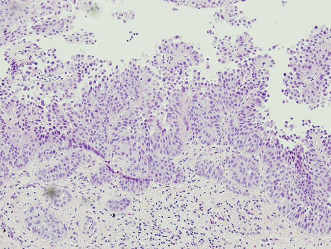
The patient’s postoperative recovery was prompt due to her undergoing minimally invasive surgery, and she resumed oral intake 12 hours postoperatively. She received a total amount of meperidine 150mg for postoperative pain relief. She was discharged after two  days. Due to a suspected urinary leak, a JJ stent was inserted to drain the right upper moiety on postoperative day 6. Final pathological examination demonstrated urothelial carcinoma of the lower moiety of the right duplex kidney, stage pT1. During two-years of follow-up, no tumor recurrence has been detected.


Discussion
Urothelial carcinoma in duplex kidneys is rare with only sporadic reports in the English literature.1,2 Nephron sparing surgery is well established for the treatment of small exophytic renal cell carcinoma.3 However, total nephroureterectomy is the treatment of choice to manage renal urothelial carcinoma. Nevertheless, nephron sparing surgery for upper tract urothelial carcinoma can still be considered in certain situations such as crossed renal ectopy or tumour in one moiety of a duplex system.4 Gur et al. demonstrated the feasibility of separating the involved kidney from its conjoint to treat patients with transitional cell carcinoma in a fused crossed ectopic kidney. Subsequent ureterectomy with bladder cuff excision was performed in our case with respect to oncological principles. We also advocate the importance of a thorough delineation of the involved renal vasculature using CT-angiography preoperatively.
Similarly, we used reconstructed 3-D CT angiography to delineate the involved renal vasculature. A radiologist also performed laparoscopic ultrasound to help decide precisely the cut-margin during the parenchymal transection. We believe these tools are extremely important in planning this type of surgery. To the best of our knowledge, this is the first case of urothelial carcinoma in one moiety of a complete duplex kidney treated by laparoscopic heminephrectomy and ureterectomy.
In our experience, laparoscopic heminephrectomy is a feasible and safe modality in treating urothelial carcinoma of the lower moiety in a duplex kidney in selected cases.


References
1. Lia-Beng Tan, Biing-Rorn Tserng, Wei-Hwang Huang, Chia-Jiuan Tarn. Synchronous bilateral carcinoma of the ureter in association with unilateral incomplete duplication of the ureter. Urol Int 56: 196-199, 1996.
2. Jenq-Daw Li, Johnny Shinn-Nan Lin, Wei-Jen Yao. Synchronous transitional cell carcinoma in both moieties of an incomplete duplex system. Urology 59: 944-945, 2002.
3. Alireza Moinzadeh, Inderbir S. Gill, Antonio Finelli, Jihad Kaouk and Mihir Desai. Laparoscopic partial nephrectomy: 3-year followup. J Urol 175: 459-462, 2006
4. Uri Gur, Ofer Yossepowitch and Jack Baniel. Transitional cell carcinoma in a fused crossed ectopic kidney. Urology 62: 748, 2003.


Date added to bjui.org: 21/07/2012
DOI: 10.1002/BJUIw-2011-108-web


© 2024 BJU International. All Rights Reserved.