Tag Archive for: Article of the Week

Posts

Video: Safety and efficacy of mirabegron as ‘add-on’ therapy in patients with OAB treated with solifenacin

Safety and efficacy of mirabegron as add-on therapy in patients with overactive bladder treated with solifenacin: a postmarketing, open-label study in Japan (MILAI study)

Osamu Yamaguchi, Hidehiro Kakizaki*, Yukio Homma, Yasuhiko Igawa, Masayuki Takeda§, Osamu Nishizawa, Momokazu Gotoh**, Masaki Yoshida††, Osamu Yokoyama‡‡, Narihito Seki§§, Akira Okitsu¶¶, Takuya Hamada¶¶, Akiko Kobayashi¶¶ and Kentarou Kuroishi¶¶


Division of Bioengineering and LUTD Research, School of Engineering, Nihon University, Koriyama, *Department of Urology, Asahikawa Medical University, Asahikawa, Department of Urology, University of Tokyo Graduate School of Medicine, Tokyo, ‡Department of Continence Medicine, University of Tokyo Graduate School of Medicine, Tokyo, §Department of Urology, Interdisciplinary Graduate School of Medicine and Engineering, University of Yamanashi, Chuo, ¶Department of Urology, Shinshu University, Matsumoto, **Department of Urology, Nagoya University Graduate School of Medicine, Nagoya, ††Department of Urology, National Centre for Geriatrics and Gerontology, Obu, ‡‡Department of Urology, University of Fukui Faculty of Medical Sciences, Fukui, §§Department of Urology, Kyushu
Central Hospital of the Mutual Aid Association of Public School Teachers, Fukuoka, and ¶¶Astellas Pharma Inc., Tokyo, Japan


OBJECTIVE

To examine the safety and efficacy of mirabegron as ‘add-on’ therapy to solifenacin in patients with overactive bladder (OAB).

PATIENTS AND METHODS

This multicentre, open-label, phase IV study enrolled patients aged ≥20 years with OAB, as determined by an OAB symptom score (OABSS) total of ≥3 points and an OABSS Question 3 score of ≥2 points, who were being treated with solifenacin at a stable dose of 2.5 or 5 mg once daily for at least 4 weeks. Study duration was 18 weeks, comprising a 2-week screening period and a 16-week treatment period. Patients meeting eligibility criteria continued to receive solifenacin (2.5 or 5 mg once daily) and additional mirabegron (25 mg once daily) for 16 weeks. After 8 weeks of treatment, the mirabegron dose could be increased to 50 mg if the patient’s symptom improvement was not sufficient, if he/she was agreeable to the dose increase, and the investigator judged that there were no safety concerns. Safety assessments included adverse events (AEs), laboratory tests, vital signs, 12-lead electrocardiogram, QT corrected for heart rate using Fridericia’s correction (QTcF) interval and post-void residual (PVR) volume. Efficacy endpoints were changes from baseline in OABSS total score, OAB questionnaire short form (OAB-q SF) score (symptom bother and total health-related quality of life [HRQL] score), mean number of micturitions/24 h, mean number of urgency episodes/24 h, mean number of urinary incontinence (UI) episodes/24 h, mean number of urgency UI episodes/24 h, mean volume voided/micturition, and mean number of nocturia episodes/night. Patients were instructed to complete the OABSS sheets at weeks −2, 0, 8 and 16 (or at discontinuation), OAB-q SF sheets at weeks 0, 8 and 16 (or at discontinuation) and patient voiding diaries at weeks 0, 4, 8, 12 and 16 (or at discontinuation).

RESULTS

Overall incidence of drug-related treatment-emergent AEs (TEAEs) was 23.3%. Almost all TEAEs were mild or moderate. The most common TEAE was constipation, with similar incidence in the groups receiving a dose increase to that observed in the groups maintained on the original dose. Changes in PVR volume, QTcF interval, pulse rate and blood pressure were not considered to be clinically significant and there were no reports of urinary retention. Significant improvement was seen for changes in efficacy endpoints from baseline to end of treatment (EOT) in all groups (patients receiving solifenacin 2.5 or 5 mg + mirabegron 25 or 50 mg).

CONCLUSIONS

Add-on therapy with mirabegron 25 mg once daily for 16 weeks, with an optional dose increase to 50 mg at week 8, was well tolerated in patients with OAB treated with solifenacin 2.5 mg or 5 mg once daily. There were significant improvements from baseline to EOT in OAB symptoms with combination therapy with mirabegron and solifenacin. Add-on therapy with mirabegron and an antimuscarinic agent, such as solifenacin, may provide an attractive therapeutic option.

Article of the Week: Nerve-sparing surgery – In vivo periprostatic nerve tracking using MPM in a rat model

Every Week the Editor-in-Chief selects an Article of the Week from the current issue of BJUI. The abstract is reproduced below and you can click on the button to read the full article, which is freely available to all readers for at least 30 days from the time of this post.

If you only have time to read one article this week, it should be this one.

Real-time in vivo periprostatic nerve tracking using multiphoton microscopy in a rat survival surgery model: a promising pre-clinical study for enhanced nerve-sparing surgery

Matthieu Durand***, Manu Jain*, Amit Aggarwal, Brian D. Robinson*‡, Abhishek Srivastava*, Rebecca Smith, Prasanna Sooriakumaran§, Joyce Loefer**, Chris Pumill††, Jean Amiel**, Daniel Chevallier**, Sushmita Mukherjee† and Ashutosh K. Tewari*


*Department of Urology, Department of Biochemistry, Department of Pathology and Laboratory Medicine, Weill Medical College of Cornell University, New York-Presbyterian Hospital, New York, NY, ††Rutgers Robert Wood Johnson Medical School, New Brunswick, NJ, USA, §Surgical Intervention Trials Unit, University of Oxford, Oxford, UK, ¶Karolinska Institute, Stockholm, Sweden, and **Department of Urology, Hopital Archet 2, Centre Hospitalier Universitaire de Nice, University of Nice Sophia-Antipolis, Nice, France


Read the full article
OBJECTIVES

To assess the ability of multiphoton microscopy (MPM) to visualise, differentiate and track periprostatic nerves in an in vivo rat model, mimicking real-time imaging in humans during RP and to investigate the tissue toxicity and reproducibility of in vivo MPM on prostatic glands in the rat after imaging and final histological correlation study.

MATERIALS AND METHODS

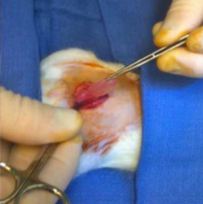
In vivo prostatic rat imaging was carried out using a custom-built bench-top MPM system generating real-time three-dimensional histological images, after performing survival surgery consisting of mini-laparotomies under xylazine/ketamine anaesthesia exteriorising the right prostatic lobe. The acquisition time and the depth of anaesthesia were adjusted for collecting multiple images in order to track the periprostatic nerves in real-time. The rats were then monitored for 15 days before undergoing a new set of imaging under similar settings. After humanely killing the rats, their prostates were submitted for routine histology and correlation studies.

RESULTS

In vivo MPM images distinguished periprostatic nerves within the capsule and the prostatic glands from fresh unprocessed prostatic tissue without the use of exogenous contrast agents or biopsy sample. Real-time nerve tracking outlining the prostate was feasible and acquisition was not disturbed by motion artefacts. No serious adverse event was reported during rat monitoring; no tissue damage due to laser was seen on the imaged lobe compared with the contralateral lobe (control) allowing comparison of their corresponding histology.

CONCLUSIONS

For the first time, we have shown that in vivo tracking of periprostatic nerves using MPM is feasible in a rat model. Development of a multiphoton endoscope for intraoperative use in humans is currently in progress and must be assessed.

Article of the Week: Trifecta and Optimal Peri-operative outcomes of Robotic and Laparoscopic Partial Nephrectomy In Surgical Treatment Of SRMs

Every Week the Editor-in-Chief selects an Article of the Week from the current issue of BJUI. The abstract is reproduced below and you can click on the button to read the full article, which is freely available to all readers for at least 30 days from the time of this post.

In addition to the article itself, there is an accompanying editorial written by a prominent member of the urological community. This blog is intended to provoke comment and discussion and we invite you to use the comment tools at the bottom of each post to join the conversation.

Finally, the third post under the Article of the Month heading on the homepage will consist of additional material or media. This week we feature a video from Dr. Jihad Kaouk discussing his paper. 

If you only have time to read one article this week, it should be this one.

Trifecta and Optimal Peri-operative outcomes of Robotic and Laparoscopic Partial Nephrectomy In Surgical Treatment Of Small Renal Masses: A Multi-Institutional Study


Homayoun Zargar*, Mohamad E. Allaf, Sam Bhayani, Michael Stifelman§, Craig Rogers, Mark W. Ball, Jeffrey Larson
, Susan Marshall§, Ramesh Kumar¶ and Jihad H. Kaouk*


*Cleveland Clinic, Glickman Urological and Kidney Institute, Cleveland, OH, The Johns Hopkins Medical Institutions, James Buchanan Brady Urological Institute, Baltimore, MD, Dept. of Urology, Washington University School of Medicine, St. Louis, MO, §Dept. of Urology, New York University School of Medicine, New York, NY, and Henry Ford Health System, Vattikuti Urology Institute, Detroit, MI, USA


Read the full article
OBJECTIVE

To compare the perioperative outcomes of robotic partial nephrectomy (RPN) with laparoscopic PN (LPN) performed for small renal masses (SRMs), in a large multi-institutional series and to define a new composite outcome measure, termed ‘optimal outcome’ for the RPN group.

PATIENTS AND METHODS

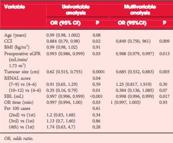
Retrospective review of 2392 consecutive cases of RPN and LPN performed in five high-volume centres from 2004 to mid-2013. We limited our study to SRMs and cases performed by surgeons with significant expertise with the technique. The Trifecta was defined as negative surgical margin, zero perioperative complications and a warm ischaemia time of ≤25 min. The ‘optimal outcome’ was defined as achievement of Trifecta with addition of 90% estimated glomerular filtration rate preservation and no chronic kidney disease stage upgrading. Univariable and multivariable analysis were used to identify factors predicting Trifecta and ‘optimal outcome’ achievement.

RESULTS

In all, 1185 RPN and 646 LPN met our inclusion criteria. Patients in the RPN group were older and had a higher median Charlson comorbidity index and higher R.E.N.A.L. nephrometry score. The RPN group had lower warm ischaemia time (18 vs 26 min), overall complication rate (16.2% vs 25.9%), and positive surgical margin rate (3.2% vs. 9.7%). There was a significantly higher Trifecta rate for RPN (70% vs 33%) and the rate of achievement of ‘optimal outcome’ for the RPN group was 38.5%.

CONCLUSIONS

In this large multi-institutional series RPN was superior to LPN for perioperative surgical outcomes measured by Trifecta. Patients in the RPN group had better outcomes for all three components of Trifecta compared with their LPN counterparts. Our more strict definition for ‘optimal outcome’ might be a better tool for assessing perioperative and functional outcomes after minimally invasive PN. This tool needs to be externally validated.


Editorial: Robotic Partial Nephrectomy: The Treatment of Choice for Minimally Invasive Nephron Sparing Surgery

Early in the adoption of robotic partial nephrectomy (RPN) as an alternative to laparoscopic PN (LPN) for the treatment of small renal masses, several of the current authors presented a similar comparison of LPN and RPN. They found RPN to result in shorter hospital stay, less blood loss, and shorter warm ischaemia time (WIT) compared with LPN [1]. They discovered that RPN outcomes were not dependent on the complexity of the tumour, which clearly impacted LPN results. They concluded that RPN is a safe and viable alternative to LPN and offered benefits even for experienced laparoscopic surgeons.

The current report in this edition of BJUI furthers the comparison of RPN and LPN and expands the assessment to include five high-volume centres of excellence in robotic surgery [2]. This retrospective, multi-institutional review of 1 185 RPN and 646 LPN represents the largest comparison to date of these two approaches for minimally invasive PN. Despite higher patient comorbidities and R.E.N.A.L. nephrometry scores in the RPN patients compared with the LPN group, there were fewer overall complications (16.2% vs 25.9%), a lower positive surgical margin rate (3.2% vs 9.7%) and a lower WIT (18 vs 26 min). They also found a much higher percentage of RPN patients (70% vs 33%) meeting the Trifecta criteria, defined as negative surgical margins, no perioperative complications, and a WIT of ≤25 min. Finally, the authors introduce a more stringent composite measure of ‘optimal outcomes’, which is the Trifecta with the addition of 90% estimated GFR preservation and no chronic kidney disease upgrading. They report 38.5% of RPN patients meeting optimal outcomes compared with 24.1% for LPN.

This study clearly demonstrates the superiority of RPN over LPN and is supported by other single-surgeon reports [3]. These results also exceed those reported for open PN with the added benefit of reduced hospital stay [4]. However, it is important to recognise that these results represent a mature experience with RPN by the leaders in the field of robotic renal surgery. Many of these authors pioneered the techniques currently used for RPN, and therefore these results may not apply to centres without the same experience or case volumes. One limitation of this report is the non-concurrent experience of LPN and RPN. The results of RPN came after an initial experience with LPN and therefore the outcomes of RPN may have benefitted from the lessons learned with LPN prior to RPN.

Reporting surgical outcomes as composite results, such as the Trifecta, allows for comparison between reports and sets an outcomes bar for future studies. Most composite measures include assessment of surgical margin status and complications, but there is no current agreement as to the optimal measure of renal functional outcomes. The current Trifecta used a WIT of ≤25 min as a measure of renal function impact, while the margin, ischaemia, and complications (MIC) score uses a WIT of <20 min [5], and others have used 90% renal parenchyma preservation as part of the Trifecta [6]. The impact of WIT on renal function has been questioned given the recently recognised importance of preserved renal parenchyma as an important predictor of renal function after PN [7]. Until there is consensus as to the best measure of renal function after nephron-sparing surgery, composite outcomes such as the Trifecta and the optimal outcomes as described by the authors will have limited utility.

Read the full article
James Porter
Robotic Surgery, Swedish Medical Center, Seattle , WA, USA
References


Video: Trifecta and Optimal Peri-operative outcomes of Robotic and Laparoscopic Partial Nephrectomy In Surgical Treatment Of SRMs

Trifecta and Optimal Peri-operative outcomes of Robotic and Laparoscopic Partial Nephrectomy In Surgical Treatment Of Small Renal Masses: A Multi-Institutional Study


Homayoun Zargar*, Mohamad E. Allaf, Sam Bhayani, Michael Stifelman§, Craig Rogers, Mark W. Ball, Jeffrey Larson, Susan Marshall§, Ramesh Kumar¶ and Jihad H. Kaouk*


*Cleveland Clinic, Glickman Urological and Kidney Institute, Cleveland, OH, The Johns Hopkins Medical Institutions, James Buchanan Brady Urological Institute, Baltimore, MD, Dept. of Urology, Washington University School of Medicine, St. Louis, MO, §Dept. of Urology, New York University School of Medicine, New York, NY, and Henry Ford Health System, Vattikuti Urology Institute, Detroit, MI, USA


Read the full article
OBJECTIVE

To compare the perioperative outcomes of robotic partial nephrectomy (RPN) with laparoscopic PN (LPN) performed for small renal masses (SRMs), in a large multi-institutional series and to define a new composite outcome measure, termed ‘optimal outcome’ for the RPN group.

PATIENTS AND METHODS

Retrospective review of 2392 consecutive cases of RPN and LPN performed in five high-volume centres from 2004 to mid-2013. We limited our study to SRMs and cases performed by surgeons with significant expertise with the technique. The Trifecta was defined as negative surgical margin, zero perioperative complications and a warm ischaemia time of ≤25 min. The ‘optimal outcome’ was defined as achievement of Trifecta with addition of 90% estimated glomerular filtration rate preservation and no chronic kidney disease stage upgrading. Univariable and multivariable analysis were used to identify factors predicting Trifecta and ‘optimal outcome’ achievement.

RESULTS

In all, 1185 RPN and 646 LPN met our inclusion criteria. Patients in the RPN group were older and had a higher median Charlson comorbidity index and higher R.E.N.A.L. nephrometry score. The RPN group had lower warm ischaemia time (18 vs 26 min), overall complication rate (16.2% vs 25.9%), and positive surgical margin rate (3.2% vs. 9.7%). There was a significantly higher Trifecta rate for RPN (70% vs 33%) and the rate of achievement of ‘optimal outcome’ for the RPN group was 38.5%.

CONCLUSIONS

In this large multi-institutional series RPN was superior to LPN for perioperative surgical outcomes measured by Trifecta. Patients in the RPN group had better outcomes for all three components of Trifecta compared with their LPN counterparts. Our more strict definition for ‘optimal outcome’ might be a better tool for assessing perioperative and functional outcomes after minimally invasive PN. This tool needs to be externally validated.


Article of the Week: External urethral sphincter electromyography and the influence of the menstrual cycle

Every Week the Editor-in-Chief selects an Article of the Week from the current issue of BJUI. The abstract is reproduced below and you can click on the button to read the full article, which is freely available to all readers for at least 30 days from the time of this post.

In addition to the article itself, there is an accompanying editorial written by a prominent member of the urological community. This blog is intended to provoke comment and discussion and we invite you to use the comment tools at the bottom of each post to join the conversation.

Finally, the third post under the Article of the Month heading on the homepage will consist of additional material or media. This week we feature a video from Chris Betts and Kate Burnett discussing their paper. 

If you only have time to read one article this week, it should be this one.

External urethral sphincter electromyography in asymptomatic women and the influence of the menstrual cycle

Cecile Tawadros*, Katherine Burnett*, Laura F. Derbyshire*, Thomas Tawadros†, Noel W. Clarke*‡ and Christopher D. Betts*


*Department of Urology, Salford Royal NHS Foundation Trust, Salford, UK, Department of Urology, Centre Hospitalier Universitaire Vaudois, Lausanne, Switzerland, and Department of Urology, Christie Hospitals NHS Foundation Trust, Manchester, UK


Read the full article
OBJECTIVE

To investigate by electromyography (EMG), the presence of complex repetitive discharges (CRDs) and decelerating bursts (DBs) in the striated external urethral sphincter during the menstrual cycle in female volunteers with no urinary symptoms and complete bladder emptying.

SUBJECTS AND METHODS

Healthy female volunteers aged 20–40 years, with regular menstrual cycles and no urinary symptoms were recruited. Volunteers completed a menstruation chart, urinary symptom questionnaires, pregnancy test, urine dipstick, urinary free flow and post-void ultrasound bladder scan. Exclusion criteria included current pregnancy, use of hormonal medication or contraception, body mass index of >35 kg/m2, incomplete voiding and a history of pelvic surgery. Eligible participants underwent an external urethral sphincter EMG, using a needle electrode in the early follicular phase and the mid-luteal phase of their menstrual cycles. Serum oestradiol and progesterone were measured at each EMG test.

RESULTS

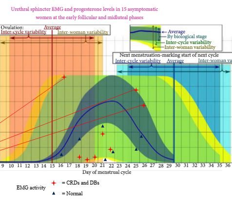
In all, 119 women enquired about the research and following screening, 18 were eligible to enter the study phase. Complete results were obtained in 15 women. In all, 30 EMG tests were undertaken in the 15 asymptomatic women. Sphincter EMG was positive for CRDs and DBs at one or both phases of the menstrual cycle in eight (53%) of the women. Three had CRDs and DBs in both early follicular and mid-luteal phases. Five had normal EMG activity in the early follicular phase and CRDs and DBs in the mid-luteal phase. No woman had abnormal EMG activity in the early follicular phase and normal activity in the luteal phase. There was no relationship between EMG activity and age, parity or serum levels of oestradiol and progesterone.

CONCLUSIONS

CRDs and DB activity in the external striated urethral sphincter is present in a high proportion of asymptomatic young women. This abnormal EMG activity has been shown for the first time to change during the menstrual cycle in individual women. CRDs and DBs are more commonly found in the luteal phase of the menstrual cycle. The importance of CRDs and DBs in the aetiology of urinary retention in young women remains uncertain. The distribution and or quantity of abnormal EMG activity in the external urethral sphincter may be important. In a woman with urinary retention the finding of CRDs and DBs by needle EMG does not automatically establish Fowler’s syndrome as the explanation for the bladder dysfunction. Urethral pressure profilometry may be helpful in establishing a diagnosis. Opiate use and psychological stress should be considered in young women with urinary retention.

Editorial: Do ‘whale noises’ help in the diagnosis of Fowler’s syndrome?

In 1985, Clare Fowler described the presence of abnormal electromyography (EMG) signals in the urethral sphincter of five women with unexplained urinary retention [1]. The presence of complex repetitive discharges (CRD) and decelerating bursts (DB) in women with urinary retention became an important diagnostic finding. Initially, it was described as an EMG finding resembling pseudomyotonia (delayed relaxation of striated muscle when deep tendon reflexes are elicited). Later, an association with polycystic ovary syndrome was hypothesized [2]. This hypothesis could not be proven, however, and the diagnostic criteria that were used at that time for diagnosing polycystic ovaries did not stand the test of time.

The main achievement of urethral sphincter EMG in women with urinary retention was the fact that, from that point onwards, women who were previously described and as being hysterical could now finally be assured that there was a measurable abnormality within their urethral sphincter. This was a major breakthrough in the management of women with urinary retention. Previously, most of these women ended up with permanent catheters or intermittent catheterization, while being stigmatized as psychiatric patients.

The presence of this EMG abnormality also seemed to correlate with the long-term outcome of sacral nerve stimulation [3]. Sacral nerve stimulation is now the standard of care for women with Fowler’s syndrome.

Many authors have questioned the value of these EMG findings. Ramm et al. [4] found the presence of CRD in 30% of healthy women and Tawadros et al. [5] also found this in 53% of healthy female volunteers. These authors showed that CRD and DB are mostly present during the luteal phase of the menstrual cycle. These results suggest at least some hormonal influence on the EMG findings. Fitzgerald et al. [6] showed that CRD correlated weakly with a history of strained voiding and urethral dilation, suggesting a higher prevalence in women with signs of obstructive voiding. Currently, however, the presence of CRD and DB is considered as non-specific.

While sphincter overactivity is generally accepted as the underlying aetiology in women with urinary retention, proving this overactivity remains a challenge. Urethral pressure profiles and urethral volume measurements by ultrasonography have a high interobserver variability and must be used with caution. Most publications originate from one centre and have not been reproduced by others [7]. MRI measurements of the urethral sphincter could possibly be helpful in the future.

While in healthy women CRD can be present, more research will be needed to establish the real value of sphincter EMG in women with pathological urinary retention. The design of good clinical trials will be difficult because of the limited number of patients with this condition. Currently, the diagnosis of Fowler syndrome remains a clinical one, based on a multimodal assessment of the patient.

Read the full article
Dirk De Ridder
Department of Urology, University Hospitals KU Leuven, Leuven, Belgium


References


2 Fowler CJ, Christmas TJ, Chapple CR, Parkhouse HF, Kirby RS, Jacobs HS. Abnormal electromyographic activity of the urethral sphincter, voiding dysfunction, and polycystic ovaries: a new syndrome? BMJ 1988; 297: 14368


4 Ramm O, Mueller ER, Brubaker L, Lowenstein L, Kenton K. Complex repetitive dischargesa feature of the urethral continence mechanism or pathological nding? J Urol 2012; 187: 21403


5 Tawadros C, Burnett K, Derbyshire LF, Tawadros T, Clarke NW, Betts CD. External urethral sphincter electromyography in asymptomatic women and the inuence of the menstrual cycle. BJU Int 2015; 42331


6 FitzGerald MP, Blazek B, Brubaker L. Complex repetitive discharges during urethral sphincter EMG: clinical correlates. Neurourol Urodyn 2000; 19: 57783


7 Wiseman OJ, Swinn MJ, Brady CM, Fowler CJ. Maximum urethral closure pressure and sphincter volume in women with urinary retention. J Urol 2002; 167: 134851; discussion 13512.


Video: External urethral sphincter electromyography and the influence of the menstrual cycle

External urethral sphincter electromyography in asymptomatic women and the influence of the menstrual cycle

Cecile Tawadros*, Katherine Burnett*, Laura F. Derbyshire*, Thomas Tawadros†, Noel W. Clarke*‡ and Christopher D. Betts*


*Department of Urology, Salford Royal NHS Foundation Trust, Salford, UK, Department of Urology, Centre Hospitalier Universitaire Vaudois, Lausanne, Switzerland, and Department of Urology, Christie Hospitals NHS Foundation Trust, Manchester, UK


Read the full article
OBJECTIVE

To investigate by electromyography (EMG), the presence of complex repetitive discharges (CRDs) and decelerating bursts (DBs) in the striated external urethral sphincter during the menstrual cycle in female volunteers with no urinary symptoms and complete bladder emptying.

SUBJECTS AND METHODS

Healthy female volunteers aged 20–40 years, with regular menstrual cycles and no urinary symptoms were recruited. Volunteers completed a menstruation chart, urinary symptom questionnaires, pregnancy test, urine dipstick, urinary free flow and post-void ultrasound bladder scan. Exclusion criteria included current pregnancy, use of hormonal medication or contraception, body mass index of >35 kg/m2, incomplete voiding and a history of pelvic surgery. Eligible participants underwent an external urethral sphincter EMG, using a needle electrode in the early follicular phase and the mid-luteal phase of their menstrual cycles. Serum oestradiol and progesterone were measured at each EMG test.

RESULTS

In all, 119 women enquired about the research and following screening, 18 were eligible to enter the study phase. Complete results were obtained in 15 women. In all, 30 EMG tests were undertaken in the 15 asymptomatic women. Sphincter EMG was positive for CRDs and DBs at one or both phases of the menstrual cycle in eight (53%) of the women. Three had CRDs and DBs in both early follicular and mid-luteal phases. Five had normal EMG activity in the early follicular phase and CRDs and DBs in the mid-luteal phase. No woman had abnormal EMG activity in the early follicular phase and normal activity in the luteal phase. There was no relationship between EMG activity and age, parity or serum levels of oestradiol and progesterone.

CONCLUSIONS

CRDs and DB activity in the external striated urethral sphincter is present in a high proportion of asymptomatic young women. This abnormal EMG activity has been shown for the first time to change during the menstrual cycle in individual women. CRDs and DBs are more commonly found in the luteal phase of the menstrual cycle. The importance of CRDs and DBs in the aetiology of urinary retention in young women remains uncertain. The distribution and or quantity of abnormal EMG activity in the external urethral sphincter may be important. In a woman with urinary retention the finding of CRDs and DBs by needle EMG does not automatically establish Fowler’s syndrome as the explanation for the bladder dysfunction. Urethral pressure profilometry may be helpful in establishing a diagnosis. Opiate use and psychological stress should be considered in young women with urinary retention.

Article of the Month: Patient reported “ever had” and “current” long term physical symptoms following prostate cancer treatments

Every Month the Editor-in-Chief selects the Article of the Month from the current issue of BJUI. The abstract is reproduced below and you can click on the button to read the full article, which is freely available to all readers for at least 30 days from the time of this post.

In addition to the article itself, there is an accompanying editorial written by a prominent member of the urological community. This blog is intended to provoke comment and discussion and we invite you to use the comment tools at the bottom of each post to join the conversation.

Finally, the third post under the Article of the Month heading on the homepage will consist of additional material or media. This week we feature a video from Dr. Anna Gavin discussing his paper. 

If you only have time to read one article this week, it should be this one.

Patient reported “ever had” and “current” long term physical symptoms following prostate cancer treatments.

Anna T. Gavin, Frances J. Drummond*, Conan Donnelly, Eamonn O’Leary*, Linda Sharp† and Heather R. Kinnear

Northern Ireland Cancer Registry, Centre for Public Health, Queen’s University Belfast, Mulhouse Building, Belfast Northern Ireland, UK, *National Cancer Registry Ireland, Building 6800, Airport Business Park Cork, Ireland, and †Institute of Health and Society, Newcastle University, Richardson Road, Newcastle upon Tyne, NE2 4AX, England, UK


Read the full article
OBJECTIVE

To investigate the prevalence of physical symptoms that were ‘ever’ and ‘currently’ experienced by survivors of prostate cancer at a population level, to assess burden and thus inform policy to support survivors.

PATIENTS AND METHODS

The study included 3 348 men surviving prostate cancer for 2–18 years after diagnosis. A cross-sectional, postal survey of 6 559 survivors diagnosed 2–18 years ago with primary, invasive prostate cancer (ICD10-C61) identified via national, population-based cancer registries in Northern Ireland and Republic of Ireland. Questions included symptoms at diagnosis, primary treatments and physical symptoms (erectile dysfunction [ED]/urinary incontinence [UI]/bowel problems/breast changes/loss of libido/hot flashes/fatigue) experienced ‘ever’ and at questionnaire completion (‘current’). Symptom proportions were weighted by age, country and time since diagnosis. Bonferroni corrections were applied for multiple comparisons.

RESULTS

Adjusted response rate 54%; 75% reported at least one ‘current’ physical symptom (‘ever’ 90%), with 29% reporting at least three. Prevalence varied by treatment. Overall, 57% reported current ED and this was highest after radical prostatectomy (RP, 76%) followed by external beam radiotherapy with concurrent hormone therapy (HT, 64%). UI (overall ‘current’ 16%) was highest after RP (‘current’ 28%; ‘ever’ 70%). While 42% of brachytherapy patients reported no ‘current’ symptoms, 43% reported ‘current’ ED and 8% ‘current’ UI. ‘Current’ hot flashes (41%), breast changes (18%) and fatigue (28%) were reported more often by patients on HT.

CONCLUSION

Symptoms after prostate cancer treatment are common, often multiple, persist long-term and vary by treatment method. They represent a significant health burden. An estimated 1.6% of men aged >45 years are survivors of prostate cancer and currently experiencing an adverse physical symptom. Recognition and treatment of physical symptoms should be prioritised in patient follow-up. This information should facilitate men and clinicians when deciding about treatment as differences in survival between radical treatments is minimal.

Editorial: Hot topic of cancer survivorship and the ‘seven deadly sins’

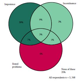
Cancer survivorship has become a hot topic as overall mortality for most cancer patients continues to decrease, the worldwide population continues to age and as patients become more information savvy [1-3]. Gavin et al. [4] provide a data-rich population-based patient survey of seven of the most common physical symptoms after prostate cancer treatment. While we, as urologists and prostate cancer providers, may not be able to recount the seven deadly sins or the seven dwarfs, we do know these seven symptoms: impotence; incontinence; bowel problems; fatigue; hot flushes; loss of libido; and breast symptoms. Urological surgeons and radiation oncologists talk to patients every day about the ‘big three’ of these: impotence, incontinence and bowel problems. Gavin et al. provide the striking statistic that ~1.6% of the male population over the age of 45 years is a prostate cancer survivor currently living with one of the seven.

The paper describes mailed survey results from a population-based cohort of 3 348 prostate cancer survivors 2–15 years after diagnosis with a response rate of 54%. The average age of respondents was 64.9 years, 64% had localized disease at presentation, 65% had Gleason 5–7 disease, and 48, 32 and 20% were surveyed 2–4.9, 5–9.9 and >10 years after diagnosis, respectively. The paper is chock full of descriptive statistics about rates of past and ongoing side effects of the various treatments and essentially has ‘something for everyone’. For example, at baseline before treatment, 51.2% of respondents reported urinary frequency, 18.8% reported impotence and 14.7% reported loss of libido. These data may be useful for estimating population-based general men’s health disease. After treatment, radical prostatectomy (RP) had the highest rates of impotence (76% current) and incontinence (current 28%; ever 70%); however, the authors examined radiation plus hormonal therapy and found impotence rates of 64% and rates of hot flushes, breast changes and bowel problems in the 20–27% range. Table 3 and Figs 3 and 4 in the paper are particularly useful to further examine the seven side effects with treatment.

On the one hand, these data could be useful in educating patients about treatment options for prostate cancer and what they might expect should they choose one treatment over another. Ideally, this education would occur in the multidisciplinary clinic setting [5]. On the other hand, these data could also be used in the wrong way. For example, an aggressive surgeon could selectively present the ‘deadly downsides’ of radiation while downplaying the ‘surgical sins’, whereas a radiation oncologist could do just the opposite to try to influence his or her patients. This highlights the limitations of the present study. While the authors are to be congratulated for a wonderful population-based survey, no control group was surveyed and, more importantly, the authors do not address satisfaction and regret. In other words, the seven side effects must be placed into the patient’s overall satisfaction regarding cancer control and the patient’s ‘trade-offs individualized internal assessment’. For example, our group examined satisfaction and regret after open and robot-assisted RP, finding an ~80–85% satisfaction rate despite levels of impotence and incontinence slightly lower but similar to those in the present population-based survey [6]. While patients who underwent open RP enjoyed more satisfaction and less regret, we attributed much of this to the ‘used car salesman’ approach to ‘selling’ robot-assisted RP in the last decade [7]. In other words, we hypothesized that patients undergoing robot-assisted RP were misled into believing the robot would lessen or eliminate the surgical sins while those undergoing open RP were counselled more realistically. Also, we found that in multivariable analysis, African-American patients exhibited more regret [6]. These data point to the fact that the present study from Ireland may not be applicable to other populations, particularly those with a mixed or different ethnic make-up. Another limitation to population-based data is the impact of centres of excellence and highly experienced treatment providers. The impact of high-volume surgeons/providers on treatment outcomes is now being recognized as a critical variable that is rarely accounted for in case series, multicentre studies or population data as seen here.

Overall, Gavin et al. are to be commended for a very rich source of side effect data for a large population-based cohort of prostate cancer survivors. The ‘seven deadly sins’ of possible side effects/complications of prostate cancer treatment should be shared openly and honestly with our patients. Furthermore, physicians and healthcare systems must be encouraged to collect provider and system-specific data to better fine-tune our pre-treatment counselling that will ultimately improve the satisfaction of our cancer survivors.

Read the full article

Judd W. Moul
Duke Cancer Institute, Durham, NC, USA


References

1 Resnick MJ, Lacchetti C, Bergman J et al. Prostate cancer survivorship care guideline: American society of clinical oncology clinical practice guideline endorsement. J Clin Oncol 2015; 33: 1078–85

2 Skolarus TA, Wolf AM, Erb NL et al. American Cancer Society prostate cancer survivorship care guidelines. CA Cancer J Clin 2014; 64: 225–49; Erratum in: CA Cancer J Clin. 2014; 64: 445

3 Gupta S, Peterson AC. Stress urinary incontinence in the prostate cancer survivor. Curr Opin Urol 2014; 24: 395–400

4 Gavin A, Drummond F, Donnelly C, O’Leary E, Sharp L, Kinnear H. Patient reported ‘ever had’ and ‘current’ long-term physical symptoms following prostate cancer treatments. BJU Int 2015.

5 Stewart SB, Ba~nez LL, Robertson CN et al. Utilization trends at a multidisciplinary prostate cancer clinic: initial 5-year experience from the Duke Prostate Center. J Urol 2012; 187: 103–8

6 Schroeck FR, Krupski TL, Sun L et al. Satisfaction and regret after open retropubic or robot-assisted laparoscopic radical prostatectomy. Eur Urol 2008; 54: 785–93

7 Schroeck FR, Krupski TL, Stewart SB et al. Pretreatment expectations of patients undergoing robotic assisted laparoscopic or open retropubic radical prostatectomy. J Urol 2012; 187: 894–8


© 2024 BJU International. All Rights Reserved.