Tag Archive for: elderly

Posts

Article of the Week: Indoor cold exposure and nocturia

Every Week the Editor-in-Chief selects an Article of the Week from the current issue of BJUI. The abstract is reproduced below and you can click on the button to read the full article, which is freely available to all readers for at least 30 days from the time of this post.

In addition to the article itself, there is an accompanying editorial written by a prominent member of the urological community. This blog is intended to provoke comment and discussion and we invite you to use the comment tools at the bottom of each post to join the conversation.

If you only have time to read one article this week, it should be this one.

Indoor cold exposure and nocturia: a cross-sectional analysis of the HEIJO-KYO study

Keigo Saeki, Kenji Obayashi and Norio Kurumatani
Department of Community Health and Epidemiology, Nara Medical University School of Medicine, Nara, Japan


Objectives

To investigate the association between indoor cold exposure and the prevalence of nocturia in an elderly population.

Subjects and Methods

The temperature in the living rooms and bedrooms of 1 065 home-dwelling elderly volunteers (aged ≥60 years) was measured for 48 h. Nocturia (≥2 voids per night) and nocturnal urine production were determined using a urination diary and nocturnal urine collection, respectively.

MayAOTW4ResultsImage

Results

The mean ± sd age of participants was 71.9 ± 7.1 years, and the prevalence of nocturia was 30.8%. A 1 °C decrease in daytime indoor temperature was associated with a higher odds ratio (OR) for nocturia (1.075, 95% confidence interval [CI] 1.026–1.126; P = 0.002), independently of outdoor temperature and other potential confounders such as basic characteristics (age, gender, body mass index, alcohol intake, smoking), comorbidities (diabetes, renal dysfunction), medications (calcium channel blocker, diuretics, sleeping pills), socio-economic status (education, household income), night-time dipping of ambulatory blood pressure, daytime physical activity, objectively measured sleep efficiency, and urinary 6-sulphatoxymelatonin excretion. The association remained significant after adjustment for nocturnal urine production rate (OR 1.095 [95% CI 1.042–1.150]; P < 0.001).

Conclusions

Indoor cold exposure during the daytime was independently associated with nocturia among elderly participants. The explanation for this association may be cold-induced detrusor overactivity. The prevalence of nocturia could be reduced by modification of the indoor thermal environment.

Editorial: Does cold exposure cause nocturia?

We have all experienced that changing from a warm environment to a colder external temperature may provoke a sudden compelling desire to void. This feeling fits quite well with part of the definition of urgency following the International Continence Society definition. Consequently, it is logical to suspect that cold exposure during daytime may influence bladder behaviour and hence contribute to nocturia episodes. These Japanese authors [1] performed a cross-sectional analysis as part of a community based cohort study in 1127 home-dwelling volunteers aged ≤60 years. Living room and bedroom temperatures were measured for 48 h and the participants completed voiding charts and nocturnal urine collection, but were excluded if >12 h were spent outside their house. The mean age of the participants was ≈72 years and nocturia was present in 30.8%. A decrease in daytime indoor temperature of 1 °C was associated with a higher odds ratio for nocturia and this was independent of outdoor temperature and other potential confounders. Furthermore, the association was independent of nocturnal urine production and hence reflect a direct effect on bladder behaviour, probably due to detrusor overactivity. However, a change of indoor temperature modified nocturia in only 29.3% of participants, and varied significantly in individuals. Nevertheless, these findings could be used as a population approach to reduce the prevalence of nocturia and hence the eventual impact on quality of life and morbidity that is known to go together with nocturia.

Philip Van Kerrebroeck, Professor of Urology
Department of Urology, Maastricht University Medical Centre, Maastricht, The Netherlands


References
1 Saeki K, Obayashi K, Kurumatani N. Indoor cold exposure and nocturia: a cross-sectional analysis of the HEIJO-KYO study. BJU Int 2016; 117: 82935


EAU 2016 Congress Day 3

Das bringt mich weiter! While the sun was shining in Munich, the 3rd day of the 31st EAU Annual Congress continued with very well attended plenary and poster sessions. And that is no wonder because the EAU Scientific Committee had created such an attractive program, including amazing plenary sessions during the morning and a plethora of informative poster sessions in the afternoon.


Professor Hendrik Borgmann (@HendrikBorgmann) has already covered highlights of the opening days 1 and 2 of this year’s Congress in his BJUI blog. We will give you some highlights of Day 3 and highly recommend you to take a look on EAU congress website, Day 3, which has archived a huge amount of material to allow you to catch up on sessions you may have missed. Indeed, lots of webcasts are available!


We focused on non-oncology plenary morning sessions and oncology poster sessions afternoon. Here are some of our highlights:

SURGERY IN THE ELDERLY – As our urological patients become older and older, surgery for octogenarians, or even nonagenarians, is increasingly common. The morning session covered various aspects on diagnosis and treatment of benign prostatic hyperplasia and other urological conditions in the ageing patient.

Professor Cosimo De Nunzio began the morning with “Highlights” on lower urinary tract symptoms and prostatic disease presented during this year’s EAU congress. Also this year, as many as every third abstract was on either prostate cancer or prostatic hyperplasia.

EAU 3-1

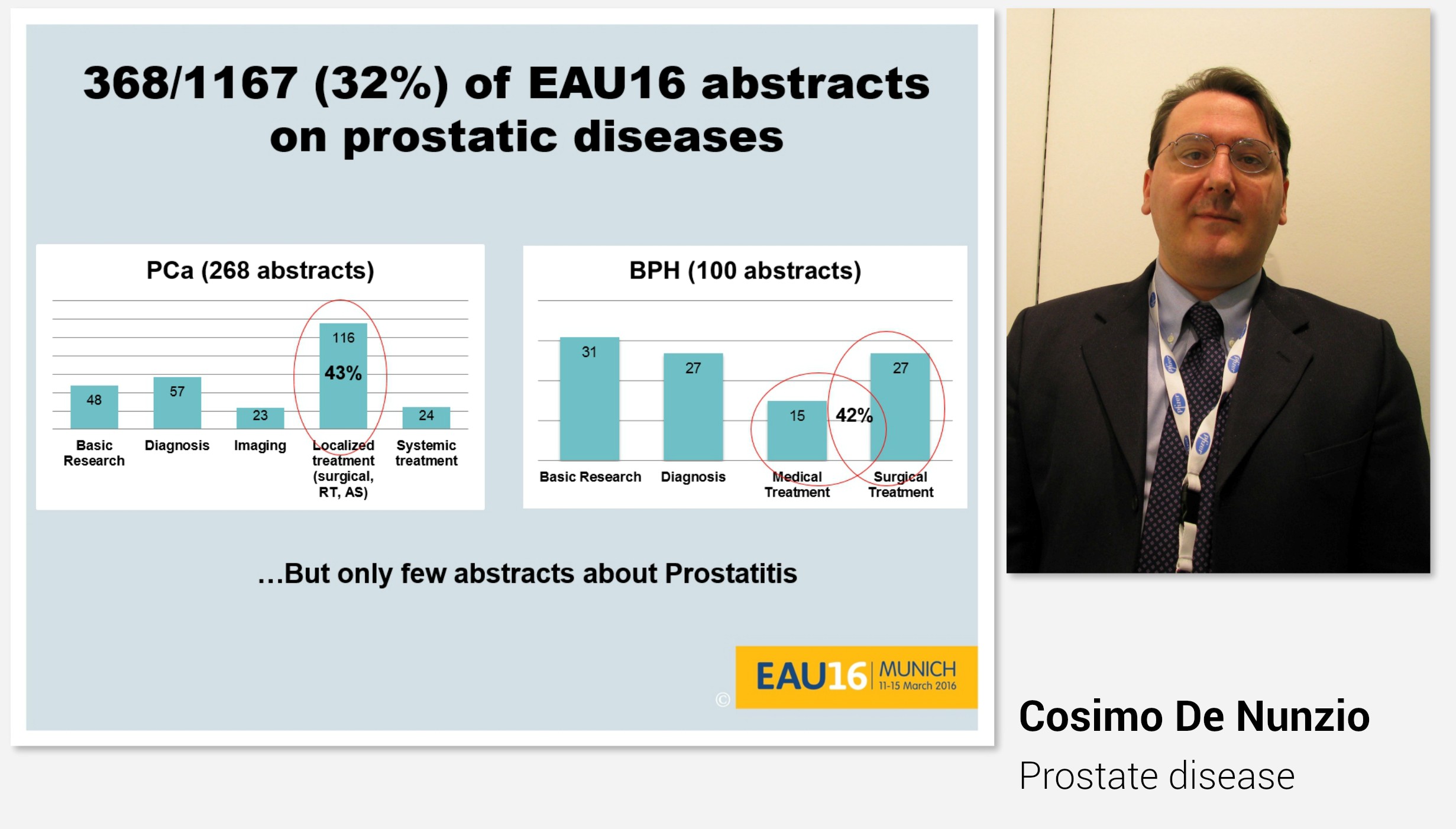
Indeed, the plenary session on Day 3 also covered prostatic disease.

Professor Alexander Bachmann talked about surgery for BPO in the elderly. He pointed out that in elderly (high-risk) patients we do not need a complete anatomical tissue removal, we do not need a (very) long-term follow-up and that we do not need tissue for prostate cancer diagnosis. Instead, we need a safe and efficient operation with individual adaptation of the technique and preferably feasibility in an ambulatory setting or local anaesthesia.

EAU 3-2

Professor Bachmann further emphasized that it would be preferable if surgery for the elderly would be performed by experienced surgeons, and that age per se is not a reason to not operate. There are several new minimally invasive operations available, and especially for elderly less is often more.

HOW AND WHEN TO STOP ANTICOAGULATION – Managing perioperative thromboprophylaxis for patients who already receive anticoagulants remains a challenge. Associate professor Daniel Eberli and Professor Per Morten Sandset covered many of these aspects in their helpful presentations.

EAU 3-3

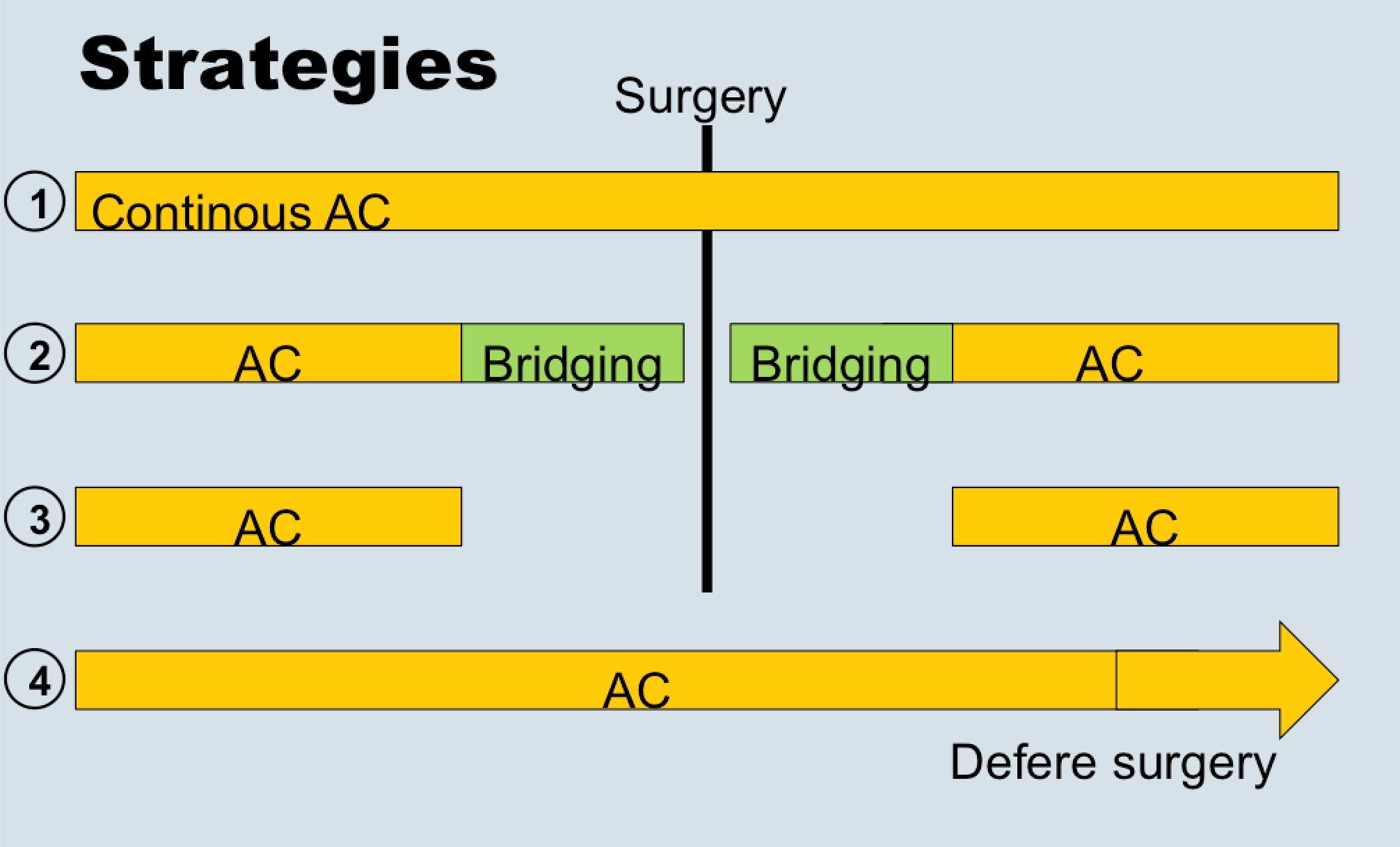
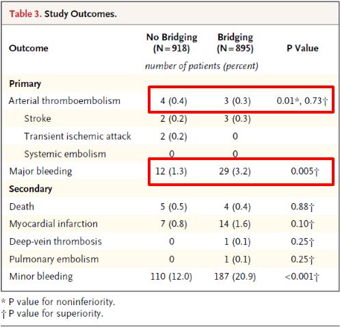
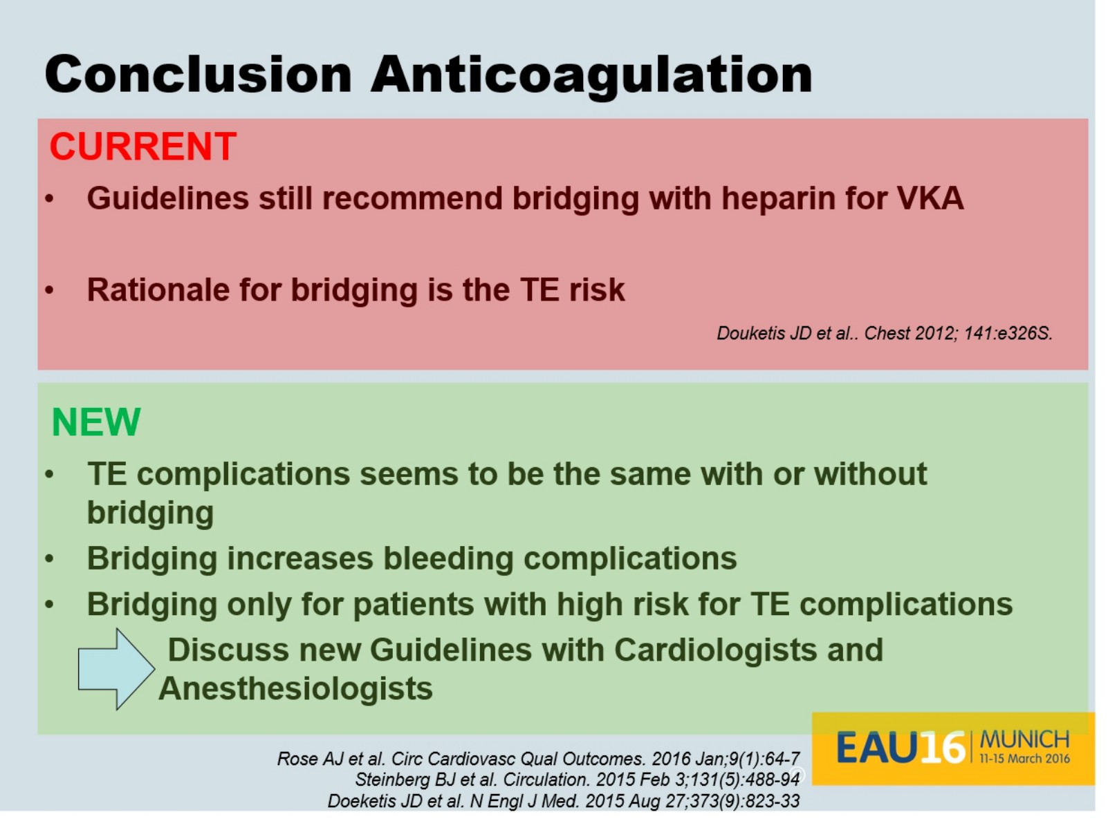
Dr. Eberli told us that bridging therapy (options for stopping or not stopping anticoagulation in the above figure) is eminence-based, as no papers exist showing benefits. He also presented data from the recent NEJM trial (BRIDGE study; see Table below), which showed that stopping anticoagulation without bridging was non-inferior to perioperative bridging for the prevention of arterial thromboembolism and decreased the risk of major bleeding.

EAU 3-4

Dr. Eberli gave us all a take home message to discuss and question our local bridging guidelines as new evidence is very likely not supporting them (concluding slide below).

EAU 3-5

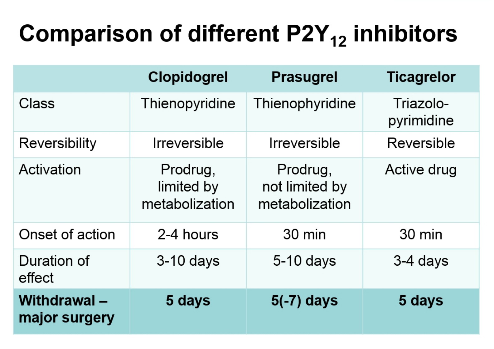
Professor Sandset recommended that during the perioperative period only use aspirin in high-risk patients, that is, those with recent thrombotic event or extensive coronary heart disease. He also informed us that stopping antiplatelet therapy 5 days before surgery (figure below) is often the way to go, and agreed with Dr. Eberli regarding bridging therapy statements.

EAU 3-6

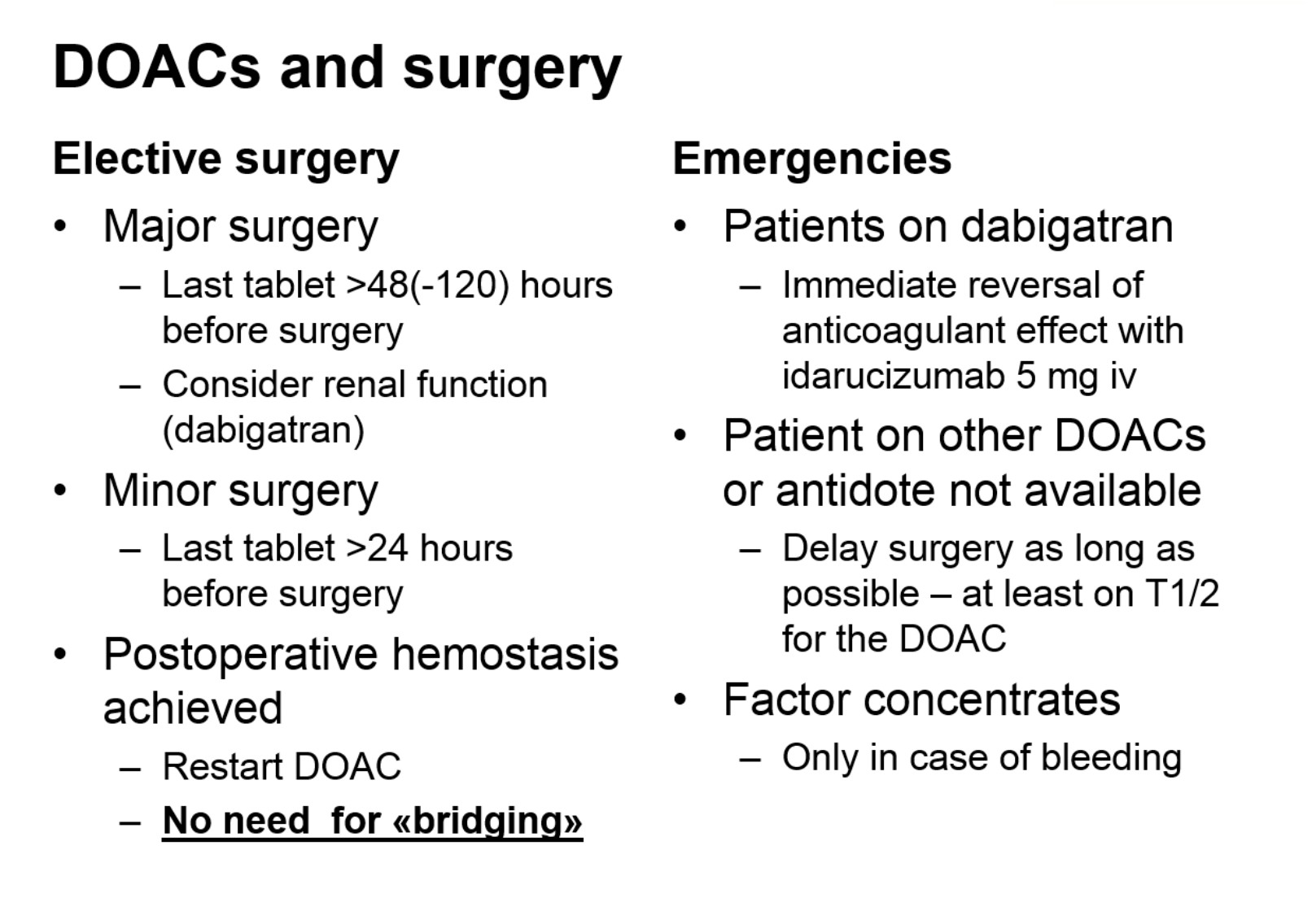
Professor Sandset also gave helpful information regarding use of direct oral anticoagulants (DOACs) in urological surgery:

EAU 3-7

There were numerous poster sessions available on Day 3, as usual, many of them on prostate cancer. We have selected some of the highlight abstracts presented.

PROSTATE CANCER – On Day 3, prostate cancer presentations dominated once again in a number of poster, abstract and thematic sessions but also kidney, bladder, testicular and penile cancer sessions, which provided new interesting data.

Molecular markers, genomic profiling and individualized risk and treatment assessments were presented and discussed in poster session 58, and summarized by Stacy Loeb (@LoebStacy). Further advances in prostate cancer biomarkers in prostate cancer were presented in poster session 84. These new tools are moving from bench to bedside and urologists can hopefully incorporate these new tools to cancer care sooner rather than later.

In sessions on prostate cancer diagnostics, more advanced risk profiling tools were highlighted. For instance, STHLM3 test combines history of the patient, clinical parameters, biochemical markers and genetic markers. It was presented earlier in the congress and on Day 3 further health, economic and clinical evaluations were presented in Thematic session 12. It is one example of the tests showing promising results to potentially decrease the number of prostate biopsies needed. Other similar risk profiling tools were also presented during the congress. In addition to PSA only, evaluation of the smart use of already available clinical and biochemical parameters and the combination of genetic markers may bring individualized risk assessment of prostate cancer to the next level.

In poster session 62 on Day 3, diagnostic proceedings in prostate cancer with co-morbidity evaluation, biopsy strategies and MRI imaging were presented.  A combination of molecular markers and imaging may be the way to proceed in future. These aspects were covered nicely in Thematic session 12.

MRIs have been heavily integrated in prostate cancer diagnostics during recent years. Image guidance in prostate biopsies seem to be making a breakthrough in prostate cancer diagnostics. Targeted biopsies with cognitive or MRI-TRUS fusion imaging were shown to be the way to enhance the results and reliability of biopsies and cut down the number of biopsies. However, as biopsies are still needed in prostate cancer diagnostics, use of the pre-biopsy MRI protocols were suggested to be done only in clinical trial setting. Many aspects of MRI diagnostics of prostate cancer were elegantly summarized in Thematic session 11.

New sophisticated imaging technologies in addition to MRI were present in several sessions during the meeting. Diagnostic enhancement has been seen also in metastatic prostate cancer. PSMA-PET seems to be replacing choline-PET-TT in evaluation of relapsing and metastatic prostate cancer (e.g. Thematic session 10). More reliable diagnostics and imaging of prostate cancer are also enhancing the treatment decision and treatment choice of patients with local prostate cancer. Finding the right patients for the active surveillance protocols is also being helped with advanced diagnostics. Indeed, finding only patients who need treatment for prostate cancer should be the ultimate goal for enhanced diagnostics as discussed in poster sessions 66 and 75 on Day 3. There are also high expectations on focal therapy (e.g. poster session 66), which at the moment is still experimental but will likely be a real option for patients with low volume prostate cancer verified by imaging.

The role of quality of life evaluations and patient reported outcomes measured were heavily discussed during the congress in all treatment modalities of both local and advanced prostate cancer. Survivorship issues are an increasingly important issue when more effective treatments both in local and advanced prostate cancer are available.

In metastatic disease, the use of early chemotherapy in combination with hormonal treatment has been implemented very rapidly to clinical use after the results of the CHAARTED and STAMPEED studies. Further evaluation of early chemo in metastatic disease is still needed and the patient selection needs still clarification. Hormonal therapy still has a very marked role in metastatic prostate cancer and new advances can also be found in new strategies of using castration therapy as presented in poster session 67. Urologists should actively follow the changing landscape of the medical treatment of metastatic prostate cancer and be active in treatment planning and treatment of these patients. At the same time with poster session 62 novel drugs and new forms of isotope radiation therapy in castration resistant prostate cancer were discussed in poster session 61. These open new possibilities for potential treatments.

The clinical and scientific content of the program of the Day 3 was of a very high standard, and reflective of the breadth of contemporary research in many areas within urology. Besides this session, it was our pleasure to meet old and new urological friends worldwide. The annual EAU meeting remains a highly effective method of knowledge translation and provides the opportunity for collaboration between surgeon scientists and other researchers in the field. As always in big congresses, there are so many interesting sessions going on at the same time, that it is hard to pick up and follow everything you would like to. We hope that this report provides some memories and take home messages of the Day 3 to the readers of the BJUI and BJUI blogs.

We look forward to future BJUI and EAU happenings!


Kari Tikkinen

Urology resident, adjunct professor of clinical epidemiology

Helsinki University Hospital, Helsinki, Finland

@KariTikkinen


Mika Matikainen

Chief of urology, adjunct professor of urology

Helsinki University Hospital, Helsinki, Finland


Epidermoid cyst of the penis

A case of a 25 year-old man with an epidermoid cyst of the penis is reported.

Authors:

TFEIL YAHYA MD (FWACS) Urologist Surgeon
Department of urology Faculty of Medicine- Nouakchott University

Corresponding Author: TFEIL YAHYA MD (FWACS) Urologist Surgeon, Department of urology Faculty of Medicine- Nouakchott University. E-mail: [email protected]


Abstract
Penile cysts are uncommon. A case of a 25 year-old man with an epidermoid cyst of the penis is reported. He was found to have an asymptomatic soft mass in the frenulum of his penis. Excision of the mass was performed, and the diagnosis of epidermoid cyst the penis was made. No recurrence has been noted within the year since excision. In such cases, clinicians should manage patients either by observation or excision of the cyst.


Introduction
Cutaneous epidermoid cysts may arise from all parts of the body, but penile epidermoid cysts are uncommon. Most are encountered in childhood [1] and are usually congenital. However, the etiology of penile epidermoid cysts in the elderly is not well understood. In previously reported cases, a penile epidermoid cyst has been described as a slowly growing mass [1, 2, and 3]. It has been reported that the period of growth varies from 2 to 8 years [2, 3]. We present a case of a rapidly growing penile epidermoid cyst that developed in a 6 month period of time.


Case Report 
A 25 year-old man was found to have an asymptomatic, slowly growing soft mass measuring 8X2X2 cm in the frenulum of his penis (Fig.1).


Figure 1.
Figure-1.-Appearance-of-the-cyst-on-the-penis


He had no history of trauma, inflammation, urinary tract infection, hematuria or dysuria. The elastic mass was non tender, freely movable within the dermis, and had a smooth surface (Fig.2).
Figure 2.

Figure2.-Appearance-of-the-cyst-after-dissection

There was neither a keratin-filled punctum nor any signs of inflammation. Excision of the cyst was performed under loco-regional anaesthesia through a semi-circular incision (Fig.3).
Figure 3.

Figure3.-Appearance-of-the-penis-after-excision-of-the-cyst-

Macroscopically, the cut surface of the mass appeared to be full of a cheesy material, and both cytology and culture results were negative. Histological examination revealed that the wall of the cyst was lined with stratified squamous epithelium and laminated keratin. It did not contain either skin appendages or germ cells. The final diagnosis was epidermoid cyst of the penis. No recurrence has been noted in the year since excision.

Discussion

Epidermal cysts are common benign tumors that may arise from the infundibular portion of the hair follicle spontaneously or subsequent to trauma, but penile epidermoid cysts are uncommon and usually congenital. The etiology is not clear. Some authors have adduced that it may develop from median raphe cyst, following an abnormal closure of the median raphe during embryogenesis [3]. Others have suggested that median raphe cysts are a different entity from epidermoid cysts [4]. These cysts may occur because of occluded hair follicles, the mechanical implantation such as that involving injection of epidermal fragments, and obstructed eccrine ducts [2]. The present case is more likely to have not originated from a median raphe cyst because of the patient’s age and the development of the cyst in a short period of time. Penile epidermoid cyst can be diagnosed by a careful examination with evaluation by ultrasonography and/or computerized tomography. Dermoid cyst, teratoma, and urethral diverticula should be considered in the differential diagnosis of the epidermoid cyst [2]. An epidermal cyst is lined by well-developed stratified epithelium, and often contains keratin, which can be expressed from the cyst, while a dermoid cyst contains skin and skin appendages, and a teratoma contains derivatives from other germ cells. Therefore, these lesions can be distinguished from an epidermoid cyst. Unlike the urethral diverticula, such cysts do not have a connection with the urethra (Fig.4).

Figure 4.

Fig.4.-retrograd-urethro-cystography

Although epidermoid cysts are benign lesions, neoplastic transformation of their epithelium has been reported to occur rarely [5, 6]. No cases of malignancy arising in the wall of an epidermoid cyst of penis have been reported previously [1, 3]. The best treatment of penile epidermoid cysts is total excision. One year after complete removal of the cyst, we did not note any local recurrence. There were no findings of malignancy in our case, similar to that reported by others [1, 3]. Although malignant transformation is very rare, it should be kept in mind and following excision of an epidermoid cyst, patients should undergo long term follow up.

Conclusion

In conclusion, epidermoid cyst of penis is rare, in the pediatric age group it can occur after a seemingly minor surgical procedure such as circumcision. The swelling may remain largely asymptomatic unless secondarily infected. Care needs to be taken while performing even minor surgical procedures to avoid this complication. The condition should to be treated by complete excision. The rare possibility of malignant transformation must be borne in mind.

References:

1-Suwa M, Takeda M, Bilim V, Takahashi K. Epidermoid cyst of the penis: A case report and review of the literature. Int J Urol 2000; 7: 431—433.
2- Khana S. Epidermoid cyst of the glans penis. Eur Urol 1991; 19: 176–177.
3-Rattan J, Rattan S, Gupta DK. Epidermoid cyst of the penis with extension into the pelvis. J Urol 1997; 158: 593–594.
4-Little Jr JS, Keating MA, Rink RC. Median raphe cyst of the genitalia. J Urol 1992; 148: 1872–1873.
5- Dini M, Innocenti A, Romano GF. Basal cell carcinoma arising from epidermoid cyst: A case report. Dermatol Surg 2001; 27: 585–586.
6- Lopez-Rios F, Rodriguez-Peralto JL, Castano E, Benito A. Squamous cell carcinoma arising in a cutaneous epidermal cyst: Case report and literature review. Am J Dermatopathol 1999; 21: 174–177.


Date added to bjui.org: 05/08/2012

DOI: 10.1002/BJUIw-2011-140-web


© 2024 BJU International. All Rights Reserved.