Archive for year: 2015

TUF Cycle India

John FThe Urology Foundation Cycle Challenge in Rajasthan

19 – 28 November 2015

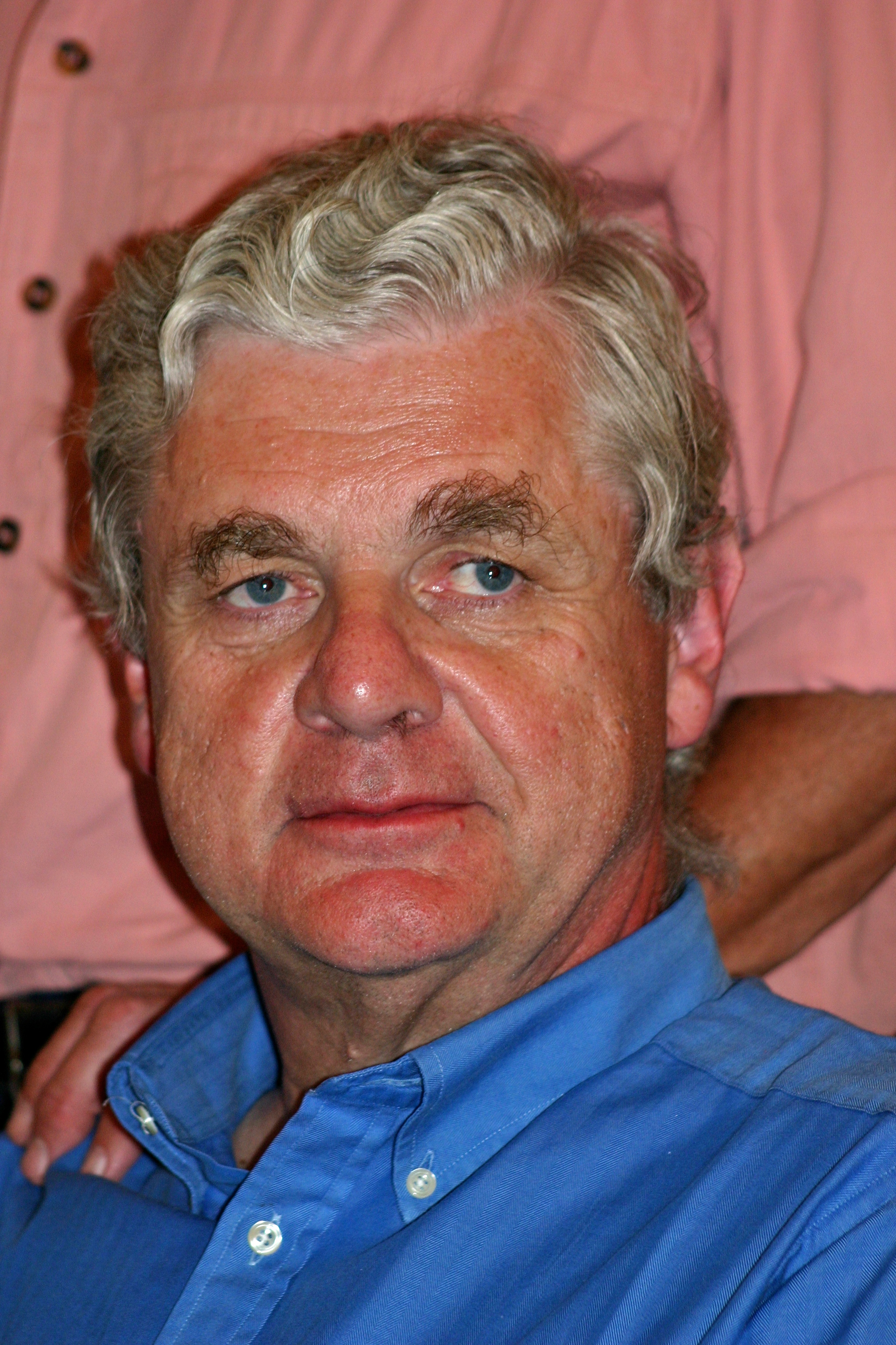
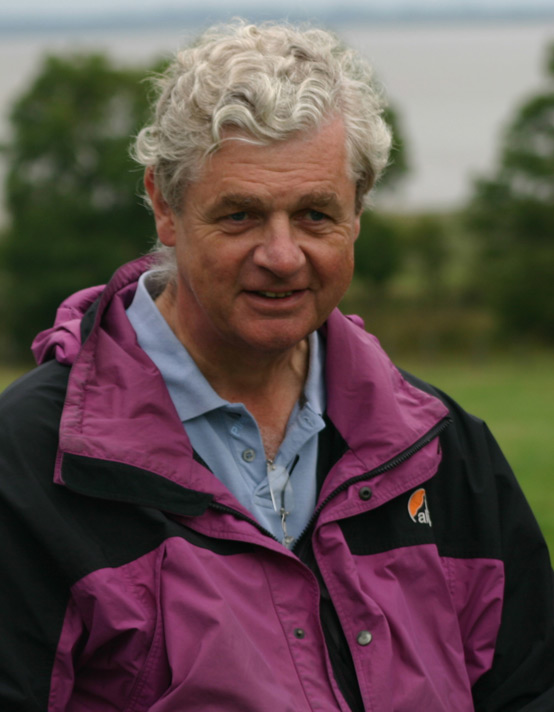
In memory of Professor John Fitzpatrick


TUF-logo-CMYK

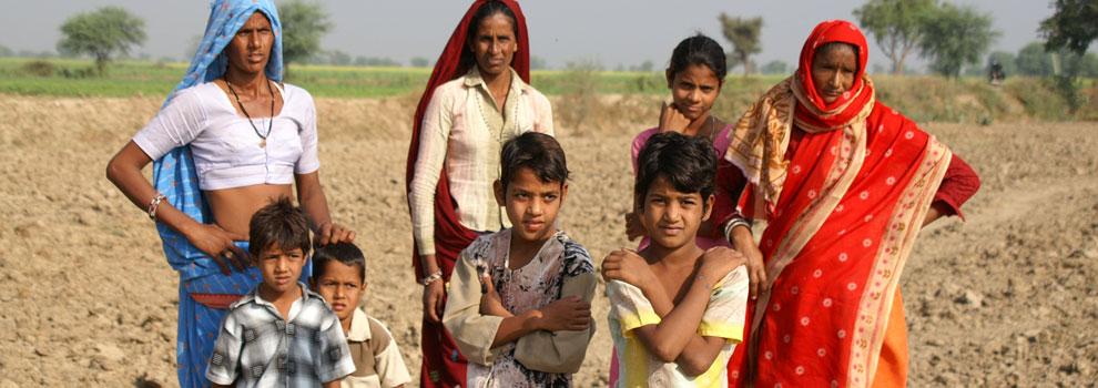
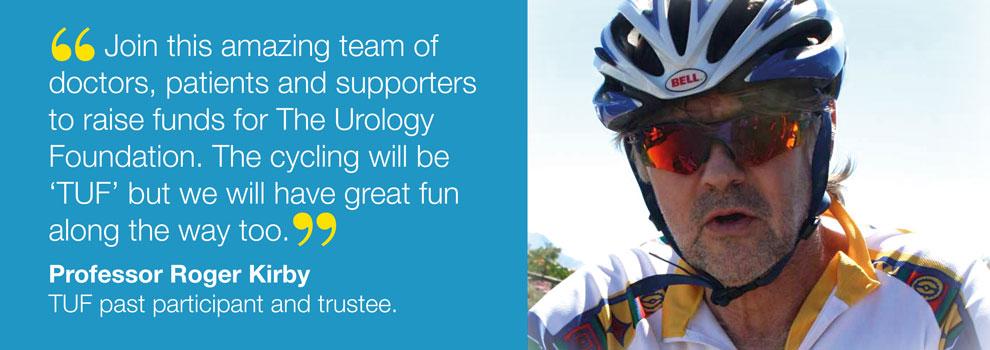
After the gruelling cycling challenges in Sicily, Malawi, Madagascar, Patagonia, and most recently South Africa, which together have raised many hundreds of thousands of pounds for The Urology Foundation (TUF), our next Challenge is a 500 Km ride through Rajasthan, India. We now have 50 intrepid cyclists signed up and ready to participate in this exciting, but very demanding, ride. Some grizzled veterans, such as Roger Plail and Andrew Etherington (80 years old next year!) will be joining us again. Peter Rimington, who led the South African challenge, will be there, but is replaced as “local knowledge team captain” by Abhay Rane, who has done a great job in recruiting and motivating participants this year.  Our wonderful CEO Louise de Winter will be bravely accompanying us on the ride, as she did in Africa.

TUF1

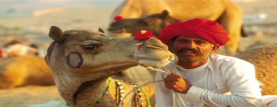
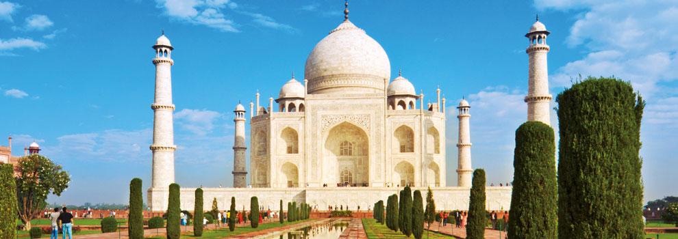
The ride commences in Bharatpur – the eastern gateway to Rajasthan.  It is most famous for the Keoladeo Ghana National Park, a world heritage site and one of the finest water-bird sanctuaries in the world.  On the first morning we will have a chance to visit the specacular Taj Mahal in Agra, one of the true wonders of the world.

TUF1_1

From there, we start our adventure by cycling through the National Park. Our first day’s cycling takes us to the Bhanwar Vilas Palace in Karauli. The following day we will ride to the famous Ranthambore National Park, which is famous for its tigers; the conservation project there is popular with wildlife buffs and professional photographers from right across the world.  With luck we may encounter some of the animals to be found in the park including sambar, cheetah, wild boar, leopard, jackal and hyena.  We will overnight at the famous “Tiger Den”.

TUF2 TUF3 TUF4

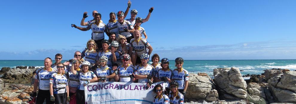
From here on it is just toil, sweat and tears, together with the ever-present risk of “Delhi Belly”! We will no doubt, just as we did before before, rise to the challenge and press on relentlessly to our final destination, the famous “pink city” Jaipur. Here the “Amber Fort” and a well-earned celebration awaits us.

TUF5 TUF6

John-F2b
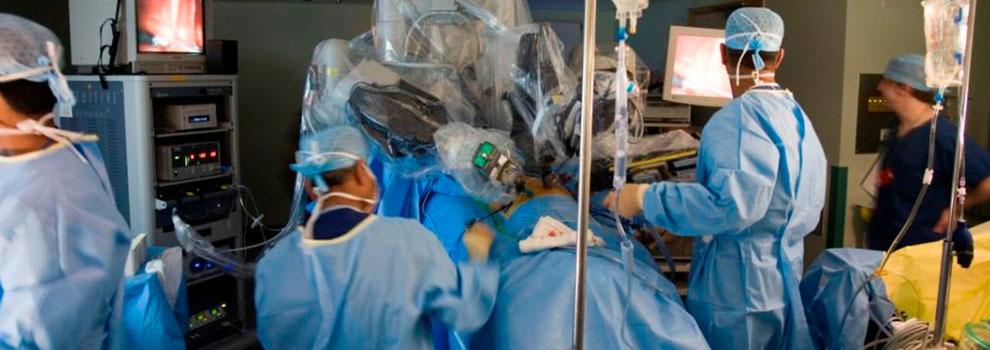
I am very much hoping that many of you will support our endeavours with a donation, and participants themselves will add their own comments, stories and photographs to this blog.  TUF is such a worthy cause, and really does an amazing job in supporting and promoting urology, not only throughout the British Isles, but in Africa and beyond. Do watch (and especially contribute to) this space! We will be posting updates to let you know how we get on.


Click here to see a short video on the challenges the TUF cyclists faced https://trendsinmenshealth.com/video/tuf-cycle-india-2016/


Cycle-Vietnam-to-Cambodia-2017-Poster

Roger Kirby, The Prostate Centre, London


Article of the Week: Trifecta and Optimal Peri-operative outcomes of Robotic and Laparoscopic Partial Nephrectomy In Surgical Treatment Of SRMs

Every Week the Editor-in-Chief selects an Article of the Week from the current issue of BJUI. The abstract is reproduced below and you can click on the button to read the full article, which is freely available to all readers for at least 30 days from the time of this post.

In addition to the article itself, there is an accompanying editorial written by a prominent member of the urological community. This blog is intended to provoke comment and discussion and we invite you to use the comment tools at the bottom of each post to join the conversation.

Finally, the third post under the Article of the Month heading on the homepage will consist of additional material or media. This week we feature a video from Dr. Jihad Kaouk discussing his paper. 

If you only have time to read one article this week, it should be this one.

Trifecta and Optimal Peri-operative outcomes of Robotic and Laparoscopic Partial Nephrectomy In Surgical Treatment Of Small Renal Masses: A Multi-Institutional Study


Homayoun Zargar*, Mohamad E. Allaf, Sam Bhayani, Michael Stifelman§, Craig Rogers, Mark W. Ball, Jeffrey Larson
, Susan Marshall§, Ramesh Kumar¶ and Jihad H. Kaouk*


*Cleveland Clinic, Glickman Urological and Kidney Institute, Cleveland, OH, The Johns Hopkins Medical Institutions, James Buchanan Brady Urological Institute, Baltimore, MD, Dept. of Urology, Washington University School of Medicine, St. Louis, MO, §Dept. of Urology, New York University School of Medicine, New York, NY, and Henry Ford Health System, Vattikuti Urology Institute, Detroit, MI, USA


Read the full article
OBJECTIVE

To compare the perioperative outcomes of robotic partial nephrectomy (RPN) with laparoscopic PN (LPN) performed for small renal masses (SRMs), in a large multi-institutional series and to define a new composite outcome measure, termed ‘optimal outcome’ for the RPN group.

PATIENTS AND METHODS

Retrospective review of 2392 consecutive cases of RPN and LPN performed in five high-volume centres from 2004 to mid-2013. We limited our study to SRMs and cases performed by surgeons with significant expertise with the technique. The Trifecta was defined as negative surgical margin, zero perioperative complications and a warm ischaemia time of ≤25 min. The ‘optimal outcome’ was defined as achievement of Trifecta with addition of 90% estimated glomerular filtration rate preservation and no chronic kidney disease stage upgrading. Univariable and multivariable analysis were used to identify factors predicting Trifecta and ‘optimal outcome’ achievement.

RESULTS

In all, 1185 RPN and 646 LPN met our inclusion criteria. Patients in the RPN group were older and had a higher median Charlson comorbidity index and higher R.E.N.A.L. nephrometry score. The RPN group had lower warm ischaemia time (18 vs 26 min), overall complication rate (16.2% vs 25.9%), and positive surgical margin rate (3.2% vs. 9.7%). There was a significantly higher Trifecta rate for RPN (70% vs 33%) and the rate of achievement of ‘optimal outcome’ for the RPN group was 38.5%.

CONCLUSIONS

In this large multi-institutional series RPN was superior to LPN for perioperative surgical outcomes measured by Trifecta. Patients in the RPN group had better outcomes for all three components of Trifecta compared with their LPN counterparts. Our more strict definition for ‘optimal outcome’ might be a better tool for assessing perioperative and functional outcomes after minimally invasive PN. This tool needs to be externally validated.


Read more articles of the week

Editorial: Robotic Partial Nephrectomy: The Treatment of Choice for Minimally Invasive Nephron Sparing Surgery

Early in the adoption of robotic partial nephrectomy (RPN) as an alternative to laparoscopic PN (LPN) for the treatment of small renal masses, several of the current authors presented a similar comparison of LPN and RPN. They found RPN to result in shorter hospital stay, less blood loss, and shorter warm ischaemia time (WIT) compared with LPN [1]. They discovered that RPN outcomes were not dependent on the complexity of the tumour, which clearly impacted LPN results. They concluded that RPN is a safe and viable alternative to LPN and offered benefits even for experienced laparoscopic surgeons.

The current report in this edition of BJUI furthers the comparison of RPN and LPN and expands the assessment to include five high-volume centres of excellence in robotic surgery [2]. This retrospective, multi-institutional review of 1 185 RPN and 646 LPN represents the largest comparison to date of these two approaches for minimally invasive PN. Despite higher patient comorbidities and R.E.N.A.L. nephrometry scores in the RPN patients compared with the LPN group, there were fewer overall complications (16.2% vs 25.9%), a lower positive surgical margin rate (3.2% vs 9.7%) and a lower WIT (18 vs 26 min). They also found a much higher percentage of RPN patients (70% vs 33%) meeting the Trifecta criteria, defined as negative surgical margins, no perioperative complications, and a WIT of ≤25 min. Finally, the authors introduce a more stringent composite measure of ‘optimal outcomes’, which is the Trifecta with the addition of 90% estimated GFR preservation and no chronic kidney disease upgrading. They report 38.5% of RPN patients meeting optimal outcomes compared with 24.1% for LPN.

This study clearly demonstrates the superiority of RPN over LPN and is supported by other single-surgeon reports [3]. These results also exceed those reported for open PN with the added benefit of reduced hospital stay [4]. However, it is important to recognise that these results represent a mature experience with RPN by the leaders in the field of robotic renal surgery. Many of these authors pioneered the techniques currently used for RPN, and therefore these results may not apply to centres without the same experience or case volumes. One limitation of this report is the non-concurrent experience of LPN and RPN. The results of RPN came after an initial experience with LPN and therefore the outcomes of RPN may have benefitted from the lessons learned with LPN prior to RPN.

Reporting surgical outcomes as composite results, such as the Trifecta, allows for comparison between reports and sets an outcomes bar for future studies. Most composite measures include assessment of surgical margin status and complications, but there is no current agreement as to the optimal measure of renal functional outcomes. The current Trifecta used a WIT of ≤25 min as a measure of renal function impact, while the margin, ischaemia, and complications (MIC) score uses a WIT of <20 min [5], and others have used 90% renal parenchyma preservation as part of the Trifecta [6]. The impact of WIT on renal function has been questioned given the recently recognised importance of preserved renal parenchyma as an important predictor of renal function after PN [7]. Until there is consensus as to the best measure of renal function after nephron-sparing surgery, composite outcomes such as the Trifecta and the optimal outcomes as described by the authors will have limited utility.

Read the full article
James Porter
Robotic Surgery, Swedish Medical Center, Seattle , WA, USA
References


Video: Trifecta and Optimal Peri-operative outcomes of Robotic and Laparoscopic Partial Nephrectomy In Surgical Treatment Of SRMs

Trifecta and Optimal Peri-operative outcomes of Robotic and Laparoscopic Partial Nephrectomy In Surgical Treatment Of Small Renal Masses: A Multi-Institutional Study


Homayoun Zargar*, Mohamad E. Allaf, Sam Bhayani, Michael Stifelman§, Craig Rogers, Mark W. Ball, Jeffrey Larson, Susan Marshall§, Ramesh Kumar¶ and Jihad H. Kaouk*


*Cleveland Clinic, Glickman Urological and Kidney Institute, Cleveland, OH, The Johns Hopkins Medical Institutions, James Buchanan Brady Urological Institute, Baltimore, MD, Dept. of Urology, Washington University School of Medicine, St. Louis, MO, §Dept. of Urology, New York University School of Medicine, New York, NY, and Henry Ford Health System, Vattikuti Urology Institute, Detroit, MI, USA


Read the full article
OBJECTIVE

To compare the perioperative outcomes of robotic partial nephrectomy (RPN) with laparoscopic PN (LPN) performed for small renal masses (SRMs), in a large multi-institutional series and to define a new composite outcome measure, termed ‘optimal outcome’ for the RPN group.

PATIENTS AND METHODS

Retrospective review of 2392 consecutive cases of RPN and LPN performed in five high-volume centres from 2004 to mid-2013. We limited our study to SRMs and cases performed by surgeons with significant expertise with the technique. The Trifecta was defined as negative surgical margin, zero perioperative complications and a warm ischaemia time of ≤25 min. The ‘optimal outcome’ was defined as achievement of Trifecta with addition of 90% estimated glomerular filtration rate preservation and no chronic kidney disease stage upgrading. Univariable and multivariable analysis were used to identify factors predicting Trifecta and ‘optimal outcome’ achievement.

RESULTS

In all, 1185 RPN and 646 LPN met our inclusion criteria. Patients in the RPN group were older and had a higher median Charlson comorbidity index and higher R.E.N.A.L. nephrometry score. The RPN group had lower warm ischaemia time (18 vs 26 min), overall complication rate (16.2% vs 25.9%), and positive surgical margin rate (3.2% vs. 9.7%). There was a significantly higher Trifecta rate for RPN (70% vs 33%) and the rate of achievement of ‘optimal outcome’ for the RPN group was 38.5%.

CONCLUSIONS

In this large multi-institutional series RPN was superior to LPN for perioperative surgical outcomes measured by Trifecta. Patients in the RPN group had better outcomes for all three components of Trifecta compared with their LPN counterparts. Our more strict definition for ‘optimal outcome’ might be a better tool for assessing perioperative and functional outcomes after minimally invasive PN. This tool needs to be externally validated.


Read more articles of the week

Functional urology is coming to you!

Dirk resizedThis month’s edition features three interesting papers in the field of functional urology. Overactive bladder (OAB) syndrome has a prevalence of 14%, prostatitis symptoms have a prevalence in the male population of 8.2% and a substantial number of all men undergoing radical prostatectomy will remain incontinent. These are clinical entities that every urologist encounters in his daily practice.

The treatment of refractory OAB symptoms with anticholinergics, can be optimized by adding mirabegron in a flexible dose scheme. This has been nicely shown in a Japanese population by Yamaguchi et al. [1]. Despite the fact that Japanese health authorities recommend starting with a lower dose of 2.5 mg of solifenacin or 25 mg of mirabegron, these data can be extrapolated to other populations as well, where 5 mg of solifenacin and 50 mg of mirabegron are used as standard doses.

Chronic bacterial prostatitis and chronic pelvic pain syndrome are difficult to deal with. As there is a lack of well-designed prospective randomized controlled studies in this field, Rees et al. [2] used the Delphi consensus methodology to draw up experience- and science-based consensus guidelines. Their Delphi panel included 58 participants consisting of GPs, urologists, pain specialists, nurse specialists, physiotherapists, cognitive behavioural specialists and sexual health specialists. The guidelines give a well-structured overview of the diagnostic and therapeutic possibilities for chronic bacterial prostatitis and chronic pelvic pain syndrome.

Post-radical prostatectomy incontinence varies widely from 3 to 87%. Artificial sphincters are still the main treatment for this complication. While the results in non-irradiated patients might be good in the long term, it remains unclear how external beam radiotherapy would affect the outcome of artificial sphincters in post-radical prostatectomy incontinence. Bates et al. [3] performed a meta-analysis on the complications occurring after the implantation of an artificial sphincter after radical prostatectomy and radiotherapy. The combination of radical prostatectomy and external beam radiotherapy increases the risk of infection and erosion and urethral atrophy and results in a greater risk of surgical revision compared with radical prostatectomy alone. Also persistent urinary incontinence is more common in this population.

These three papers highlight important and relevant problems in urology. It is clear from these papers that we need more insight and more research into the underlying mechanisms of these highly prevalent entities. With an ageing population that wants to remain active as long as possible, we need to invest more time, people and money in this field to improve the quality of life of these patients. Basic science and clinical science need to work together to improve our knowledge and understanding.

Functional urology is coming to you! You will not escape from this growing population.


References


Dirk De Ridder
Department of Urology, University Hospitals KU Leuven, Leuven, Belgium


RE: Prostate Carcinoma With Positive Margins at Radical Prostatectomy: Role of Tumour Zonal Origin in Biochemical Recurrence

Sir,

With great interest, we read the recent article by O’Neil et al. [1], in which the authors investigated the relation between the tumour zonal origin (transition zone vs peripheral zone), positive surgical margins (PSM) after radical prostatectomy and the risk of biochemical recurrence (BCR). Clinicopathological data for 323 patients with PSM after prostatectomy were analysed, of which tumours arising in the prostate transition zone (TZ) were 13%, while tumours in the peripheral zone (PZ) were 87%. The data showed that the percentage of PSM was higher for TZ compared with PZ tumours, with frequent involvement of the bladder neck margins for TZ tumours, without significant difference in time to BCR. In this cohort of patients, adjuvant radiotherapy (ART) was performed in 41% and 53% of TZ and PZ tumours, respectively. Contrary to expectation, BCR was earlier and at higher rate in patients who underwent radiotherapy for TZ rather than PZ tumours.

In the authors’ opinion, these data represent a selection bias, probably due to the higher incidence of bladder neck positive margins in the ART group. This is a novel finding that warrants further investigation.

As radiation oncologists, we make the following comments:

ART is recommended in patients with adverse pathologic features in order to reduce the risk of BCR, local recurrence and clinical progression [2]. Despite the use of a multimodal approach for radiotherapy planning, approximately 40% of patients experienced BCR long-term with an higher risk of developing metastases or cancer-related death [3,4]. It is plausible that the reasons of local failure may include inadequate radiation dose and target coverage, particularly at the anastomotic site, and this probably represents another bias to be considered as well as patient selection, as suggested by O’Neil and colleagues. To date, four different guidelines for clinical target volume (CTV) delineation in the post-prostatectomy setting are available, although there is little data to guide radiation oncologists on appropriate margins selection [5]. The proposed CTVs differed significantly among these consensus guidelines with respect to target border, especially in anterior and cranial directions. In particular, the European Organization for Research and Treatment of Cancer (EORTC) CTV was smaller in comparison with other consensus volumes, with more limited prostate bed coverage and with less irradiation of surrounding healthy tissue, to reduce radiation therapy side effects[5].

In our opinion, for the majority of cancer patients to be treated effectively, the risk of marginal misses is greater than normal tissue complications, and could affect the clinical outcome; thus, target coverage should generally not be compromised [6]. We would also like to add that small treatment margins may be more sensitive to geometrical uncertainties, including set-up errors and inter- and intra-fraction target motions of the prostate bed, and daily image guidance is required [7]. Finally, we believe that to make definitive recommendations regarding the magnitude of margin reduction with improvement of therapeutic ratio, an accurate selection of patients is required.

Read the article

Maria Grazia Ruo Redda1 MD, Alessia Reali1 MD, Roberta Verna1 MD, and Simona Allis1 MD.

1Department of Oncology, Radiation Oncology, University of Turin, S. Luigi Gonzaga Hospital, Orbassano, Turin, Italy.

References

  1. O’Neil LM, Walsh S, Cohen RJ, Lee S. Prostate carcinoma with positive margins at radical prostatectomy: role of tumour zonal origin in biochemical recurrence. BJUI 2015, Jul 27. [Epub ahead of print]
  2.  

  3. Thompson IM, Valicenti RK, Albertsen P, et al. Adjuvant and Salvage Radiotherapy after Prostatectomy: AUA/ASTRO Guideline. J Urol 2013;190:441-9.
  4.  

  5. Bolla M, van Poppel H, Tombal B, et al. Postoperative radiotherapy after radical prostatectomy for high-risk prostate cancer: long-term results of a randomised controlled trial (EORTC trial 22911). Lancet 2012;380:2018-27.
  6.  

  7. Thompson IM, Tangen CM, Paradelo J, et al. Adjuvant radiotherapy for pathological T3N0M0 prostate cancer significantly reduces risk of metastases and improves survival: long term follow-up of arandomized trial. J Urol 2009;181:956–62.
  8.  

  9. Malone S, Croke J, Roustan-Delatour N, et al. Postoperative radiotherapy for prostate cancer: a comparison of four consensus guidelines and dosimetric evaluation of 3D-CRT versus tomotherapy IMRT. Int J Radiat Oncol Biol Phys 2012;84:725-32.
  10.  

  11. Marks LB, Yorke ED, Jackson A, et al. Use of normal tissue complication probability models in the clinic. Int J Radiat Oncol Biol Phys 2010;76(3):S10-9.
  12.  

  13. Gill S, Isiah R, Adams R, et al. Conventional margins not sufficient for post-prostatectomy prostate bed coverage: An analysis of 477 cone-beam computed tomography scans. Radiother Oncol 2014 Feb;110(2):235-9.
  14.  

Article of the Week: External urethral sphincter electromyography and the influence of the menstrual cycle

Every Week the Editor-in-Chief selects an Article of the Week from the current issue of BJUI. The abstract is reproduced below and you can click on the button to read the full article, which is freely available to all readers for at least 30 days from the time of this post.

In addition to the article itself, there is an accompanying editorial written by a prominent member of the urological community. This blog is intended to provoke comment and discussion and we invite you to use the comment tools at the bottom of each post to join the conversation.

Finally, the third post under the Article of the Month heading on the homepage will consist of additional material or media. This week we feature a video from Chris Betts and Kate Burnett discussing their paper. 

If you only have time to read one article this week, it should be this one.

External urethral sphincter electromyography in asymptomatic women and the influence of the menstrual cycle

Cecile Tawadros*, Katherine Burnett*, Laura F. Derbyshire*, Thomas Tawadros†, Noel W. Clarke*‡ and Christopher D. Betts*


*Department of Urology, Salford Royal NHS Foundation Trust, Salford, UK, Department of Urology, Centre Hospitalier Universitaire Vaudois, Lausanne, Switzerland, and Department of Urology, Christie Hospitals NHS Foundation Trust, Manchester, UK


Read the full article
OBJECTIVE

To investigate by electromyography (EMG), the presence of complex repetitive discharges (CRDs) and decelerating bursts (DBs) in the striated external urethral sphincter during the menstrual cycle in female volunteers with no urinary symptoms and complete bladder emptying.

SUBJECTS AND METHODS

Healthy female volunteers aged 20–40 years, with regular menstrual cycles and no urinary symptoms were recruited. Volunteers completed a menstruation chart, urinary symptom questionnaires, pregnancy test, urine dipstick, urinary free flow and post-void ultrasound bladder scan. Exclusion criteria included current pregnancy, use of hormonal medication or contraception, body mass index of >35 kg/m2, incomplete voiding and a history of pelvic surgery. Eligible participants underwent an external urethral sphincter EMG, using a needle electrode in the early follicular phase and the mid-luteal phase of their menstrual cycles. Serum oestradiol and progesterone were measured at each EMG test.

RESULTS

In all, 119 women enquired about the research and following screening, 18 were eligible to enter the study phase. Complete results were obtained in 15 women. In all, 30 EMG tests were undertaken in the 15 asymptomatic women. Sphincter EMG was positive for CRDs and DBs at one or both phases of the menstrual cycle in eight (53%) of the women. Three had CRDs and DBs in both early follicular and mid-luteal phases. Five had normal EMG activity in the early follicular phase and CRDs and DBs in the mid-luteal phase. No woman had abnormal EMG activity in the early follicular phase and normal activity in the luteal phase. There was no relationship between EMG activity and age, parity or serum levels of oestradiol and progesterone.

CONCLUSIONS

CRDs and DB activity in the external striated urethral sphincter is present in a high proportion of asymptomatic young women. This abnormal EMG activity has been shown for the first time to change during the menstrual cycle in individual women. CRDs and DBs are more commonly found in the luteal phase of the menstrual cycle. The importance of CRDs and DBs in the aetiology of urinary retention in young women remains uncertain. The distribution and or quantity of abnormal EMG activity in the external urethral sphincter may be important. In a woman with urinary retention the finding of CRDs and DBs by needle EMG does not automatically establish Fowler’s syndrome as the explanation for the bladder dysfunction. Urethral pressure profilometry may be helpful in establishing a diagnosis. Opiate use and psychological stress should be considered in young women with urinary retention.

Read more articles of the week

Editorial: Do ‘whale noises’ help in the diagnosis of Fowler’s syndrome?

In 1985, Clare Fowler described the presence of abnormal electromyography (EMG) signals in the urethral sphincter of five women with unexplained urinary retention [1]. The presence of complex repetitive discharges (CRD) and decelerating bursts (DB) in women with urinary retention became an important diagnostic finding. Initially, it was described as an EMG finding resembling pseudomyotonia (delayed relaxation of striated muscle when deep tendon reflexes are elicited). Later, an association with polycystic ovary syndrome was hypothesized [2]. This hypothesis could not be proven, however, and the diagnostic criteria that were used at that time for diagnosing polycystic ovaries did not stand the test of time.

The main achievement of urethral sphincter EMG in women with urinary retention was the fact that, from that point onwards, women who were previously described and as being hysterical could now finally be assured that there was a measurable abnormality within their urethral sphincter. This was a major breakthrough in the management of women with urinary retention. Previously, most of these women ended up with permanent catheters or intermittent catheterization, while being stigmatized as psychiatric patients.

The presence of this EMG abnormality also seemed to correlate with the long-term outcome of sacral nerve stimulation [3]. Sacral nerve stimulation is now the standard of care for women with Fowler’s syndrome.

Many authors have questioned the value of these EMG findings. Ramm et al. [4] found the presence of CRD in 30% of healthy women and Tawadros et al. [5] also found this in 53% of healthy female volunteers. These authors showed that CRD and DB are mostly present during the luteal phase of the menstrual cycle. These results suggest at least some hormonal influence on the EMG findings. Fitzgerald et al. [6] showed that CRD correlated weakly with a history of strained voiding and urethral dilation, suggesting a higher prevalence in women with signs of obstructive voiding. Currently, however, the presence of CRD and DB is considered as non-specific.

While sphincter overactivity is generally accepted as the underlying aetiology in women with urinary retention, proving this overactivity remains a challenge. Urethral pressure profiles and urethral volume measurements by ultrasonography have a high interobserver variability and must be used with caution. Most publications originate from one centre and have not been reproduced by others [7]. MRI measurements of the urethral sphincter could possibly be helpful in the future.

While in healthy women CRD can be present, more research will be needed to establish the real value of sphincter EMG in women with pathological urinary retention. The design of good clinical trials will be difficult because of the limited number of patients with this condition. Currently, the diagnosis of Fowler syndrome remains a clinical one, based on a multimodal assessment of the patient.

Read the full article
Dirk De Ridder
Department of Urology, University Hospitals KU Leuven, Leuven, Belgium


References


2 Fowler CJ, Christmas TJ, Chapple CR, Parkhouse HF, Kirby RS, Jacobs HS. Abnormal electromyographic activity of the urethral sphincter, voiding dysfunction, and polycystic ovaries: a new syndrome? BMJ 1988; 297: 14368


4 Ramm O, Mueller ER, Brubaker L, Lowenstein L, Kenton K. Complex repetitive dischargesa feature of the urethral continence mechanism or pathological nding? J Urol 2012; 187: 21403


5 Tawadros C, Burnett K, Derbyshire LF, Tawadros T, Clarke NW, Betts CD. External urethral sphincter electromyography in asymptomatic women and the inuence of the menstrual cycle. BJU Int 2015; 42331


6 FitzGerald MP, Blazek B, Brubaker L. Complex repetitive discharges during urethral sphincter EMG: clinical correlates. Neurourol Urodyn 2000; 19: 57783


7 Wiseman OJ, Swinn MJ, Brady CM, Fowler CJ. Maximum urethral closure pressure and sphincter volume in women with urinary retention. J Urol 2002; 167: 134851; discussion 13512.


Video: External urethral sphincter electromyography and the influence of the menstrual cycle

External urethral sphincter electromyography in asymptomatic women and the influence of the menstrual cycle

Cecile Tawadros*, Katherine Burnett*, Laura F. Derbyshire*, Thomas Tawadros†, Noel W. Clarke*‡ and Christopher D. Betts*


*Department of Urology, Salford Royal NHS Foundation Trust, Salford, UK, Department of Urology, Centre Hospitalier Universitaire Vaudois, Lausanne, Switzerland, and Department of Urology, Christie Hospitals NHS Foundation Trust, Manchester, UK


Read the full article
OBJECTIVE

To investigate by electromyography (EMG), the presence of complex repetitive discharges (CRDs) and decelerating bursts (DBs) in the striated external urethral sphincter during the menstrual cycle in female volunteers with no urinary symptoms and complete bladder emptying.

SUBJECTS AND METHODS

Healthy female volunteers aged 20–40 years, with regular menstrual cycles and no urinary symptoms were recruited. Volunteers completed a menstruation chart, urinary symptom questionnaires, pregnancy test, urine dipstick, urinary free flow and post-void ultrasound bladder scan. Exclusion criteria included current pregnancy, use of hormonal medication or contraception, body mass index of >35 kg/m2, incomplete voiding and a history of pelvic surgery. Eligible participants underwent an external urethral sphincter EMG, using a needle electrode in the early follicular phase and the mid-luteal phase of their menstrual cycles. Serum oestradiol and progesterone were measured at each EMG test.

RESULTS

In all, 119 women enquired about the research and following screening, 18 were eligible to enter the study phase. Complete results were obtained in 15 women. In all, 30 EMG tests were undertaken in the 15 asymptomatic women. Sphincter EMG was positive for CRDs and DBs at one or both phases of the menstrual cycle in eight (53%) of the women. Three had CRDs and DBs in both early follicular and mid-luteal phases. Five had normal EMG activity in the early follicular phase and CRDs and DBs in the mid-luteal phase. No woman had abnormal EMG activity in the early follicular phase and normal activity in the luteal phase. There was no relationship between EMG activity and age, parity or serum levels of oestradiol and progesterone.

CONCLUSIONS

CRDs and DB activity in the external striated urethral sphincter is present in a high proportion of asymptomatic young women. This abnormal EMG activity has been shown for the first time to change during the menstrual cycle in individual women. CRDs and DBs are more commonly found in the luteal phase of the menstrual cycle. The importance of CRDs and DBs in the aetiology of urinary retention in young women remains uncertain. The distribution and or quantity of abnormal EMG activity in the external urethral sphincter may be important. In a woman with urinary retention the finding of CRDs and DBs by needle EMG does not automatically establish Fowler’s syndrome as the explanation for the bladder dysfunction. Urethral pressure profilometry may be helpful in establishing a diagnosis. Opiate use and psychological stress should be considered in young women with urinary retention.

Read more articles of the week
© 2024 BJU International. All Rights Reserved.