Archive for category: Videos

Video: Risk prediction tool for grade re-classification in men with favourable-risk prostate cancer on active surveillance

Risk prediction tool for grade re-classification in men with favourable-risk prostate cancer on active surveillance

Abstract

Objective

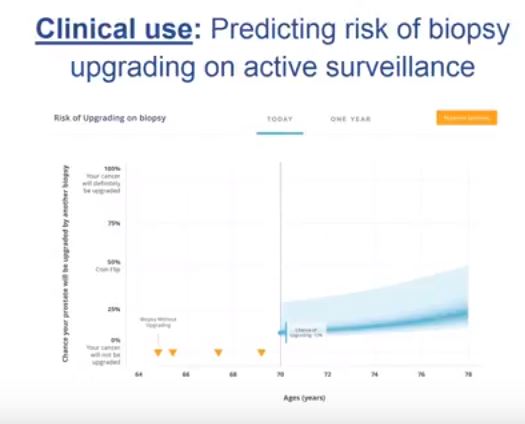
To create a nomogram for men on active surveillance (AS) for prediction of grade re-classification (GR) above Gleason score 6 (Grade group >2) at surveillance biopsy.

Patients and Methods

From a cohort of men enrolled in an AS programme, a multivariable model was used to identify clinical and pathological parameters predictive of GR. Nomogram performance was assessed using receiver operating characteristic curves, calibration, and decision curve analysis.

Results

Of 1 374 men, 254 (18.50%) were re-classified to Gleason ≥7 on surveillance prostate biopsy. Variables predictive of GR were earlier year of diagnosis [≤2004 vs ≥2005; odds ratio (OR) 2.16, P < 0.001], older age (OR 1.05, P < 0.001), higher prostate-specific antigen density [OR 1.19 (per 0.1 unit increase), P = 0.04], bilateral disease (OR 2.86, P < 0.001), risk strata (low-risk vs very-low-risk, OR 1.79, P < 0.001), and total number of biopsies without GR (OR 0.68, P < 0.001). On internal validation, a nomogram created using the multivariable model had an area under the curve of 0.757 (95% confidence interval 0.730–0.797) for predicting GR at the time of next surveillance biopsy.

Conclusion

The nomogram described is currently being used at each return visit to assess the need for a surveillance biopsy, and could increase retention in AS.

Video: Decision-Making by PCa Physicians During AS

Qualitative study on decision-making by prostate cancer physicians during active surveillance


Read the full article

Abstract

Objective

To explore and identify factors that influence physicians’ decisions while monitoring patients with prostate cancer on active surveillance (AS).

Subjects and Methods

A purposive sampling strategy was used to identify physicians treating prostate cancer from diverse clinical backgrounds and geographic areas across the USA. We conducted 24 in-depth interviews from July to December 2015, until thematic saturation was reached. The Applied Thematic Analysis framework was used to guide data collection and analysis. Interview transcripts were reviewed and coded independently by two researchers. Matrix analysis and NVivo software were used for organization and further analysis.

Results

Eight key themes emerged to explain variation in AS monitoring: (i) physician comfort with AS; (ii) protocol selection; (iii) beliefs about the utility and quality of testing; (iv) years of experience and exposure to AS during training; (v) concerns about inflicting ‘harm’; (vi) patient characteristics; (vii) patient preferences; and (viii) financial incentives.

Conclusion

These qualitative data reveal which factors influence physicians who manage patients on AS. There is tension between providing standardized care while also considering individual patients’ needs and health status. Additional education on AS is needed during urology training and continuing medical education. Future research is needed to empirically understand whether any specific protocol is superior to tailored, individualized care.

Video: Value of 111In-PSMA-RGS for salvage lymphadenectomy in recurrent PCa

Value of 111In-prostate-specific membrane antigen (PSMA)-radioguided surgery for salvage lymphadenectomy in recurrent prostate cancer: correlation with histopathology and clinical follow-up


Read the full article


Abstract

Objectives

To evaluate the use of 111In-labelled prostate-specific membrane antigen (PSMA)-I&T-based radioguided surgery (111In-PSMA-RGS) for salvage surgery in recurrent prostate cancer (PCa) using comparison of intra-operative gamma probe measurements with histopathological results of dissected specimens. In addition, to determine the success of 111In-PSMA-RGS with regard to postoperative prostate-specific antigen (PSA) responses, PCa-specific treatment-free survival rates and postoperative complication rates.

Patients and Methods

A total of 31 consecutive patients with localized recurrent PCa undergoing salvage surgery with PSMA-targeted radioguided surgery using a 111In-labelled PSMA ligand between April 2014 and July 2015 were retrospectively included in this study. The preoperative (interquartile range; range) median PSA level was 1.3 (0.57–2.53 ng/mL; 0.2–13.9 ng/mL). Results of ex vivo radioactivity rating (positive vs negative) of resected tissue specimens were compared with findings of postoperative histological analysis. Best PSA response without additional treatment was determined after 111In-PSMA-RGS, and salvage-surgery-related postoperative complications and PCa-specific additional treatments were recorded.

Results

In 30/31 patients, 111In-PSMA-RGS allowed intra-operative identification of metastatic lesions. In total, 145 surgical specimens were removed and 51 showed metastatic involvement at histological analysis. According to 111In-PSMA-RGS ex vivo measurements, 48 specimens were correctly classified as metastatic and 87 as cancer-free, four were false-negative and six were false-positive compared with histological evaluation. Follow-up information was available for 30/31 patients. PSA declines of >50% and >90% were observed in 23/30 patients and in 16/30 patients, respectively. In 18/30 patients, a PSA decline to <0.2 ng/mL was observed. In 10/30 patients further PCa-specific treatment was given after a median (range) of 125 (48–454) days post-111In-PSMA-RGS. The remaining 20 patients remained treatment-free at a median (range) follow-up of 337 (81–591) days. Of 30 patients, 10 presented with surgery-related complications (Clavien–Dindo grade 1, n = 6, Clavien–Dindo grade 3b, n = 4).

Conclusion

111In-PSMA-RGS proved to be of high value for intra-operative detection of even small metastatic lesions in patients with PCa scheduled for salvage lymphadenectomy. It allows the exact localization and resection of metastatic tissue during 111In-PSMA-RGS and is therefore anticipated to have a beneficial influence on further disease progression; however, identification of suitable patients on the basis of PSMA-positron-emission tomography imaging as well as clinical variables is essential for satisfactory results to be obtained.

Video: Treatment of ED after RP using nerve grafts and end-to-side somatic-autonomic neurorraphy

Long-term follow-up of treatment of erectile dysfunction after radical prostatectomy using nerve grafts and end-to-side somatic-autonomic neurorraphy: a new technique

Read the full article

Abstract

Objective

To study a novel penile reinnervation technique using four sural nerve grafts and end-to-side neurorraphies connecting bilaterally the femoral nerve and the cavernous corpus and the femoral nerve and the dorsal penile nerves.

Patients and Methods

Ten patients (mean [± sd; range] age 60.3 [± 4.8; 54–68] years), who had undergone radical prostatectomy (RP) at least 2 years previously, underwent penile reinnervation in the present study. Four patients had undergone radiotherapy after RP. All patients reported satisfactory sexual activity prior to RP. The surgery involved bridging of the femoral nerve to the dorsal nerve of the penis and the inner part of the corpus cavernosum with sural nerve grafts and end-to-side neurorraphies. Patients were evaluated using the International Index of Erectile Function (IIEF) questionnaire and pharmaco-penile Doppler ultrasonography (PPDU) preoperatively and at 6, 12 and 18 months postoperatively, and using a Clinical Evolution of Erectile Function (CEEF) questionnaire, administered after 36 months.

Results

The IIEF scores showed improvements with regard to erectile dysfunction (ED), satisfaction with intercourse and general satisfaction. Evaluation of PPDU velocities did not reveal any difference between the right and left sides or among the different time points. The introduction of nerve grafts neither caused fibrosis of the corpus cavernosum, nor reduced penile vascular flow. CEEF results showed that sexual intercourse began after a mean of 13.7 months with frequency of sexual intercourse varying from once daily to once monthly. Acute complications were minimal. The study was limited by the small number of cases.

Conclusions

A total of 60% of patients were able to achieve full penetration, on average, 13 months after reinnervation surgery. Patients previously submitted to radiotherapy had slower return of erectile function. We conclude that penile reinnervation surgery is a viable technique, with effective results, and could offer a new treatment method for ED after RP.

Video: Silicone renal models and complex tumour resections prior to RALPN

Utility of patient-specific silicone renal models for planning and rehearsal of complex tumour resections prior to robot-assisted laparoscopic partial nephrectomy

Read the full article

Abstract

Objective

To describe our experience using patient-specific tissue-like kidney models created with advanced three-dimensional (3D)-printing technology for preoperative planning and surgical rehearsal prior to robot-assisted laparoscopic partial nephrectomy (RALPN).

Patients and Methods

A feasibility study of 10 patients with solid renal masses who underwent RALPN after preoperative rehearsal using 3D-print kidney models. A single surgeon performed all surgical rehearsals and procedures. Using standard preoperative imaging and 3D reconstruction, we generated pre-surgical models using a silicone-based material. All surgical rehearsals were performed using the da Vinci® robotic system (Intuitive Surgical Inc., Sunnyvale, CA, USA) before the actual procedure. To determine construct validity, we compared resection times between the model and actual tumour in a patient-specific manner. Using 3D laser scanning in the operating room, we quantified and compared the shape and tumour volume resected for each model and patient tumour.

Results

We generated patient-specific models for 10 patients with complex tumour anatomy. R.E.N.A.L. nephrometry scores were between 7 and 11, with a mean maximal tumour diameter of 40.6 mm. The mean resection times between model and patient (6:58 vs 8:22 min, P = 0.162) and tumour volumes between the computer model, excised model, and excised tumour (38.88 vs 38.50 vs 41.79 mm3, P = 0.98) were not significantly different.

Conclusions

We have developed a patient-specific pre-surgical simulation protocol for RALPN. We demonstrated construct validity and provided accurate representation of enucleation time and resected tissue volume. This simulation platform can assist in surgical decision-making, provide preoperative rehearsals, and improve surgical training.

Video: Comparing FG, USG and CG for renal access in mini-PCNL

A prospective and randomised trial comparing fluoroscopic, total ultrasonographic, and combined guidance for renal access in mini-percutaneous nephrolithotomy

Read the full article

Abstract

Objective

To compare the safety and efficacy of fluoroscopic guidance (FG), total ultrasonographic guidance (USG), and combined ultrasonographic and fluoroscopic guidance (CG) for percutaneous renal access in mini-percutaneous nephrolithotomy (mini-PCNL).

Patients and methods

The present study was conducted between July 2014 and May 2015 as a prospective randomised trial at the First Affiliated Hospital of Guangzhou Medical University. In all, 450 consecutive patients with renal stones of >2 cm were randomised to undergo FG, USG, or CG mini-PCNL (150 patients for each group). The primary endpoints were the stone-free rate (SFR) and blood loss (haemoglobin decrease during the operation and transfusion rate). Secondary endpoints included access failure rate, operating time, and complications. S.T.O.N.E. score was used to document the complexity of the renal stones. The study was registered at https://clinicaltrials.gov/ (NCT02266381).

Results

The three groups had similar baseline characteristics. With S.T.O.N.E. scores of 5–6 or 9–13, the SFRs were comparable between the three groups. For S.T.O.N.E. scores of 7–8, FG and CG achieved significantly better SFRs than USG (one-session SFR 85.1% vs 88.5% vs 66.7%, P = 0.006; overall SFR at 3 months postoperatively 89.4% vs 90.2% vs 69.8%, P = 0.002). Multiple-tracts mini-PCNL was used more frequently in the FG and CG groups than in the USG group (20.7% vs 17.1% vs 9.5%, P = 0.028). The mean total radiation exposure time was significantly greater for FG than for CG (47.5 vs 17.9 s, P < 0.001). The USG had zero radiation exposure. There was no significant difference in the haemoglobin decrease, transfusion rate, access failure rate, operating time, nephrostomy drainage time, and hospital stay among the groups. The overall operative complication rates using the Clavien–Dindo grading system were similar between the groups.

Conclusions

Mini-PCNL under USG is as safe and effective as FG or CG in the treatment of simple kidney stones (S.T.O.N.E. scores 5–6) but with no radiation exposure. FG or CG is more effective for patients with S.T.O.N.E. scores of 7–8, where multiple percutaneous tracts may be necessary.

Video: Comparing VEILND with OILND for Penile Cancer

Prospective study comparing video-endoscopic radical inguinal lymph node dissection (VEILND) with open radical ILND (OILND) for penile cancer over an 8-year period

Read the full article

Abstract

Objective

To compare the complications and oncological outcomes between video-endoscopic inguinal lymph node dissection (VEILND) and open ILND (OILND) in men with carcinoma of the penis.

Patients and methods

A prospectively collected institutional database was used to determine the outcomes in 42 consecutive patients undergoing ILND between 2008 and 2015 in a centre for treating penile cancer. Before 2013 all procedures were OILNDs. Since 2013 we have performed VEILND on all patients in need of ILND. The wound-related and non-wound-related complications, length of stay, and oncological safety between OILND and VEILND groups were compared. The mean duration of follow-up was 71 months for OILND and 16 months for the VEILND groups.

Results

In the study period 42 patients underwent 68 ILNDs (OILND 35, VEILND 33). The patients’ demographics, primary stage and grade, and indications were comparable in both groups. There were no intraoperative complications in either group. The wound complication rate was significantly lower in the VEILND group at 6% compared to 68% in the OILND group. Lymphocoele rates were similar in both the groups (27% and 20%). The VEILND group had a better or the same lymph node yield, mean number of positive lymph nodes, and lymph node density confirming oncological safety. There were no groin recurrences in either group of patients. VEILND significantly reduced the mean length of stay by 4.8 days (P < 0.001).

Conclusion

VEILND is an oncologically safe procedure with considerably low morbidity and reduced length of stay, at a mean (range) follow-up of 16 (4–35) months.

Video: Accuracy of ultrasonography for renal stone detection and size determination: is it good enough for management decisions?

Accuracy of ultrasonography for renal stone detection and size determination: is it good enough for management decisions?


Read the full article

Abstract

Objectives

To determine the sensitivity and specificity of ultrasonography (US) for detecting renal calculi and to assess the accuracy of US for determining the size of calculi and how this can affect counselling decisions.

Materials and Methods

We retrospectively identified all patients at our institution with a diagnosis of nephrolithiasis who underwent US followed by non-contrast computed tomography (CT) within 60 days. Data on patient characteristics, stone size (maximum axial diameter) and stone location were collected. The sensitivity, specificity and size accuracy of US was determined using CT as the standard.

Results

A total of 552 US and CT examinations met the inclusion criteria. Overall, the sensitivity and specificity of US was 54 and 91%, respectively. There was a significant association between sensitivity of US and stone size (P < 0.001), but not with stone location (P = 0.58). US significantly overestimated the size of stones in the 0–10 mm range (P < 0.001). Assuming patients with stones 0–4 mm in size will be selected for observation and those with stones ≥5 mm could be counselled on the alternative of intervention, we found that in 14% (54/384) of cases where CT would suggest observation, US would lead to a recommendation for intervention. By contrast, when CT results would suggest intervention as management, US would suggest observation in 39% (65/168) of cases. An average of 22% (119/552) of patients could be inappropriately counselled. Stones classified as 5–10 mm according to US had the highest probability (43% [41/96]) of having their management recommendation changed when CT was performed. The use of plain abdominal film of kidney, ureter and bladder and US increases sensitivity (78%), but 37% (13/35) of patients may still be counselled inappropriately to undergo observation.

Conclusions

Using US to guide clinical decision-making for residual or asymptomatic calculi is limited by low sensitivity and inability to size the stone accurately. As a result, one in five patients may be inappropriately counselled when using US alone.

Video: International Consultation on Urological Diseases and European Association of Urology International Consultation on Minimally Invasive Surgery in Urology: laparoscopic and robotic adrenalectomy

International Consultation on Urological Diseases and European Association of Urology International Consultation on Minimally Invasive Surgery in Urology: laparoscopic and robotic adrenalectomy

Mark W. Ball*, Ashok K. Hemal† and Mohamad E. Allaf*


*James Buchanan Brady Urological Institute and Department of Urology, Johns Hopkins University School of Medicine, Baltimore, MD, and Department of Urology, Wake Forest School of Medicine, Winston-Salem, NC, USA


Read the full article

Abstract

The aim of this study was to provide an evidence-based systematic review of the use of laparoscopic and robotic adrenalectomy in the treatment of adrenal disease as part of the International Consultation on Urological Diseases and European Association of Urology consultation on Minimally Invasive Surgery in Urology. A systematic literature search (January 2004 to January 2014) was conducted to identify comparative studies assessing the safety and efficacy of minimally invasive adrenal surgery. Subtopics including the role of minimally invasive surgery for pheochromocytoma, adrenocortical carcinoma (ACC) and large adrenal tumours were examined. Additionally, the role of transperitoneal and retroperitoneal approaches, as well as laparoendoscopic single-site (LESS) and robotic adrenalectomy were reviewed. The major findings are presented in an evidence-based fashion. Large retrospective and prospective data were analysed and a set of recommendations provided by the committee was produced. Laparoscopic surgery should be considered the first-line therapy for benign adrenal masses requiring surgical resection and for patients with pheochromocytoma. While a laparoscopic approach may be feasible for selected cases of ACC without adjacent organ involvement, an open surgical approach remains the ‘gold standard’. Large adrenal tumours without preoperative or intra-operative suspicion of ACC may be safely resected via a laparoscopic approach. Both transperitoneal and retroperitoneal approaches to laparoscopic adrenalectomy are safe. The approach should be chosen based on surgeon training and experience. LESS and robotic adrenalectomy should be considered as alternatives to laparoscopic adrenalectomy but require further study.


aotwjan3-reults
Read more articles of the week


Video: Step by Step. Evolution of robot-assisted orthotopic ileal neobladder formation

Evolution of robot-assisted orthotopic ileal neobladder formation: a step-by-step update to the University of Southern California (USC) technique

Sameer Chopra, Andre Luis de Castro Abreu, Andre K. Berger, Shuchi SehgalInderbir Gill, Monish Aron and Mihir M. Desai


USC Institute of Urology, Keck School of Medicine, University of Southern California, Los Angeles, CA, USA


Read the full article

Objective

To describe our, step-by-step, technique for robotic intracorporeal neobladder formation.

Patients and Methods

The main surgical steps to forming the intracorporeal orthotopic ileal neobladder are: isolation of 65 cm of small bowel; small bowel anastomosis; bowel detubularisation; suture of the posterior wall of the neobladder; neobladder–urethral anastomosis and cross folding of the pouch; and uretero-enteral anastomosis. Improvements have been made to these steps to enhance time efficiency without compromising neobladder configuration.

Results

Our technical improvements have resulted in an improvement in operative time from 450 to 360 min.

Conclusion

We describe an updated step-by-step technique of robot-assisted intracorporeal orthotopic ileal neobladder formation.


© 2024 BJU International. All Rights Reserved.