Re: The Origins of Urinary Stone Disease: Upstream mineral formations initiate downstream Randall’s plaque
Letter to the Editor
The Origins of Urinary Stone Disease: Upstream mineral formations initiate downstream Randall’s plaque
Sir,
We have read with great interest the paper by Hsi et al.[5] and we would like to comment on this paper with two aims: Firstly, to congratulate the authors on a new observation that could transform our understanding of mineralization processes in the renal papilla, but secondly to voice caution concerning the new hypothesis that they have put forth to explain the formation of Randall’s (interstitial) plaque.
Hsi et al.[5] took renal papillae from non-stone formers undergoing nephrectomy, and analyzed mineral content using micro CT. This means that they were able to visualize mineral throughout each papilla without using the laborious method of serial section. They found intratubular mineral in the outer medulla of all 12 patient papillae that they examined.
Our own studies [1-3], have focused on biopsies of the papilla tip, so we have little data on the outer medulla. However, we have examined the entire medulla in four patients (non-stone formers with no family history of stones) undergoing nephrectomy (two for renal cell carcinoma and two for benign disease) and we have not seen mineral deposits such as Hsi et al.[5] describe but we did not carry out micro CT on those specimens. A more recent report on mineralization in the renal medulla did state that intratubular mineral was seen in the majority of specimens, but no details on these were provided in that study [4]. The presence of microscopic mineral deposits in tubules of the outer medulla by Hsi et al.[5] is an interesting finding, but since the patients studied were not stone formers the implications of such deposits on nephrocalcinosis and renal stones is unclear. Further work on this is certainly required.
In the meantime, we would caution readers that the connection that Hsi et al.[5] make in their paper between mineral deposits in the outer medulla and the formation of Randall’s plaque at the papillary tip is still quite hypothetical. First of all, mineral in kidneys from cancer patients could reflect that disease more than it would necessarily provide data applicable to kidney stones. Secondly, the linking of intratubular mineral in the outer medulla with interstitial mineral at the papilla tip is based solely on the fact that when Hsi et al.[5] observed Randall’s plaque, the intratubular mineral in the outer medulla was especially prevalent. This coincidence, of course, could simply reflect greater crystallization at both locations due to a shared risk factor (such as increased calcium excretion). In particular, the association between calcifications in both locations they observed need not imply a causal link whereby mineral in the outer medulla leads to mineral at the papilla tip. Finally, the pressure model used by Hsi et al.[5] to explain the deposition of Randall’s plaque at the papilla tip is one that ignores the normal, homeostatic mechanisms controlling blood flow and nephron filtration rates, which are likely to have more control over flow in the tubules of the medulla than would blockage of peripheral nephrons.
In summary, we recognize the findings of Hsi et al.[5] as novel, but urge appropriate caution toward some of their conclusions. The title of their paper notwithstanding, there is much to do to establish that these observations are relevant to mechanisms of kidney stone formation.
James E. Lingeman
Amy E. Krambeck
Department of Urology, Indiana University School of Medicine, Indianapolis, IN, USA
Tarek M. El-Achkar
Division of Nephrology, Indiana University School of Medicine, Indianapolis, IN, USA
Andrew P. Evan
James C. Williams, Jr.
Department of Anatomy and Cell Biology, Indiana University School of Medicine, Indianapolis, IN, USA
John C. Lieske
Department of Medicine, Mayo Clinic, Rochester, MN, USA
Elaine M. Worcester
Fredric L. Coe
Renal Section, University of Chicago School of Medicine, Chicago, IL, USA
References
- Evan AP, Lingeman JE, Coe FL, Parks JH, Bledsoe SB, Shao Y, Sommer AJ, Paterson RF, Kuo RL and Grynpas M. Randall’s plaque of patients with nephrolithiasis begins in basement membranes of thin loops of henle. J Clin Invest 2003; 111:607-616
- Coe FL, Evan AP, Lingeman JE and Worcester EM. Plaque and deposits in nine human stone diseases. Urol Res 2010; 38:239-247
- Evan AP, Lingeman JE, Worcester EM, Sommer AJ, Phillips CL, Williams JC, Jr. and Coe FL. Contrasting histopathology and crystal deposits in kidneys of idiopathic stone formers who produce hydroxyapatite, brushite, or calcium oxalate stones. Anat Rec 2014; 297:731-748
- Verrier C, Bazin D, Huguet L, Stéphan O, Gloter A, Verpont M-C, Frochot V, Haymann J-P, Brocheriou I, Traxer O, Daudon M and Letavernier E. Topography, composition and structure of incipient randall’s plaque at the nanoscale level. J Urol 2016; 196:1566-1574
Reply by the authors
Intratubular minerals that were previously unappreciated have been identified in the proximal regions of the renal papilla using non-invasive high resolution X-ray microscopy/tomography. As also observed by Verrier et al., J Urol., 2016 [1], these proximal intratubular minerals do exist and are real. Many groups focus their research exclusively on the distal interstitial papillary minerals, the classic Randall’s plaque (RP). The proximal intratubular minerals cannot be seen endoscopically in contrast to the distal papillary minerals as illustrated in our manuscript.
It is valid to ask if, and how the intratubular biominerals are related to interstitial biominerals; collectively, are they related to stone pathogenesis? Our proposed model was formulated on consistently observed patterns from over 30 renal papillae excised from human kidneys (from cancer and non-cancer non-stone formers, and stone formers). Intratubular minerals were observed in the absence of interstitial biominerals. Conversely, interstitial biominerals were never found without proximal intratubular biominerals. In the absence of a valid animal model, our observations led us to hypothesize that there could be a temporal evolution of papillary biomineralization starting first in the proximal intratubular regions, and progressively mature into the interstitial minerals which are endoscopically visible and often documented/investigated.
What are Randall plaques? This question often is asked by scientists from different disciplines that hear about kidney stones. How are they formed? Are they precursors to kidney stones? The etiology of RP formation has been asked in urology for close to a century. The constant interrogation of the renal tip may not provide all the critical insights into the initiation of stone disease. Several hypotheses have been proposed regarding stone pathogenesis by others. Intratubular formations are based on fundamentals of diffusion and pressure gradients, and physical chemistry approaches. Fundamentally, physical chemistry can explain the sequestration of inorganic on organic and subsequent aggregation of small nanoparticles forming into larger particles. While these approaches can provide insights into interstitial biomineral formations, they also can be applied to explain the uniquely different intratubular biominerals.
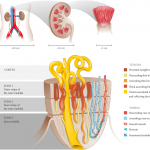
Form and function can be used to help explain the formation of intratubular minerals at the levels of the papilla and the nephron as described by Jean Oliver 1968 [2]. Analogous to a stream, where leaves gather along the edges, particulates within the filtrate gather along the sides of the nephrons while maintaining fluid flow at the center of the nephron. This fundamental related to fluid flow can be leveraged at several length scales. We have applied it to the nephron and clusters of nephrons that form a pyramidal-shaped papilla. The collective aspects of linking intratubular with the commonly known interstitial biominerals unite the fields of two distinct yet overlapping disciplines – urology and nephrology. Urologists center their attention on the renal papilla and nephrologists on the nephron of the same papilla. Merging the structure of the nephron to that of the renal papilla will help understand stone pathogenesis, and this forms the basis for the title of our manuscript.
We thank you for providing this opportunity to further elaborate on our recent findings.
Sunita P. Ho, PhD
Division of Biomaterials and Bioengineering
School of Dentistry, UCSF
Ryan Hsi, MD
Urologic Surgery
School of Medicine
Vanderbilt University
Marshall Stoller, MD
Department of Urology, UCSF
References
- Verrier C, Bazin D, Huguet L, Stéphan O, Gloter A, Verpont M-C, Frochot V, Haymann J-P, Brocheriou I, Traxer O, Daudon M and Letavernier E. Topography, composition and structure of incipient Randall’s plaque at the nanoscale level. J Urol 2016; 196:1566-1574
- Oliver J. Nephrons and kidneys: a quantitative study of development and evolutionary mammalian renal architectonics. New York: Harper & Row; 1968.


