Tag Archive for: image fusion

Posts

Article of the Week: Evaluation of targeted and systematic biopsies using MRI and US image-fusion guided transperineal prostate biopsy

Every Week the Editor-in-Chief selects an Article of the Week from the current issue of BJUI. The abstract is reproduced below and you can click on the button to read the full article, which is freely available to all readers for at least 30 days from the time of this post.

In addition to the article itself, there is an accompanying editorial written by a prominent member of the urological community. This blog is intended to provoke comment and discussion and we invite you to use the comment tools at the bottom of each post to join the conversation.

If you only have time to read one article this week, it should be this one.

Multicentre evaluation of targeted and systematic biopsies using magnetic resonance and ultrasound image-fusion guided transperineal prostate biopsy in patients with a previous negative biopsy


Nienke L. Hansen*†‡, Claudia Kesch§, Tristan Barrett, Brendan Koo, Jan P. Radtke§**, David Bonekamp** , Heinz-Peter Schlemmer**, Anne Y. Warren‡††, Kathrin Wieczorek‡‡Markus Hohenfellner§, Christof Kastner§§ and Boris Hadaschik§


*Department of Diagnostic and Interventional Radiology, University Hospital RWTH Aachen, Aachen, Germany, CamPARI Clinic, Addenbrookes Hospital and University of Cambridge, Cambridge, UK, Department of Diagnostic and Interventional Radiology, University Hospital Cologne, Cologne§Department of Urology, University Hospital Heidelberg, Heidelberg, Germany, Department of Radiology, Addenbrookes Hospital and University of Cambridge, Cambridge, UK, **Department of Radiology, DKFZ, Heidelberg, Germany, ††Department of Pathology, AddenbrookeHospital and University of Cambridge, Cambridge, UK, ‡‡Institute of Pathology, University of Heidelberg, Heidelberg, Germany, and §§Department of Urology, Addenbrookes Hospital and University of Cambridge, Cambridge, UK


Abstract

Objectives

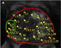
To evaluate the detection rates of targeted and systematic biopsies in magnetic resonance imaging (MRI) and ultrasound (US) image-fusion transperineal prostate biopsy for patients with previous benign transrectal biopsies in two high-volume centres.

Patients and Methods

A two centre prospective outcome study of 487 patients with previous benign biopsies that underwent transperineal MRI/US fusion-guided targeted and systematic saturation biopsy from 2012 to 2015. Multiparametric MRI (mpMRI) was reported according to Prostate Imaging Reporting and Data System (PI-RADS) Version 1. Detection of Gleason score 7–10 prostate cancer on biopsy was the primary outcome. Positive (PPV) and negative (NPV) predictive values including 95% confidence intervals (95% CIs) were calculated. Detection rates of targeted and systematic biopsies were compared using McNemar’s test.

Results

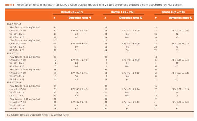
The median (interquartile range) PSA level was 9.0 (6.7–13.4) ng/mL. PI-RADS 3–5 mpMRI lesions were reported in 343 (70%) patients and Gleason score 7–10 prostate cancer was detected in 149 (31%). The PPV (95% CI) for detecting Gleason score 7–10 prostate cancer was 0.20 (±0.07) for PI-RADS 3, 0.32 (±0.09) for PI-RADS 4, and 0.70 (±0.08) for PI-RADS 5. The NPV (95% CI) of PI-RADS 1–2 was 0.92 (±0.04) for Gleason score 7–10 and 0.99 (±0.02) for Gleason score ≥4 + 3 cancer. Systematic biopsies alone found 125/138 (91%) Gleason score 7–10 cancers. In patients with suspicious lesions (PI-RADS 4–5) on mpMRI, systematic biopsies would not have detected 12/113 significant prostate cancers (11%), while targeted biopsies alone would have failed to diagnose 10/113 (9%). In equivocal lesions (PI-RADS 3), targeted biopsy alone would not have diagnosed 14/25 (56%) of Gleason score 7–10 cancers, whereas systematic biopsies alone would have missed 1/25 (4%). Combination with PSA density improved the area under the curve of PI-RADS from 0.822 to 0.846.

Conclusion

In patients with high probability mpMRI lesions, the highest detection rates of Gleason score 7–10 cancer still required combined targeted and systematic MRI/US image-fusion; however, systematic biopsy alone may be sufficient in patients with equivocal lesions. Repeated prostate biopsies may not be needed at all for patients with a low PSA density and a negative mpMRI read by experienced radiologists.

Editorial: Getting to the right biopsy in the right patient at the right time

Guidelines now recommend performing multiparametric MRI (mpMRI) and targeted prostate biopsies in men with a history of prior negative biopsy and continued concern for significant cancer. This new approach to prostate re-biopsy is aimed at improving prostate cancer detection. However, several important clinical factors may help clinicians’ fine-tune the process of repeated prostate biopsy. In this month’s issue of the BJUI, Hansen et al. [1] present a multicentre study of patients with prior negative TRUS biopsy undergoing MRI/TRUS-fusion transperineal biopsy.

In the study, 487 men undergo mpMRI and transperineal biopsy with detection of clinically significant (Gleason score 7–10) cancer as the primary outcome. Several factors are evaluated to compare cancer detection rates, including systematic biopsies, targeted biopsies, PSA density (PSAD), and Prostate Imaging Reporting and Data System (PI-RADS) version 1 score. From their cohort, a suspicious lesion (PIRADS 3–5) was identified in 343 (70%) patients. Prostate cancer was detected in 249 (51%), with 149 (31%) having Gleason score 7–10 cancer. Potentially missed significant cancers from the anterior prostate were found in 27% (40/149). Cancer was detected in 28% (40/144) of patients with PI-RADS 1–2 lesions, with 8% (11/144) being Gleason score 7–10. For patients with PI-RADS 3–5 lesions, cancer was identified in 61% (209/343) with 40% (138/343) being Gleason score 7–10. For patients with PI-RADS 3–5 lesions, systematic biopsies alone failed to detect 13/138 significant cancers, while targeted biopsies missed 24/138 cancers. The combination of systematic and targeted biopsies was significantly better for Gleason score 7–10 prostate cancer detection than either alone. The addition of a PSAD threshold of 0.15 ng/mL/mL for the detection of Gleason score 7–10 resulted in a significant improvement in the area under the curve (0.846) of the receiver operating characteristic curve for PSAD groups and PI-RADS score.

Getting the right biopsy: In this study [1], patients with a prior negative TRUS biopsy underwent TRUS-fusion transperineal biopsy. Having two approaches to prostate biopsy can be advantageous when evaluating men with prior negative biopsies. Historical studies have found comparable prostate cancer detection between transrectal and transperineal biopsies for men undergoing both initial biopsy [2] and saturation re-biopsy [3]. However, the detection of anterior lesions has remained a persistent challenge from the transrectal approach. As in the current study [1], use of transperineal biopsy can detect cancer in up to 30% of tumours that would otherwise be missed on extended template TRUS biopsy [4]. Although attempts to reach anterior lesions from the transrectal approach may be feasible [5], the transperineal approach is felt to provide better sampling in comparison [6].

Getting the right patient: Patient-specific factors such as PI-RADS lesions 3–5 and PSAD have become increasing utilised for stratifying patients who may benefit from additional biopsies using image guidance. As the authors suggest, patients with negative imaging may consider deferring repeat biopsy, particularly those with reassuring PSADs (<0.15 ng/mL/mL). In their study [1], only 4% (6/144) of men with negative mpMRI and a PSAD of <0.15 ng/mL/mL harboured clinically significant cancer (five Gleason score 3 + 4 and one Gleason score 8). Patients with concerning PSAD, but negative mpMRI and those with lesions identified in the peripheral zone could have the option to undergo repeated, fusion-directed TRUS or transperineal biopsy. For patients with lesions identified in the anterior prostate, a transperineal prostate biopsy may provide the highest detection rate.

At the right time: Now that high quality prostate MRI is becoming more widely available; men with a prior negative biopsy should strongly consider the benefit of repeated biopsy after prostate imaging. In addition to identifying suspicious lesions, calculating PSAD has been found to improve the likelihood of detecting clinically significant prostate cancer. Without additional testing, a personalised biopsy plan can be created.

A thorough discussion of the prescribed biopsy approach and the likelihood of detecting a significant cancer is the final step to the right biopsy in the right patient at the right time.

Kelly Stratton


Department of Urology, University of Oklahoma College of Medicine, Oklahoma City, OK, USA


1 Hansen NL, Kesch C, Barrett T et al. Multicentre evaluation of target and systematic biopsies using magnetic resonance and ultrasound image-fusion guided transperineal prostate biopsy in patients with a previous negative biopsy. BJU Int 2016; 120: 6318


2 Hara R, Jo Y, Fujii T et al. Optimal approach for prostate cancer detection as initial biopsy: prospective randomized study comparing transperineal versus transrectal systematic 12-core biopsy. Urology 2008; 71: 1915


3 Abdollah F, Novara G, Briganti A et al. Trans-rectal versus trans- perineal saturation rebiopsy of the prostate: is there a difference in cancer detection rate? Urology 2011; 77: 9215


4 KomaiY, Numao N, Yoshida S et al. High diagnostic ability of multiparametric magnetic re onance imaging to detect anterior prostate cancer miss ed by transrectal 1 2-core biopsy. JUrol2013; 190: 867 7


5 Volkin D, Turkbey B, Hoang AN et al. Multiparametric magnetic resonance imaging (MRI) and subsequent MRI/ultrasonography fusion-guided biopsy increase the detection of anteriorly located prostate cancers. BJU Int 2014; 114: E439


6 Borkowetz A, Platzek I, Toma M et al. Direct comparison of multiparametric magnetic resonance imaging (MRI) results with nal histopathology in patients with proven prostate cancer in MRI/ ultrasonography-fusion biopsy. BJU Int 2016; 118: 21320


© 2024 BJU International. All Rights Reserved.