Tag Archive for: laparoscopy

Posts

Laparoscopic and robot-assisted continent urinary diversions

Video: Double Yang-Monti ileal conduit

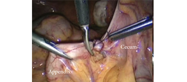
Video: Mitrofanoff appendicovesicostomy

Laparoscopic and robot-assisted continent urinary diversions (Mitrofanoff and Yang-Monti conduits) in a consecutive series of 15 adult patients: the Saint Augustin technique

Denis Rey*, Elie Helou*, Marco Oderda*, Jacopo Robbiani*, Laurent Lopez* and Pierre-Thierry Piechaud*

*Department of Urology, Clinique Saint Augustin, Bordeaux, France, Saint Joseph University, Beirut, Lebanon and Department of Urology, University of Turin, Turin, Italy

OBJECTIVE

• To present a series of 15 laparoscopic and robot-assisted Mitrofanoff and Yang-Monti vesicostomies in an adult population, and to assess the feasibility and safety of these minimally invasive approaches.

PATIENTS AND METHODS

• Between 2009 and 2012, 15 patients underwent laparoscopic (n = 11) or robot-assisted (n = 4) construction of vesicostomy by a single surgeon (D.R.): Mitrofanoff appendicovesicostomy (n = 11) or double Yang-Monti ileal conduit (n = 4). Fourteen patients underwent concomitant augmentation enterocystoplasty.

• Indications for surgery included neurogenic bladder (n = 11) and urethral dysfunction (n = 4).

• The patients were evaluated postoperatively using cystography. Quality of life (QoL) was evaluated using an internally developed questionnaire.

RESULTS

• All surgeries were successfully completed with no conversions. Operating time was always <5 h. The mean estimated blood loss was 150 mL and the mean follow-up was 22 months.

• Early postoperative complications included deep retrovesical abscess (n = 2) and upper urinary tract infections (n = 4), and one patient had peri-operative cardiac failure.

• Late postoperative complications included stomal stenosis (n = 2), persistent low-pressure bladder incontinence (n = 1) and recurrent infections (n = 1). Surgical excision of the conduit was necessary in one patient.

• Postoperatively, patients showed complete bladder emptying and no leak on follow-up cystography. According to our QoL questionnaire, 13/15 patients did not regret the surgery.

CONCLUSION

• While a longer follow-up is needed to assess the durability of our results, this series shows that the laparoscopic and robot-assisted approaches for the construction of continent urinary diversions are feasible and safe in an adult population.

Single-port transvesical LRP for organ-confined prostate cancer

Click here for the extended video.

Single-port transvesical laparoscopic radical prostatectomy for organ-confined prostate cancer: technique and outcomes

Xin Gao, Jun Pang, Jie Si-tu, Yun Luo, Hao Zhang, Liao-yuan Li and Yan Zhang

Department of Urology, The Third Affiliated Hospital, Sun Yat-sen University, Guangzhou, China
X. G. and J. P. contributed equally to this work.

Read the full article
OBJECTIVE

• To report a novel technique for performing single-port transvesical laparoscopic radical prostatectomy (STLRP) and to evaluate the oncological and functional outcomes in 16 patients with organ-confined prostate cancer.

PATIENTS AND METHODS

• In total, 16 consecutive patients with clinical stage T1-2aN0M0 were scheduled for STLRP, and their continence and erectile status were investigated preoperatively.

• The patients’ mean age was 62 years, mean prostate volume 42 mL and mean prostate-specific antigen (PSA) 7.5 ng/mL.

• The STLRP procedures were performed by a single surgeon, and all the operating procedures were conducted transvesically and laparoscopically.

• Intra-operative and postoperative complications, assessed according to the modified Clavien system, were recorded and peri-operative and functional outcome data were analysed.

• All patients were followed up for a minimum of 12 months postoperatively through PSA detection, daily pads, the International Index of Erectile Function (IIEF)-6 score and urography.

RESULTS

• All of the 16 STLRP procedures were successfully completed. The mean (range) operation duration was 105 (75–180) min, and the mean (range) estimated blood loss was 130 (75–500) mL. No patients had positive surgical margins. Postoperative complications occurred in five patients, including three cases of urinary infection and two cases of haematuria (grade II). Catheters were removed after a mean (range) time of 11.2 (9–14) days with cystography. The mean (range) hospital stay was 12.7 (10–15) days.

• Of the 16 patients, 13 were immediately continent (0 pads/day), and three had mild incontinence (2–3 pads/day) after catheter removal. All patients were observed as continent 3 months postoperatively.

• In total, 10/16 and 12/16 patients achieved a satisfactory erection at 6 and 12 months follow-up postoperatively, respectively, with an IIEF-6 score ≥ 18.

• The mean postoperative PSA levels at 3, 6 and 12 months were 0.015 ng/mL, 0.017 ng/mL and 0.016 ng/mL, respectively. No patients were identified with biochemical recurrence in this series. No patients demonstrated vesico-urethral stricture during follow-up for 12–24 months.

CONCLUSION

• We conclude that STLRP is technically feasible for patients with low-risk organ-confined prostate cancer and demonstrates promising functional outcomes regarding continence and potency.

Article of the week: Robotic surgery training methods: take your pick

Every week the Editor-in-Chief selects the Article of the Week from the current issue of BJUI. The abstract is reproduced below and you can click on the button to read the full article, which is freely available to all readers for at least 30 days from the time of this post.

In addition to the article itself, there is an accompanying editorial written by a prominent member of the urological community. This blog is intended to provoke comment and discussion and we invite you to use the comment tools at the bottom of each post to join the conversation.

Finally, the third post under the Article of the Week heading on the homepage will consist of additional material or media. This week we feature a video of Dr. Goh discussing standardized robotic surgery training methods.

If you only have time to read one article this week, it should be this one.

Comparative assessment of three standardized robotic surgery training methods

Andrew J. Hung, Isuru S. Jayaratna, Kara Teruya, Mihir M. Desai, Inderbir S. Gill and Alvin C. Goh*

USC Institute of Urology, Hillard and Roclyn Herzog Center for Robotic Surgery, Keck School of Medicine, University of Southern California, Los Angeles, CA, and *Department of Urology, Methodist Institute for Technology, Innovation and Education, The Methodist Hospital, Houston, TX, USA

Read the full article
OBJECTIVES

• To evaluate three standardized robotic surgery training methods, inanimate, virtual reality and in vivo, for their construct validity.

• To explore the concept of cross-method validity, where the relative performance of each method is compared.

MATERIALS AND METHODS

• Robotic surgical skills were prospectively assessed in 49 participating surgeons who were classified as follows: ‘novice/trainee’: urology residents, previous experience <30 cases (n = 38) and ‘experts’: faculty surgeons, previous experience ≥30 cases (n = 11).

• Three standardized, validated training methods were used: (i) structured inanimate tasks; (ii) virtual reality exercises on the da Vinci Skills Simulator (Intuitive Surgical, Sunnyvale, CA, USA); and (iii) a standardized robotic surgical task in a live porcine model with performance graded by the Global Evaluative Assessment of Robotic Skills (GEARS) tool.

• A Kruskal–Wallis test was used to evaluate performance differences between novices and experts (construct validity).

• Spearman’s correlation coefficient (ρ) was used to measure the association of performance across inanimate, simulation and in vivo methods (cross-method validity).

RESULTS

• Novice and expert surgeons had previously performed a median (range) of 0 (0–20) and 300 (30–2000) robotic cases, respectively (P < 0.001).

• Construct validity: experts consistently outperformed residents with all three methods (P < 0.001).

• Cross-method validity: overall performance of inanimate tasks significantly correlated with virtual reality robotic performance (ρ = −0.7, P < 0.001) and in vivo robotic performance based on GEARS (ρ = −0.8, P < 0.0001).

• Virtual reality performance and in vivo tissue performance were also found to be strongly correlated (ρ = 0.6, P < 0.001).

CONCLUSIONS

• We propose the novel concept of cross-method validity, which may provide a method of evaluating the relative value of various forms of skills education and assessment.

• We externally confirmed the construct validity of each featured training tool.


Read Previous Articles of the Week


Editorial: Three robotic surgery training methods: is there a clear winner?

All training adds value. A craft-based specialty such as surgery has always recognised this. The advent of advanced minimally invasive surgical technology and techniques has provided both new challenges and new opportunities for surgical performance and for the delivery of training. Conceptually, we have moved from the Halstedian model of ‘See one, do one, teach one’ [1] to an environment where skills are acquired away from the operating room in simulator, inanimate and in vivo (animal) laboratory training sessions. Increased scrutiny of credentialling and medico-legal aspects of robotic surgery have reinforced the importance of training and have led to a number of papers outlining pathways to facilitate this [2, 3].

In the present paper, Hung et al. evaluate the construct validity of three standardised training methods (inanimate, simulator and in vivo) and also compare the three different platforms for cross-method training value. As others have shown, the latest generation of robotic surgery simulators have high face, content and construct validity [4, 5] and the present paper confirms the value of both inanimate and simulator training for novice surgeons. In addition, the authors confirmed the construct validity of a simple in vivo exercise using the daVinci© surgical system by demonstrating that experts outperformed novices. Using Spearman’s rank correlation coefficient, the authors compared the three training methods under evaluation and concluded that they were strongly correlated for construct validity between exert and novice surgeons. While construct validation of these exercises may be established, are they useful for experts? Until realistic virtual reality surgical simulations are available, only a novice, an inexperienced or an occasional robot-assisted surgeon may benefit from virtual reality exercises.

What are we therefore to conclude from this? For certain, the advent of excellent surgical simulators and structured inanimate exercises has provided tools for novice surgeons to acquire console skills in a safe and structured environment. This will enhance their operating performance and reduce aspects of the learning curve such as operating time; however, the lack of availability of in vivo training opportunities greatly limits the applicability of this method of surgical training. In many countries (including Australia and the UK), this type of training is illegal or not available. The robotic surgery industry has strongly recommended that in vivo training should be undertaken in one of their official training facilities before surgeons are given the credentials to use this technology; however, even in the USA where most of these facilities are located, key leaders within the AUA have called for the awarding of credentials for robotic surgery ‘not to be an industry driven process, but one that is a result of a standardized, competency based, peer evaluation system’ [2]. Notably, the current AUA Standard Operating Practices (guidelines) for the awarding of credentials for robotic surgery list in vivo training as being optional.

Our view is that although all training has value, there is not enough evidence that in vivo training (particularly on an animal with a rudimentary prostate), which requires international travel and considerable expense, adds sufficient value to be mandatory in any credentialling process. In fact, we have dropped the requirement to complete in vivo training from our requirements at major robotic surgery centres in Australia in favour of structured Mini-Fellowship training [6]. Hung et al. have confirmed what we already knew, which is that all training adds value; however it is likely that only simulator and inanimate training adds enough value to be incorporated into standardised training in robotic surgery.

The multi-disciplinary ‘Fundamentals of Robotic Surgery’ (FRS) curriculum being created by Dr Richard Satava and associates is working on psychomotor skills tasks that include inanimate models as well as corresponding virtual reality exercises. Multi-institutional validation of the FRS or similar curricula will allow the establishment of training milestones and proficiency benchmarks. We must continue to strive for further development of robotic and surgical simulation to change the training paradigm so that surgical training does not need to be at the expense, however minor, of increased operating time or adverse patient outcome.

Declan G. Murphy* and Chandru P. Sundaram
*Peter MacCallum Cancer Centre, Division of Cancer Surgery, University of Melbourne, Australian Prostate Cancer Research Centre, Epworth Richmond Hospital, Melbourne, Australia, and Department of Urology, Indiana University, Indianapolis, IN, USA

Read the full article

References

  1. Halsted WS. The training of the surgeon. Bull Johns Hop Hosp 1904; XV: 8
  2. Lee JY, Mucksavage P, Sundaram CP, McDougall EM. Best practices for robotic surgery training and credentialingJ Urol 2011;185: 1191–1197
  3. Zorn KC, Gautam G, Shalhav AL et al. Training, credentialing, proctoring and medicolegal risks of robotic urological surgery: recommendations of the society of urologic robotic surgeonsJ Urol 2009; 182: 1126–1132
  4. Finnegan KT, Meraney AM, Staff I, Shichman SJ. da Vinci Skills Simulator construct validation study: correlation of prior robotic experience with overall score and time score simulator performanceUrology 2012; 80: 330–335
  5. Abboudi H, Khan MS, Aboumarzouk O et al. Current status of validation for robotic surgery simulators – a systematic reviewBJU Int 2013; 111: 194–205
  6. Melbourne Uro-Oncology Training Program. Robotic surgery training. Available at: https://www.declanmurphy.com.au/training. Accessed 28 February 2013

Video: Take three: assessing robotic surgery training methods

Comparative assessment of three standardized robotic surgery training methods

Andrew J. Hung, Isuru S. Jayaratna, Kara Teruya, Mihir M. Desai, Inderbir S. Gill and Alvin C. Goh*

USC Institute of Urology, Hillard and Roclyn Herzog Center for Robotic Surgery, Keck School of Medicine, University of Southern California, Los Angeles, CA, and *Department of Urology, Methodist Institute for Technology, Innovation and Education, The Methodist Hospital, Houston, TX, USA

Read the full article
OBJECTIVES

• To evaluate three standardized robotic surgery training methods, inanimate, virtual reality and in vivo, for their construct validity.

• To explore the concept of cross-method validity, where the relative performance of each method is compared.

MATERIALS AND METHODS

• Robotic surgical skills were prospectively assessed in 49 participating surgeons who were classified as follows: ‘novice/trainee’: urology residents, previous experience <30 cases (n = 38) and ‘experts’: faculty surgeons, previous experience ≥30 cases (n = 11).

• Three standardized, validated training methods were used: (i) structured inanimate tasks; (ii) virtual reality exercises on the da Vinci Skills Simulator (Intuitive Surgical, Sunnyvale, CA, USA); and (iii) a standardized robotic surgical task in a live porcine model with performance graded by the Global Evaluative Assessment of Robotic Skills (GEARS) tool.

• A Kruskal–Wallis test was used to evaluate performance differences between novices and experts (construct validity).

• Spearman’s correlation coefficient (ρ) was used to measure the association of performance across inanimate, simulation and in vivo methods (cross-method validity).

RESULTS

• Novice and expert surgeons had previously performed a median (range) of 0 (0–20) and 300 (30–2000) robotic cases, respectively (P < 0.001).

• Construct validity: experts consistently outperformed residents with all three methods (P < 0.001).

• Cross-method validity: overall performance of inanimate tasks significantly correlated with virtual reality robotic performance (ρ = −0.7, P < 0.001) and in vivo robotic performance based on GEARS (ρ = −0.8, P < 0.0001).

• Virtual reality performance and in vivo tissue performance were also found to be strongly correlated (ρ = 0.6, P < 0.001).

CONCLUSIONS

• We propose the novel concept of cross-method validity, which may provide a method of evaluating the relative value of various forms of skills education and assessment.

• We externally confirmed the construct validity of each featured training tool.

Step-by-Step: Percutaneous suprapubic tube bladder drainage in RARP

Percutaneous suprapubic tube bladder drainage after robot-assisted radical prostatectomy: a step-by-step guide

Khurshid R. Ghani, Quoc-Dien Trinh, Jesse D. Sammon, Wooju Jeong, Andrea Simone, Ali Dabaja, Stacey Dusik, James O. Peabody and Mani Menon

Vattikuti Urology Institute, Henry Ford Health System, Detroit, MI, USA

Read the full article
OBJECTIVE

• To describe our technique of maintaining bladder drainage after robot-assisted radical prostatectomy (RARP) using a percutaneous suprapubic tube (PST) in place of a urethral catheter.

METHODS

• A watertight anastomosis permits placement of the PST. Contraindications include morbid obesity, concomitant inguinal hernia mesh repair, anticoagulation therapy, limited hand dexterity in the patient, bladder neck reconstruction and extensive adhesiolysis at RARP.

• The necessary equipment includes a 14-F PST balloon catheter set, a three-way connector, a connecting tube, a suture passer, 1/0 polypropylene sutures on a CT1 needle, a sterile plastic button, adhesive and steri-strips.

RESULTS

• The important steps for PST placement are: Step 1: robot-assisted placement of a bladder wall anchor suture; Step 2: transferring the bladder wall suture to anterior abdominal skin; Step 3: guided placement of the PST under robotic vision; Step 4: securing the PST within the bladder and abdominal wall; Step 5. postoperative care: clamping the PST on postoperative day 5, recording each void and post-void residual urine volumes in a patient diary, removal of the PST on postoperative day 7 after 48 h of voiding with residual urine <100 mL per void.

CONCLUSION

• We provide a concise step-by-step guide for placement of a PST during RARP as well as important management aspects for the successful adoption of this technique.

Step-by-Step: Robot-assisted AUS insertion


Robot-assisted laparoscopic artificial urinary sphincter insertion in men with neurogenic stress urinary incontinence

David R. Yates, Véronique Phé, Morgan Rouprêt, Christophe Vaessen, Jérôme Parra, Pierre Mozer and Emmanuel Chartier-Kastler

Academic department of Urology, Pitié-Salpétrière Hospital, Assistance Publique-Hôpitaux de Paris (AP-HP), Faculté de Médecine Pierre et Marie Curie, University Paris 6, Paris, France

The first two authors contributed equally to this article.

Read the full article
OBJECTIVES

• To describe for the first time the technique of robot-assisted artificial urinary sphincter (R-AUS) insertion in male patients with neurogenic incontinence.

MATERIALS AND METHODS

• From January 2011 to the present date, six patients with spinal cord injury have undergone R-AUS insertion at our academic institution and we have prospectively collected data on pre-, peri- and early postoperative outcomes.

• A transperitoneal five-port approach was used using a three-arm standard da Vinci® robot (Intuitive Surgical, Sunnyvale, CA, USA) in a 30° reverse Trendelenburg position.

• The artificial urinary sphincter (AUS) cuff was placed circumferentially around the bladder neck, the reservoir was left intra-abdominally in a lateral vesicular space and the pump was placed in a classic scrotal position.

RESULTS

• All six patients had successful robotic implantation of the AUS.

• The median patient age was 51.5 years, the median (range) operating time was 195 (175–250) min with no significant blood loss or intra-operative complications. The median (range) length of hospital stay was 4 (4–6) days.

• At a median (interquartile range) follow-up of 13 (6–21) months, all six patients had a functioning device with complete continence.

• To date, we have observed no incidence of early erosion, device infection or device malfunction.

CONCLUSIONS

• Allowing for the preliminary nature of our data, R-AUS insertion appears safe and technically feasible.

• Larger studies with long-term follow-up and comparison with open AUS insertion are necessary before definitive statements can be made for R-AUS in respect of complications and functional outcomes.

Procedure: Robot-assisted laparoscopic PN

A prospective comparison of surgical and pathological outcomes obtained after robot-assisted or pure laparoscopic partial nephrectomy in moderate to complex renal tumours: results from a French multicentre collaborative study

Alexandra Masson-Lecomte1,2,3, Karim Bensalah5,6, Elise Seringe2,3, Christophe Vaessen1,2, Alexandre de la Taille4,7, Nicolas Doumerc8,9, Pascal Rischmann8,9, Franck Bruyère10,11, Laurent Soustelle12,13, Stéphane Droupy12,13 and Morgan Rouprêt1,2

1Department of Urology, Pitié Salpétrière, Assistance Publique – Hôpitaux de Paris, Paris, 2Université Paris 6, Paris, 3Department of Statistics, Pitié Salpétrière, Assistance Publique – Hôpitaux de Paris, Paris, 4Department of Urology, Henri Mondor, Assistance Publique – Hôpitaux de Paris, Paris, 5Department of Urology, CHU de Reims, Reims, 6Université de Reims Champagnes-Ardenne, Marne, 7Université Paris-Est Creteil, Marne, 8Department of Urology, CHU Rangueil, Toulouse, 9Université Toulouse 3, Toulouse, 10Department of Urology, CHU Bretonneau, Tours, 11Université François-Rabelais, Tours, 12Department of Urology, CHU Caremeau, Nimes, 13Université Montpellier 1, Montpellier, France

Read the full article
OBJECTIVE

• To prospectively compare the surgical and pathological outcomes obtained with robot-assisted laparoscopic partial nephrectomy (RAPN) or laparoscopic PN (LPN) for renal cell carcinoma in a multicentre cohort.

PATIENTS AND METHODS

• Between 2007 and 2011, 265 nephron-sparing surgeries were performed at six French urology departments. The patients underwent either RAPN (n = 220) or LPN (n = 45) procedures.

• The operative data included operative duration, warm ischaemia time (WIT) and estimated blood loss (EBL). The postoperative outcomes included length of stay (LOS), creatinine variation (Modification of Diet in Renal Disease group), Clavien complications and pathological results.

• The complexity of the renal tumour was classified using the R.E.N.A.L. nephrometry scoring system. Student’s t-test and chi-squared tests were used to compare variables.

RESULTS

• The median follow-ups for the RAPN and LPN groups were 7 and 18 months, respectively (P < 0.001).

• Age and American Society of Anesthesiology score were significantly higher in the LPN group (P = 0.02 and P = 0.004, respectively).

• These variables were lower in the RAPN group: WIT [mean (SD) 20.4 (9.7) vs 24.3 (15.2) min; P = 0.03], operative duration [mean (SD) 168.1 (55.5) vs 199.7 (51.2) min; P < 0.001], operating room occupation time [mean (SD) 248.3 (66.7) vs 278.2 (71.3) min; P = 0.008], EBL [mean (SD) 244.8 (365.4) vs 268.3 (244.9) mL; P = 0.01], use of haemostatic agents [used in 78% of RAPNs and 100% of LPNs; P < 0.001] and LOS [mean (SD) 5.5 (4.3) vs 6.8 (3.2) days; P = 0.05).

• There were no significant differences between pre- and postoperative creatinine levels, pathology report or complication rates between the groups. The main limitation was due to the study’s non-randomised design.

CONCLUSION

• RAPN is not inferior to pure LPN for perioperative outcomes (i.e. EBL, operative duration, WIT, LOS). Only a randomised study with a longer follow-up can now provide further insight into oncological outcomes.


Laparoscopic heminephrectomy and ureterectomy for lower moiety urothelial carcinoma in a complete duplex right kidney

Here, we present a rare case in which a urothelial carcinoma was located at the lower moiety of a complete duplex right kidney. 

Authors: PEI-YU LIN1, VICTOR CHIA-HSIANG LIN1,2, RENG-HONG WU3,  TSAN-JUNG YU1,2

Department of Urology 1, E-Da Hospital/I-Shou University, Kaohsiung City 2, Department of Radiology3, Chi-Mei Medical Center, Tainan City, Taiwan

Corresponding Author: Victor Chia-Hsiang Lin, M.D., Division of Urology, Department of Surgery, Minimally Invasive Surgical Center, E-Da Hospital/I-Shou University, Kaohsiung City 824,  Taiwan 824.   E-mail: [email protected]


Abstract
Total nephroureterectomy is usually considered to be the gold standard treatment for urothelial carcinoma involving the kidney, where nephron sparing surgery is seldom considered because of the concerns of tumor spillage. However, heminephrectomy with ipsilateral ureterectomy may be performed when the malignancy occurs in a patient with a renal fusion anomaly or in a single moiety of a complete duplex kidney. Here, we present a unique case with urothelial carcinoma in the lower moiety of a complete duplex right kidney. Laparoscopic right heminephrectomy with ureterectomy was performed, after preoperative evaluation with reconstructed 3-dimensional computed tomography angiography and intraoperative navigation with laparoscopic ultrasound.
Complete duplication of the collecting system is an uncommon congenital anomaly, occurring in approximately 1 in 125 individuals. Duplex kidneys can be associated with ectopic ureters, ureteroceles, and vesicoureteric reflux, and cause various clinical manifestations including incontinence, voiding dysfunction and urinary tract infection. A common disease variant in this entity is a ectopic ureteric orifice, associated with a dysplastic poorly functioning upper-pole renal moiety. Heminephrectomy is considered to be the treatment of choice for patients with certain pathologies. Here, we present a rare case in which a urothelial carcinoma was located at the lower moiety of a complete duplex right kidney. The patient underwent laparoscopic heminephrectomy with ureterectomy after thorough preoperative evaluation.


Case Report
An 82-year-old female presented with the chief complaint of intermittent painless gross hematuria for 3 months. Physical examination was unremarkable. Cystoscopy with retrograde pyelography revealed complete duplication of the right collecting system and a 2×2 cm filling defect located over the lower moiety of the duplex right kidney (Fig. 1 A & B).


Figure 1. Retrograde pyelography revealed duplication of the right collecting system. A. Hydronephrosis of the right upper moiety due to external compression indicated by a black arrow. B. The white arrow indicates a filling defect over the lower calyx of the right duplex kidney.


Reconstructed 3-dimensional computed tomography (3-D CT) angiography demonstrated a tumor in the lower moiety renal pelvis of the right duplex kidney and several parapelvic renal cysts compressing the upper moiety, causing hydronephrosis and hydrocalycosis. (Fig. 2 A & B).


Figure 2. A. Coronal view of a computed tomography (CT) scan showing a tumor in the duplex right renal pelvis as indicated by the white arrow. B. Reconstruction CT clearly demonstrated a soft tissue mass over the lower calyx of the duplex right kidney, as indicated by the white arrow.


After discussing the available options with the patient and her family, they agreed for her to undergo laparoscopic right heminephrectomy and ureterectomy.


Diagnostic ureteroscopy showed a large nodular tumor in the lower moiety of the duplex right kidney. Subsequently, a transperitoneal laparoscopic technique was adopted, taking down the mesocolon of the ascending colon from the hepatic flexure to the level of the bifurcation of the right iliac vessels. After deroofing the parapelvic renal cysts, the right renal hilum was carefully exposed and the supplying branches to the lower moiety were skeletonized individually. The arterial branch which supplied the lower moiety was confirmed and clamped using a surgical bulldog clamp. The lower moiety appeared cyanotic when its supplying branch was clamped correctly. The venous branch of the lower moiety was accompanied by the arterial branch and was positively identified. By first clipping and dividing the branches of the right renal artery supplying the lower moiety, a clear line of demarcation between the ischaemic lower moiety and the normal upper moiety was seen. Laparoscopic ultrasound further confirmed the upper margin of the lower moiety calyces, and cutting and division of the moieties were performed precisely and efficiently with electrocautery and ultrasonic shears. Hemostasis of the bare surface of the upper moiety was achieved with laparoscopic free-hand suture and knot-tying. The ureter of the lower moiety was skeletonized to the level of the right iliac vessels, and clips were put on the distal end to prevent extravasation of tumor cells. The specimen was then placed in an endobag. The extravesical bladder cuff excision was performed using an open approach through a right Gibson’s incision. When dissecting into the retroperitoneum, the distal ureter was identified, the endobag in which the lower moiety of the right kidney and its proximal ureteral segment were placed was removed thereafter. Retracting the distal ureter cephalad, dissection was continued distally until the bladder was reached. The bladder was incised just anterior to the ureter and the ureteric lumen was identified. The posterior portion of the ureter was excised and detached from the bladder cuff carefully under vision. The bladder defect was sutured with 1-0 chromic gut. There was no injury to the ureteric orifice of the upper moiety of the right kidney.
The patient’s postoperative recovery was prompt due to her undergoing minimally invasive surgery, and she resumed oral intake 12 hours postoperatively. She received a total amount of meperidine 150mg for postoperative pain relief. She was discharged after two  days. Due to a suspected urinary leak, a JJ stent was inserted to drain the right upper moiety on postoperative day 6. Final pathological examination demonstrated urothelial carcinoma of the lower moiety of the right duplex kidney, stage pT1. During two-years of follow-up, no tumor recurrence has been detected.


Discussion
Urothelial carcinoma in duplex kidneys is rare with only sporadic reports in the English literature.1,2 Nephron sparing surgery is well established for the treatment of small exophytic renal cell carcinoma.3 However, total nephroureterectomy is the treatment of choice to manage renal urothelial carcinoma. Nevertheless, nephron sparing surgery for upper tract urothelial carcinoma can still be considered in certain situations such as crossed renal ectopy or tumour in one moiety of a duplex system.4 Gur et al. demonstrated the feasibility of separating the involved kidney from its conjoint to treat patients with transitional cell carcinoma in a fused crossed ectopic kidney. Subsequent ureterectomy with bladder cuff excision was performed in our case with respect to oncological principles. We also advocate the importance of a thorough delineation of the involved renal vasculature using CT-angiography preoperatively.
Similarly, we used reconstructed 3-D CT angiography to delineate the involved renal vasculature. A radiologist also performed laparoscopic ultrasound to help decide precisely the cut-margin during the parenchymal transection. We believe these tools are extremely important in planning this type of surgery. To the best of our knowledge, this is the first case of urothelial carcinoma in one moiety of a complete duplex kidney treated by laparoscopic heminephrectomy and ureterectomy.
In our experience, laparoscopic heminephrectomy is a feasible and safe modality in treating urothelial carcinoma of the lower moiety in a duplex kidney in selected cases.


References
1. Lia-Beng Tan, Biing-Rorn Tserng, Wei-Hwang Huang, Chia-Jiuan Tarn. Synchronous bilateral carcinoma of the ureter in association with unilateral incomplete duplication of the ureter. Urol Int 56: 196-199, 1996.
2. Jenq-Daw Li, Johnny Shinn-Nan Lin, Wei-Jen Yao. Synchronous transitional cell carcinoma in both moieties of an incomplete duplex system. Urology 59: 944-945, 2002.
3. Alireza Moinzadeh, Inderbir S. Gill, Antonio Finelli, Jihad Kaouk and Mihir Desai. Laparoscopic partial nephrectomy: 3-year followup. J Urol 175: 459-462, 2006
4. Uri Gur, Ofer Yossepowitch and Jack Baniel. Transitional cell carcinoma in a fused crossed ectopic kidney. Urology 62: 748, 2003.


Date added to bjui.org: 21/07/2012
DOI: 10.1002/BJUIw-2011-108-web


© 2024 BJU International. All Rights Reserved.