Tag Archive for: laser lithotripsy

Posts

Article of the week: Deep learning computer vision algorithm for detecting kidney stone composition

Every week, the Editor-in-Chief selects an Article of the Week from the current issue of BJUI. The abstract is reproduced below and you can click on the button to read the full article, which is freely available to all readers for at least 30 days from the time of this post.

If you only have time to read one article this week, we recommend this one. 

Deep learning computer vision algorithm for detecting kidney stone composition

Kristian M. Black*, Hei Law, Ali Aldoukhi*, Jia Deng and Khurshid R. Ghani*

*Department of Urology, University of Michigan, Ann Arbor, MI, and Department of Computer Science, Princeton University, Princeton, NJ, USA

Abstract

Objectives

To assess the recall of a deep learning (DL) method to automatically detect kidney stones composition from digital photographs of stones.

Materials and Methods

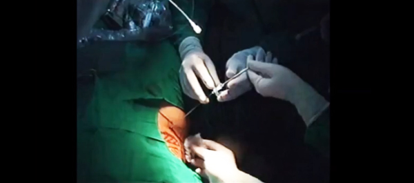
A total of 63 human kidney stones of varied compositions were obtained from a stone laboratory including calcium oxalate monohydrate (COM), uric acid (UA), magnesium ammonium phosphate hexahydrate (MAPH/struvite), calcium hydrogen phosphate dihydrate (CHPD/brushite), and cystine stones. At least two images of the stones, both surface and inner core, were captured on a digital camera for all stones. A deep convolutional neural network (CNN), ResNet‐101 (ResNet, Microsoft), was applied as a multi‐class classification model, to each image. This model was assessed using leave‐one‐out cross‐validation with the primary outcome being network prediction recall.

Fig.2. Representative samples for each stone composition prior to cropping. A total of 63 stones were used in this study including: 17 UA, 21 COM, seven struvite, four cystine, and 14 brushite stones consisting of a total of 127 images. Automatic stone composition recall was highest for UA stones at 94%.

Results

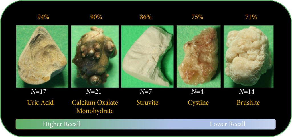
The composition prediction recall for each composition was as follows: UA 94% ( = 17), COM 90% ( = 21), MAPH/struvite 86% ( = 7), cystine 75% ( = 4), CHPD/brushite 71% ( = 14). The overall weighted recall of the CNNs composition analysis was 85% for the entire cohort. Specificity and precision for each stone type were as follows: UA (97.83%, 94.12%), COM (97.62%, 95%), struvite (91.84%, 71.43%), cystine (98.31%, 75%), and brushite (96.43%, 75%).

Conclusion

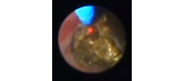
Deep CNNs can be used to identify kidney stone composition from digital photographs with good recall. Future work is needed to see if DL can be used for detecting stone composition during digital endoscopy. This technology may enable integrated endoscopic and laser systems that automatically provide laser settings based on stone composition recognition with the goal to improve surgical efficiency.

Article of the week: Smaller is better? Microperc comes of age

Micropercutaneous nephrolithotomy (microperc) vs retrograde intrarenal surgery for the management of small renal calculi: a randomized controlled trial

Ravindra B. Sabnis, Raguram Ganesamoni, Amit Doshi, Arvind P. Ganpule, Jitendra Jagtap and Mahesh R. Desai

Department of Urology, Muljibhai Patel Urological Hospital, Nadiad, Gujarat, India

Read the full article

OBJECTIVE

• To compare micropercutaneous nephrolithotomy (microperc) and retrograde intrarenal surgery (RIRS) for the management of renal calculi <1.5 cm with regard to stone clearance rates and surgical characteristics, complications and postoperative recovery.

PATIENTS AND METHODS

• Seventy patients presenting with renal calculi <1.5 cm were equally randomized to a microperc or a RIRS group between February 2011 and August 2012 in this randomized controlled trial. Randomization was based on centralized computer-generated numbers. Patients and authors assessing the outcomes were not blinded to the procedure.

• Microperc was performed using a 4.85-F (16-gauge) needle with a 272-μm laser fibre. RIRS was performed using a uretero-renoscope.

• Variables studied were stone clearance rates, operating time, need for JJ stenting, intra-operative and postoperative complications (according to the Clavien–Dindo classification system), surgeon discomfort score, postoperative pain score, analgesic requirement and hospital stay.

• Stone clearance was assessed using ultrasonography and X-ray plain abdominal film of kidney, ureter and bladder at 3 months.

RESULTS

• There were 35 patients in each group. All the patients were included in the final analysis.

• The stone clearance rates in the microperc and RIRS groups were similar (97.1 vs 94.1%, P = 1.0).

• The mean [sd] operating time was similar between the groups (51.6 [18.5] vs 47.1 [17.5], P = 0.295). JJ stenting was required in a lower proportion of patients in the microperc group (20 vs 62.8%, P < 0.001). Intra-operative complications were a minor pelvic perforation in one patient and transient haematuria in two patients, all in the microperc group. One patient in each group required conversion to miniperc.

• One patient in the microperc group needed RIRS for small residual calculi 1 day after surgery. The decrease in haemoglobin was greater in the microperc group (0.96 vs 0.56 g/dL, P < 0.001). The incidence of postoperative fever (Clavien I) was similar in the two groups (8.6 vs 11.4%, P = 1.0). None of the patients in the study required blood transfusion.


Read Previous Articles of the Week


Editorial: Micro-PNL vs RIRS: dealer’s choice? The devil is in the details

Advances in minimally invasive endourological techniques continue to provide the Urologist a myriad of options for the management of symptomatic renal calculi. Previously, shock wave lithotripsy (SWL) or standard percutaneous nephrolithotomy (PNL) were the only two endourological options available. Yet, limitations of these two ‘standard’ techniques result from hard or dependent stones (for SWL) or the potential of increased morbidity during the treatment of small renal calculi (for PNL). Now the introduction of smaller fibre-optic needle-scopes combined with laser stone fragmentation (micro-PNL or ‘microperc’) provides access to difficult-to-reach renal calculi with minimal patient morbidity. Moreover, newer flexible ureteroscopes, along with nitinol baskets and graspers (retrograde intra-renal surgery or ‘RIRS’) allow another minimally invasive option for hard-to-reach renal calculi.

In the present issue of BJUI, Sabnis et al. present a well performed, randomised, prospective trial comparing microperc to RIRS for the management of renal calculi of <1.5 cm in diameter. They determined that both procedures were essentially identical in their ability to remove small-to-moderate sized renal stones with minimal patient morbidity/complications. Yet, both of these procedures have inherent limitations that are unique to the instrumentation used for each technique. Microperc has limited applicability for stones located anteriorly within the kidney, while RIRS is an ideal technique to access symptomatic renal stones within an anterior calyx. RIRS may not be able to target lower pole calculi in those patients with an acute infundibular angle or stones in a calyceal diverticulum, whereas a microperc can be used to reach hard-to-access calculi.

One must accept that both of these innovative procedures are inefficient in their ability for removal of large volume stones and neither technique offers an efficient method of clearing multiple stone fragments. Ideally, both microperc and RIRS should only be used for small volume renal calculi.

I would offer that both procedures are safe and effective alternatives for the management of small renal calculi. Yet, we must be realistic in offering these techniques to our patients, pairing each procedure with the most appropriate situation. Even in 2013, large volume renal calculi are best managed by standard or mini-PNL, where devices such as ultrasonic lithotripsy or dual ultrasonic/pneumatic lithotripsy offer efficient methods of stone removal. SWL is still a reasonable option for the treatment of renal calculi of ≤1 cm in diameter.

Now RIRS and microperc can be added to the list of treatment options for managing symptomatic renal calculi. In patients with large renal calculi, along with multiple medical comorbidities or bleeding diatheses/on anti-coagulation, RIRS provides another, yet inefficient, alternative for stone removal, often requiring multiple procedures to clear the stone. In those situations where the flexible ureteroscope cannot target a renal calculus of <1.5 cm, microperc provides the option of accessing the calculus, yet offers no method of efficiently clearing stone fragments. If we set reasonable expectations for the use of these two minimally invasive endourological techniques, our patients will surely benefit.

Glenn M. Preminger
Duke University Medical Center, Durham, NC, USA

Read the full article

Video: Small renal calculi can be effectively managed with microperc

Micropercutaneous nephrolithotomy (microperc) vs retrograde intrarenal surgery for the management of small renal calculi: a randomized controlled trial

Ravindra B. Sabnis, Raguram Ganesamoni, Amit Doshi, Arvind P. Ganpule, Jitendra Jagtap and Mahesh R. Desai

Department of Urology, Muljibhai Patel Urological Hospital, Nadiad, Gujarat, India

Read the full article

OBJECTIVE

• To compare micropercutaneous nephrolithotomy (microperc) and retrograde intrarenal surgery (RIRS) for the management of renal calculi <1.5 cm with regard to stone clearance rates and surgical characteristics, complications and postoperative recovery.

PATIENTS AND METHODS

• Seventy patients presenting with renal calculi <1.5 cm were equally randomized to a microperc or a RIRS group between February 2011 and August 2012 in this randomized controlled trial. Randomization was based on centralized computer-generated numbers. Patients and authors assessing the outcomes were not blinded to the procedure.

• Microperc was performed using a 4.85-F (16-gauge) needle with a 272-μm laser fibre. RIRS was performed using a uretero-renoscope.

• Variables studied were stone clearance rates, operating time, need for JJ stenting, intra-operative and postoperative complications (according to the Clavien–Dindo classification system), surgeon discomfort score, postoperative pain score, analgesic requirement and hospital stay.

• Stone clearance was assessed using ultrasonography and X-ray plain abdominal film of kidney, ureter and bladder at 3 months.

RESULTS

• There were 35 patients in each group. All the patients were included in the final analysis.

• The stone clearance rates in the microperc and RIRS groups were similar (97.1 vs 94.1%, P = 1.0).

• The mean [sd] operating time was similar between the groups (51.6 [18.5] vs 47.1 [17.5], P = 0.295). JJ stenting was required in a lower proportion of patients in the microperc group (20 vs 62.8%, P < 0.001). Intra-operative complications were a minor pelvic perforation in one patient and transient haematuria in two patients, all in the microperc group. One patient in each group required conversion to miniperc.

• One patient in the microperc group needed RIRS for small residual calculi 1 day after surgery. The decrease in haemoglobin was greater in the microperc group (0.96 vs 0.56 g/dL, P < 0.001). The incidence of postoperative fever (Clavien I) was similar in the two groups (8.6 vs 11.4%, P = 1.0). None of the patients in the study required blood transfusion.

• The mean [sd] postoperative pain score at 24 h was slightly higher in the microperc group (1.9 [1.2] vs 1.6 [0.8], P = 0.045). The mean [sd] analgesic requirement was higher in the microperc group (90 [72] vs 40 [41] mg tramadol, P < 0.001). The mean [sd] hospital stay was similar in the two groups (57 [22] vs 48 [18] h, P = 0.08).

CONCLUSIONS

• Microperc is a safe and effective alternative to RIRS for the management of small renal calculi and has similar stone clearance and complication rates when compared to RIRS.

• Microperc is associated with higher haemoglobin loss, increased pain and higher analgesic requirements, while RIRS is associated with a higher requirement for JJ stenting.

© 2024 BJU International. All Rights Reserved.