Tag Archive for: renal tumour

Posts

Highlights from #BAUS15

Photo 22-06-2015 22 14 18

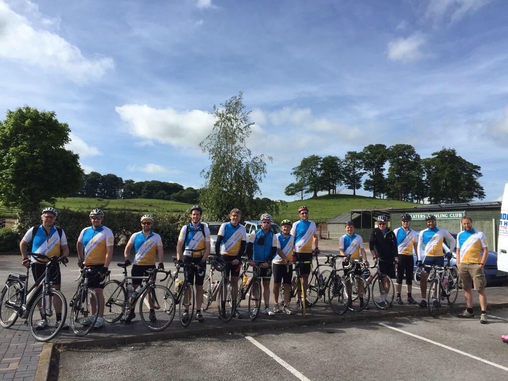
#BAUS15 started to gain momentum from as early as the 26th June 2014 and by the time we entered the Manchester Central Convention Complex well over 100 tweets had been made. Of course it wasn’t just Twitter that started early with a group of keen urologists cycling 210 miles to conference in order to raise money for The Urology Foundation.

Photo 22-06-2015 22 06 04

Monday 15th June 2015

By the time the cyclists arrived conference was well under way with the andrology, FNUU and academic section meetings taking place on Monday morning:

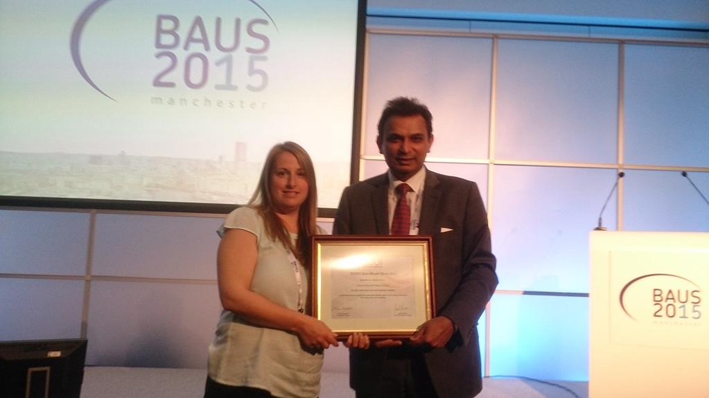
  • The BJU International Prize for the Best Academic Paper was awarded to Richard Bryant from the University of Oxford for his work on epithelial-to-mesenchymal transition changes found within the extraprostatic extension component of locally invasive prostate cancers.
  • Donna Daly from the University of Sheffield received the BJUI John Blandy prize for her work on Botox, demonstrating reductions in afferent bladder signaling and urothelial ATP release.

Photo 22-06-2015 22 06 22Photo 22-06-2015 22 06 18


  • Professor Reisman’s talk on ‘Porn, Paint and Piercing’ as expected drew in the crowds and due to a staggering 44% complication rate with genital piercings it is important for us to try to manage these without necessarily removing the offending article as this will only serve to prevent those in need from seeking medical attention.
  • With the worsening worldwide catastrophe of antibiotic resistance, the cycling of antibiotics for prevention of recurrent UTIs is no longer recommended. Instead, Tharani Nitkunan provided convincing evidence for the use of probiotics and D-Mannose.

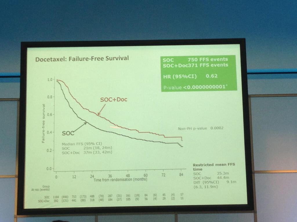
The afternoon was dominated by the joint oncology and academic session with Professor Noel Clarke presenting the current data from the STAMPEDE trial. Zolendronic acid conferred no survival benefit over hormones alone and consequently has been removed from the trial (stampede 1). However, Docetaxal plus hormones has shown benefit, demonstrated significantly in M1 patients with disease-free survival of 65 months vs. 43 months on hormones alone (Hazard ratio 0.73) (stampede 2). This means that the control arm of M1 patients who are fit for chemotherapy will now need to be started on this treatment as the trial continues to recruit in enzalutamide, abiraterone and metformin arms.

Photo 22-06-2015 22 21 00Photo 22-06-2015 22 21 07


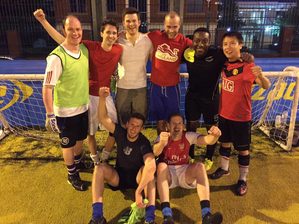
The evening was rounded off with the annual BAUS football tournament won this year by team Manchester (obviously a rigged competition!), whilst some donned the

Photo 22-06-2015 22 23 58Photo 22-06-2015 22 25 07

lycra and set out for a competition at the National Cycle Centre. For those of us not quite so energetic, it was fantastic to catch up with old friends at the welcome drinks reception.


Tuesday 16th June 2015

Photo 22-06-2015 22 29 49

Tuesday kicked off bright and early with Professor John Kelly presenting results from the BOXIT clinical trial, which has shown some benefit over standard treatment of non-muscle invasive bladder cancer, but with significant cardiovascular toxicity.

The new NICE bladder cancer guidelines were presented with concerns voiced by Professor Marek Babjuk over discharging low-risk bladder cancer at 12 months given a quoted 30-50% five-year recurrence risk. Accurate risk stratification, it would seem, is going to be key.

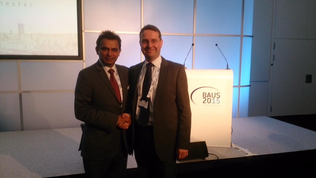
The President’s address followed along with the presentation of the St. Peter’s medal for notable contribution to the advancement of urology, which was presented to Pat Malone from Southampton General Hospital. Other medal winners included Adrian Joyce who received the BAUS Gold Medal, and the St. Paul’s medal went to Mark Soloway.

Photo 22-06-2015 22 33 42

A plethora of other sessions ensued but with the help of the new ‘native’ BAUS app my programme was already conveniently arranged in advance:

  •     ‘Heartsink Conditions’ included pelvic and testicular pain and a fascinating talk by Dr Gareth Greenslade highlighted the importance of early and motivational referral to pain management services once no cause has been established and our treatments have been exhausted. The patient’s recovery will only start once we have said no to further tests: ‘Fix the thinking’
  • Poster sessions are now presented as ‘e-posters’, abolishing the need to fiddle with those little pieces of Velcro and allowing for an interactive review of the posters.


Photo 22-06-2015 22 36 07Pravisha Ravindra from Nottingham demonstrated that compliance with periodic imaging of patients with asymptomatic small renal calculi (n=147) in primary care is poor, and indeed, these patients may be better managed with symptomatic imaging and re-referral as no patients required intervention based on radiograph changes alone.

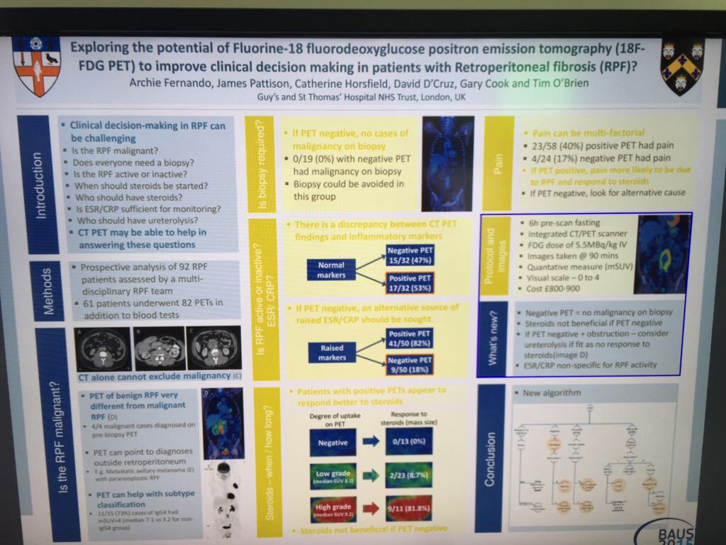
Archana Fernando from Guy’s presented a prospective study demonstrating the value of CTPET in the diagnosis of malignancy in  patients with retroperitoneal fibrosis (n=35), as well as demonstrating that those with positive PET are twice as likely to respond to steroids.


Wednesday 17th June 2015

Another new addition to the programme this year was the Section of Endourology ‘as live surgery’ sessions. This was extremely well received and allowed delegates to benefit from observing operating sessions from experts in the field whilst removing the stressful environment and potential for risk to patient associated with live surgery. This also meant that the surgeon was present in the room to answer questions and talk through various steps of the operation allowing for a truly interactive session.
Wednesday saw multiple international speakers dominating the Exchange Auditorium:

Photo 22-06-2015 22 39 51Photo 17-06-2015 14 46 29

  • The BJU International guest lecture was given by Professor Hendrik Van Poppel: a heartfelt presentation describing what he believes to be the superiority of surgery over radiotherapy for high-risk localised prostate cancer.
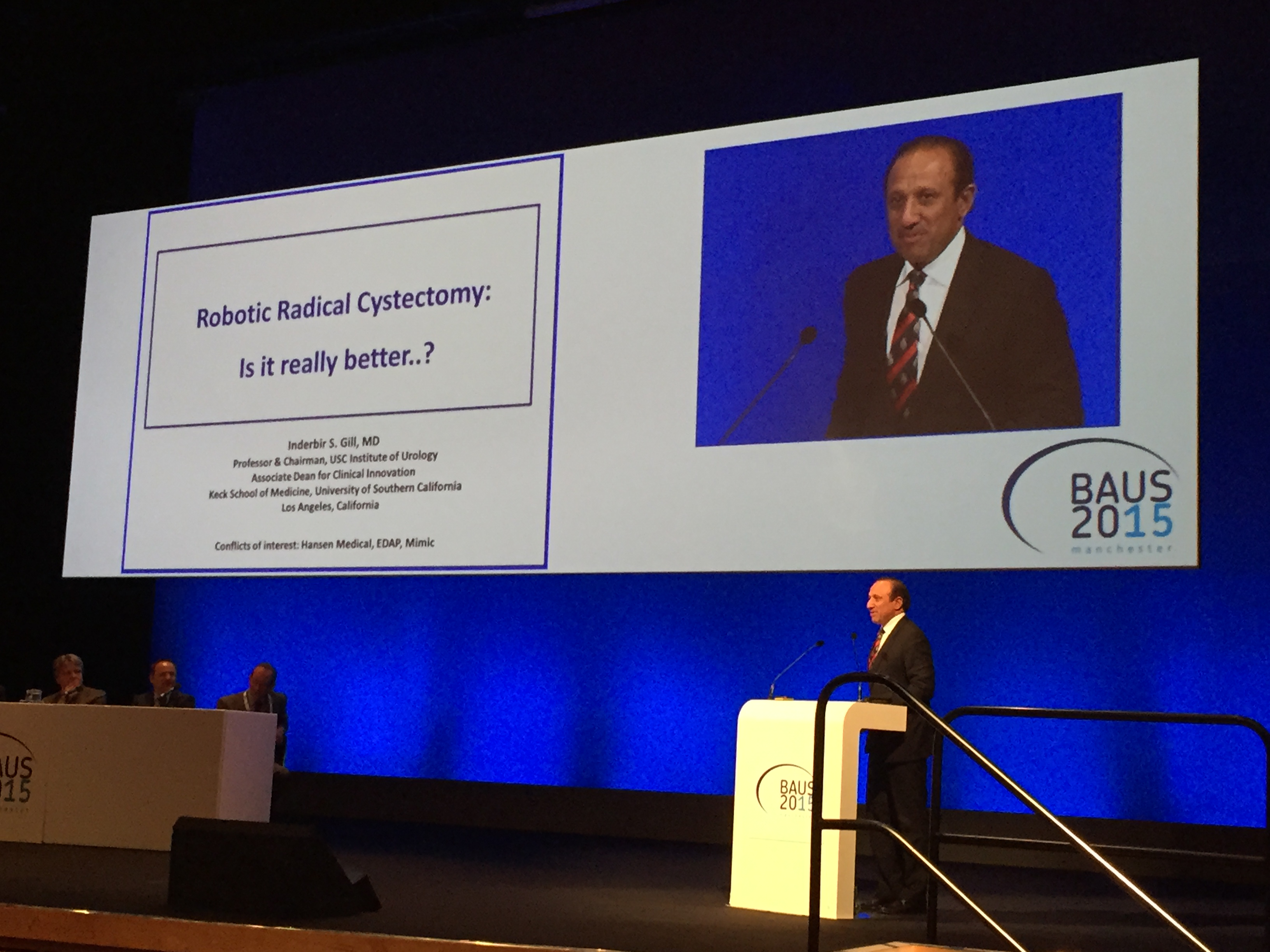
  • The Urology Foundation presented the Research Scholar Medal to Ashwin Sachdeva from Freeman Hospital, Newcastle for his work on the ‘Role of mitochondrial DNA mutations in prostate carcinogenesis’. This was followed by an inspiring guest lecture by Inderbir Gill on ‘Robotic Urologic Oncology: the best is yet to come’ with the tag line ‘the only thing that should be open in 2015 is our minds’
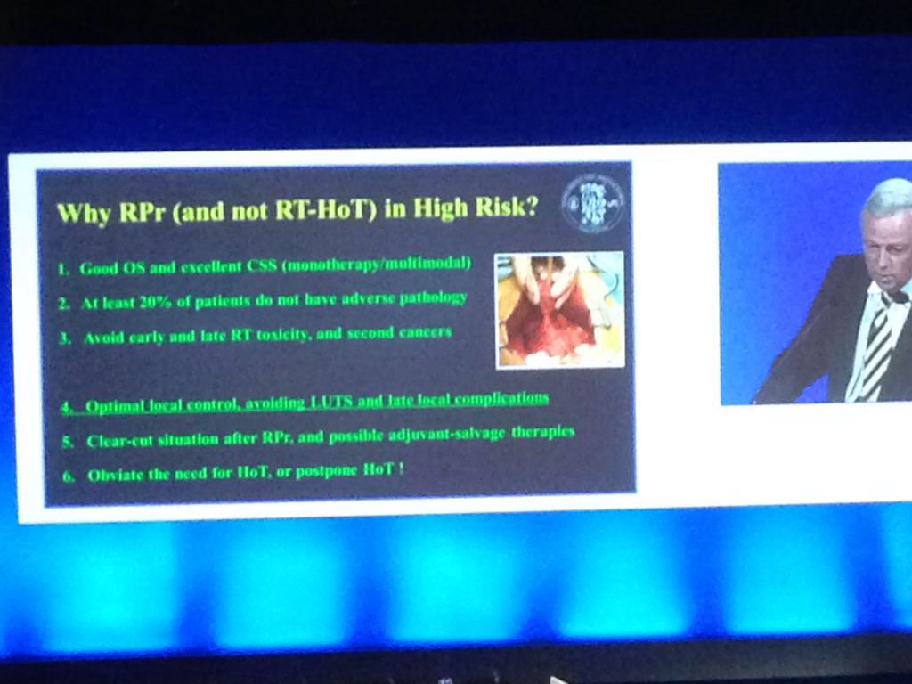
  • Robotic Surgery in UK Urology: Clinical & Commissioning Priorities was a real highlight in the programme with talks from Jim Adshead and Professor Jens-Uwe Stolzenburg focussing on the fact that only 40% of T1a tumours in the UK were treated with partial (as opposed to radical) nephrectomy, and that the robot really is the ‘game-changer’ for this procedure. Inderbir Gill again took to the stage to stress that all current randomised trials into open vs. robotic cystectomy have used extracorporeal reconstruction and so do not reflect the true benefits of the robotic procedure as the dominant driver of complications is in the open reconstruction.

These lectures were heard by James Palmer, Clinical Director of Specialised Commissioning for NHS England who then discussed difficulties in making decisions to provide new technologies, controlling roll out and removing them if they show no benefit. Clinical commissioning policies are currently being drafted for robotic surgery in kidney and bladder cancer. This led to a lively debate with Professor Alan McNeill having the last word as he pointed out that what urologists spend on the robot to potentially cure cancer is a drop in the ocean compared with what the oncologists spend to palliate!


Thursday 18th June 2015

The BJU International session on evidence-based urology highlighted the need for high-quality evidence, especially in convincing commissioners to spend in a cash-strapped NHS. Professor Philipp Dahm presented a recent review in the Journal of Urology indicated that the quality of systematic reviews in four major urological journals was sub-standard. Assistant Professor Alessandro Volpe then reviewed the current evidence behind partial nephrectomy and different approaches to this procedure.

Another fantastic technology, which BAUS adopted this year, was the BOD-POD which allowed delegates to catch-up on sessions in the two main auditoria that they may have missed due to perhaps being in one of the 21 well designed teaching courses that were available this year. Many of these will soon be live on the BAUS website for members to view.

The IBUS and BAUS joint session included a lecture from Manoj Monga from The Cleveland Clinic, which led to the question being posed on Twitter: ‘Are you a duster or a basketer?’The audience was also advised to always stent a patient after using an access sheath unless the patient was pre-stented.

Photo 23-06-2015 20 28 55


The updates session is always valuable especially for those studying for the FRCS (Urol) exam with far too many headlines to completely cover:

  • Endourology: The SUSPEND trial published earlier this year was a large multi-centre RCT that showed no difference in terms of rates of spontaneous passage of ureteric stone, time to stone passage or analgesic use between placebo, tamsulosin and nifedipine. There was a hot debate on this: should we be waiting for the meta-analysis or should a trial of this size and design be enough to change practice?
  • Oncology-Prostate: The Klotz et al., paper showed active surveillance can avoid over treatment, with 98% prostate cancer survival at 10 years.
  • Oncology-Kidney: Ellimah Mensah’s team from Imperial College London (presented at BAUS earlier in the week) demonstrated that over a 14-year period there were a higher number of cardiovascular-related admissions to hospital in patients who have had T1 renal tumours resected than the general population, but no difference between those who have had partial or radical nephrectomy.
  • Oncology-Bladder: Arends’s team presented at EAU in March on the favourable results of hyperthermic mitomycin C vs. BCG in the treatment of intermediate- and high-risk bladder cancer.
  • Female and BPH: The BESIDE study has demonstrated increased efficacy with combination solifenacin and mirabegron.
  • Andrology: Currently recruiting in the UK is the MASTER RCT to evaluate synthetic sling vs. artificial sphincter in men with post-prostatectomy urinary incontinence.


Overall BAUS yet again put on a varied and enjoyable meeting. The atmosphere was fantastic and the organisers should be proud of the new additions in terms of allowing delegates to engage with new technologies, making for a memorable week. See you all in Liverpool!


Photo 18-06-2015 12 15 03

Rebecca Tregunna, Urological Trainee, West Midlands Deanery @rebeccatregunna


Dominic Hodgson, Consultant Urologist, Portsmouth @hodgson_dominic


Papillary Renal Cell Carcinoma and Clear Cell Renal Cell Carcinoma Arising in a Single Kidney

We report a case of clear cell RCC and papillary RCC arising in the same kidney.

Authors: Tait, Laura; Coleman, Pamela; Ahaghotu, Chiledum
Corresponding Author: Tait, Laura Department of Surgery-Division of Urology, Howard University Hospital Washington, DC


Introduction
Renal cell carcinoma (RCC) is responsible for 80 percent of all primary renal neoplasms and approximately 65,000 Americans are diagnosed with RCC each year (1). Clear cell RCC comprises a majority of all RCC tumors diagnosed histologically. They arise from the proximal tubule and macroscopically appearsolid or cystic (2). Clear cell RCC occurs most commonly as a sporadic disease. Papillary RCC are diagnosed in 10 to 15 percent of patients with RCC (3, 4, 5, 6). As with clear cell cancers, papillary RCCs originate from the proximal tubule, but they are morphologically and genetically distinct malignancies. Papillary carcinomas are frequently multifocal and bilateral, and commonly present as small, early stage tumors (7). For localized disease, radical nephrectomy has been the most widely used approach and remains the preferred procedure for cases suggestive of renal malignancy. The diagnosis of clear cell RCC has been established by identifying distinct morphological and histologic features of this tumor following nephrectomy. These unique qualities include neoplastic cells with clear cytoplasm in an acinar growth pattern. These findings separate it from papillary RCC in which psammoma bodies and a strong positive staining for CK7 are characteristic. This case is unique in that there were no prior available reports describing two RCC tumors of separate origin found within a single kidney. We report a case of clear cell RCC and papillary RCC arising in the same kidney.

Case report
A 55-year-old African American male presented to his PCP with a two-week history of hematuria. The patient’s past medical history included hypertension, hepatitis c, and a hernia repair. He was referred to urology and a computed tomography scan demonstrateda complex heterogenous cyst within the right kidney with characteristics suggestive of malignancy and the presence of a left pelvic kidney. Right nephrectomy was performed. Two renal tumors were noted upon gross examination. The main tumor mass was a well-circumscribed, lobulated, homogeneous yellow tumor at the inferior pole of the kidney measuring 4.2×4.0x3.6 cm and demonstrated microscopic invasion of the renal sinus. The satellite tumor was found to be compressing the renal parenchyma at the inferior pole and was a 0.6 cm, firm, well-circumscribed nodule with homogeneous, tan/grey cut surface with characteristic hilar vessel wall invasion. Renal tumor tissues obtained at operation wasfixed in formalin and embedded in paraffin. The specimens were serially sectioned using three-micrometer thick cuts. The sections were stained with hematoxylin-eosin and immunohistochemical studies were undertaken. CK7 staining was performed with the satellite papillary renal cell carcinoma showing strong positive staining. CK7 staining was negative within the main tumor mass. The pathology results provide evidence that clear cell RCC and papillary RCC can simultaneously arise within a single kidney.

Pathology
Macroscopically, the kidney showed no gross abnormalities. The tumor measuring 4.2 x 4.0 x 3.6 cm and the tumor measuring 0.6 cm were observed in the inferior pole. The cut surface of the main tumor showed a yellowish color while the satellite tumor had a tan/grey cut surface appearance. At the posterior/inferior pole it was noted that the renal capsule was expanded by a gelatinous blood clot. The cut surface is shown (Fig. 1).

Microscopically, the main tumor located at the inferior pole showed a proliferation of neoplastic cells with clear cytoplasm and an acinar growth pattern. The nuclear characteristics were a Fuhrman nuclear grade 4 (Fig. 2). Given this tumor’s microscopic characteristics, a diagnosis of clear cell carcinoma was made.

The satellite tumor showed a proliferation of neoplastic cells with foamy macrophages and psammoma bodies (Fig. 3). This satellite tumor also demonstrated strong positive staining for CK7, a well-known tumor marker for papillary renal cell carcinoma. Based on the microscopic and immunohistologic findings, a diagnosis of papillary renal cell carcinoma was made.

Discussion
This case report describes a unique finding of clear cell and papillary RCC within a single kidney. The pathology of this conditionis described to illustrate how both tumors were morphologically different from one another. This particular anomaly has not been previously described in the literature.
The risk factors for RCC have been well-documented and include smoking, hypertension, occupational exposure to toxic compounds, obesity, acquired cystic disease of the kidney, analgesic abuse nephropathy, and genetic predisposition (8,9). These documented findings are similar to our case report in that the patient had a history of hypertension. The literature suggests that hypertension predisposes to the development of RCC. Although our patient had high blood pressure for many years, the incidence of RCC seems to be independent of anti-hypertensive medications. Therefore, hypertension may have played a role in the development of clear cell and papillary RCC seen in this patient.
Another risk factor our patient had for developing RCC waschronic hepatitis c infection. An epidemiologic study of over 67,000 patients found that chronic infection with the hepatitis C virus was associated with a significantly increased risk of RCC (10). This risk factor combined with hypertension may have put our patient at increased risk for RCC.
Although no clear cell RCC and papillary RCC have been found simultaneously before, a coexisting RCC can be identified in 10 to 32 percent of patients with oncocytoma (11). This evidence demonstrates that two unique RCC tumors can exist in a single kidney. Our findings of two RCC within the same kidney is consistent with past evidence of simultaneous tumors but are new in that none have reported clear cell and papillary RCC. Therefore, in our case, we were able to determine the possible risk factors predisposing our patient to RCC and report the new discovery of two RCC tumors within the same kidney.

Conclusions
We present a unique example of clear cell and papillary RCC within the same kidney. Histology of the tissues at the time of nephrectomy confirmed the diagnosis of two distinct renal tumors. Risk factors for RCC seen in this patient include hypertension and chronic hepatitis c. Although there are reports of oncomytoma and coexisting RCC, there have been no observed findings of clear cell and papillary RCC simultaneously.

060Fig1

Fig. 1

060Fig2

Fig. 2

060Fig3

Fig.3

060Image1

Image 1

References
1. Siegel R, Naishadham D, Jemal A. Cancer statistics, 2012. CA Cancer J Clin 2012; 62:10.
2. Presti JC Jr, Rao PH, Chen Q, et al. Histopathological, cytogenetic, and molecular characterization of renal cortical tumors. Cancer Res 1991; 51:1544.
3. Tannenbaum M. Ultrastructural pathology of human renal cell tumors.PatholAnnu 1971; 6:249.
4. Thoenes W, Störkel S, Rumpelt HJ. Histopathology and classification of renal cell tumors (adenomas, oncocytomas and carcinomas).The basic cytological and histopathological elements and their use for diagnostics.Pathol Res Pract 1986; 181:125.
5. Störkel S, van den Berg E. Morphological classification of renal cancer. World J Urol 1995; 13:153.
6. Patard JJ, Leray E, Rioux-Leclercq N, et al. Prognostic value of histologic subtypes in renal cell carcinoma: a multicenter experience. J ClinOncol 2005; 23:2763.
7. Beck SD, Patel MI, Snyder ME, et al. Effect of papillary and chromophobe cell type on disease-free survival after nephrectomy for renal cell carcinoma. Ann SurgOncol 2004; 11:71.
8. Mandel JS, McLaughlin JK, Schlehofer B, et al. International renal-cell cancer study. IV. Occupation. Int J Cancer 1995; 61:601.
9. Wolk A, Gridley G, Niwa S, et al. International renal cell cancer study. VII. Role of diet. Int J Cancer 1996; 65:67.
10. Gordon SC, Moonka D, Brown KA, et al. Risk for renal cell carcinoma in chronic hepatitis C infection. Cancer Epidemiol Biomarkers Prev 2010; 19:1066.
11. Chao DH, Zisman A, Pantuck AJ, et al. Changing concepts in the management of renal oncocytoma. Urology 2002; 59:635.


Date added to bjui.org: 12/12/2012

DOI: 10.1002/BJUIw-2012-060-web


Port–Site recurrence following Laparoscopic Radical Nephrectomy for Chromophobe Renal Cell Carcinoma

Port-site metastasis following laparoscopic radical nephrectomy is being increasingly recognized as a complication following laparoscopic surgery, especially when correct surgical principles are violated. All previously reported cases have been of either the clear cell or papillary variant of renal cell carcinoma. Herein we report a case of chromophobe renal cell carcinoma with port-site recurrence 10 months after laparoscopic radical nephrectomy.

Authors: Javali, Tarun; Dogra, Premnath; Singh, Prabhjot
Corresponding Author: Javali, Tarun


Introduction
Port-site metastasis following laparoscopic radical nephrectomy is being increasingly recognized as a complication following laparoscopic surgery, especially when correct surgical principles are violated. All previously reported cases have been of either the clear cell or papillary variant of renal cell carcinoma. Herein we report a case of chromophobe renal cell carcinoma with port-site recurrence 10 months after laparoscopic radical nephrectomy.

Case History
A 44 year old lady presented with a 6 month history of left flank pain and hematuria. Computed tomography revealed a 12×5×5 cms heterogeneous enhancing left renal upper pole mass. [Fig.1]. There was no evidence of lymph node enlargement or liver metastasis, and no tumor thrombus. The CT scan also revealed a multiloculated right adnexal cyst of size 6×4×4 cms. The patient underwent laparoscopic transperitoneal left radical nephrectomy and right oophorectomy. Additional ports were placed for the oophorectomy. The radical nephrectomy was performed first because it was deemed to be more technically challenging than the oophorectomy. Both the specimens were entrapped separately in retrieval bags and removed through a Pfannensteil incision. The specimen was not morcellated.
Histopathology revealed a chromophobe renal cell carcinoma. On immunohistochemical analysis, the tumor was positive for cytokeratin and negative for CD10 and vimentin. There was no evidence of any capsular breach. The tumor had reached the renal sinus but had not infiltrated it. Histopathology of the right oophorectomy specimen showed an endometrial cyst.
The patient was reviewed on a three monthly basis with a chest x-ray, liver and kidney function tests and a full blood count. No abdominal imaging was done at this time. Ten months after surgery, the patient presented with pain and swelling at one of the port sites. Examination revealed a hard subcutaneous nodule of size 2×1.5 cm at the 12mm working port site above and medial to the left anterior superior iliac spine [Fig 2]. The surgical incision site for specimen removal had no evidence of recurrence. The patient had already had fine needle aspiration cytology (F.N.A.C.) performed, which was reported as showing ‘malignant cells suspicious for renal cell carcinoma’. A contrast enhanced CT scan of her abdomen revealed an irregular enhancing subcutaneous soft tissue lesion in left anterior abdominal wall measuring 1.8 cms, thought likely to be a metastasis. The liver and right kidney were normal and there were no retroperitoneal nodes or masses. Positron emission tomography-computed tomography (PET-CT) revealed increased 18-fluro deoxy glucose (18FDG) uptake in the soft tissue density lesion in left lower abdominal wall [Fig 3]. The patient underwent wide local excision of the port-site nodule. Histology revealed chromophobe renal cell carcinoma with the same histomorphology as the primary tumour [Fig 4,5].
At one year post excision of the port-site nodule, patient is well, and has had no recurrences, either local or systemic.

Discussion
Factors which have been implicated in port-site recurrence include natural tumor factors, local wound factors, immune response and laparoscopic related factors such as direct wound contamination, either instrument contamination or during specimen extraction, specimen morcellation, use of specimen retrieval bags and pneumoperitoneum pressure [1,2]. Poorly differentiated transitional cell carcinomas have accounted for most of the cases of port site recurrence in laparoscopic uro-oncologic case series [1,2,3]. Only a few cases of port site recurrence following laparoscopic nephrectomy for renal cell carcinoma have been reported [4,5,6,7,8,9,10]. In all of these cases the histopathology was clear cell carcinoma. Russo et al reported a case of papillary renal cell carcinoma with port site metastasis following laparoscopic partial nephrectomy [11]. To the best of our knowledge this the first case of chromophobe renal cell carcinoma with port site recurrence. In the present case the tumor was organ confined (pT2) and well differentiated. Most studies report more favorable prognosis for chromophobe compared to conventional renal cell carcinoma. This goes against the commonly held notion that adverse tumor biology or aggressive nature of the tumor is the most important causative factor in port site recurrence [1]. Greco et al have further elaborated on the factors that accelerate the development of port-site metastasis [12]. These conditions include performing laparoscopic surgery in the presence of ascites, lack of trocar fixation to prevent dislodgement and gas leakage around the trocars, inadequate laparoscopic equipment and technique and tumour boundary violation. Factors which have deemed to reduce the incidence of port-site metastases include use of a bag for specimen retrieval, placement of drainage before desufflation, povidone-iodine irrigation of instruments, trocars and port-site wounds and suturing trocar wounds ≥10mm in size.
In the present case, all possible precautions were taken during the course of surgery, including changing the instruments, once the radical nephrectomy was over. A probable etiologic factor in this case may have been the prolonged operating time as laparoscopic nephrectomy was followed by laparoscopic oophorectomy and the gynaecologists also used the left sided 12mm working port. Microleakage around ports (“chimney effect”) has been postulated to play a role in port site metastasis [13]. Ports used by the main operating surgeon have been proved to have more contamination by tumor cells than either those used by the assistants or the camera port [14]. It is a matter of conjecture that whether performing the oophorectomy first followed by radical nephrectomy could have altered the result.
We wish to highlight two main points through this case report. Firstly, that tumor histology may not be predictive of port site recurrence. Chromophobe renal cell carcinoma is biologically a tumor of low malignant potential. Metastasis of chromophobe tumour constitute less than 1% of all metastatic renal cell carcinoma [15]. Advanced pathological T stage, tumor necrosis and sarcomatoid change have been purported to predict an aggressive phenotype of chromophobe renal cell carcinoma [16]. However none of these features were present in this case. Hence following laparoscopic radical nephrectomy patients need to be carefully examined at each visit with particular attention to the port sites. This should be done regardless of the grade, stage or histology of the tumor.
The second point we wish to highlight is that PET-CT could be a useful adjunct in diagnosing port-site recurrence in equivocal cases. This may be especially relevant in patients who return within a short span of time following laparoscopic radical nephrectomy, wherein induration due to surgical factors at scar site may be confused vis-a vis port site recurrence.

035fig1


Fig. 1 – CT KUB (Kidney-ureter-bladder) showing left upper pole tumor.

035fig2


Fig. 2 – Port-site nodule

035fig3


Fig. 3 – PET-CT showing port-site metastasis

035fig4


Fig. 4 – Hematoxylin and Eosin staining of excised port-site specimen. Sheets of cells with round to oval nuclei and perinuclear halo with prominent cell membranes.

035fig5


Fig. 5 – Immunohistochemistry. Negative for CD10 and vimentin.

References
1. Rassweiler J, Tsivian A, Kumar AV, Lymberakis C, Schulze M, Seeman O, et al. Oncological safety of laparoscopic surgery for urological malignancy: experience with more than 1,000 operations. J Urol 2003; 169:2072–5.
2. Tsivian A, Sidi AA. Port site metastases in urological laparoscopic surgery. J Urol 2003; 169:1213–8.
3. Micali S, Celia A, Bove P, et al: Tumor seeding in urological laparoscopy: an international survey. J Urol 2004; 171: 2151–4.
4. Fentie DD, Barret PH, and Taranger LA: Metastatic renal cell carcinoma after laparoscopic radical nephrectomy: long term follow-up. J Endourol 2000; 14: 407–11.
5. Landman J, and Clayman R: Re: port site tumor recurrences of renal cell carcinoma after videolaparoscopic radical nephrectomy (letter). J Urol 2001; 166: 629–30..
6. Castilho LN, Fugita OEH, Mitre AI, et al: Port site tumor recurrences of renal cell carcinoma after videolaparoscopic radical nephrectomy. J Urol 2001; 165: 519.
7. Chen YT, Yang SSD, Hsieh CH, et al: Hand port-site metastasis of renal-cell carcinoma following hand-assisted laparoscopic radical nephrectomy: case report. J Endourol 2003; 17: 771–4.
8. Iwamura M, Tsumura H, Matsuda D, et al: Port site recurrence of renal cell carcinoma following retroperitoneoscopic radical nephrectomy with manual extraction without using entrapment sac or wound protector. J Urol 2004; 171: 1234–5.
9. Dhobada S, Patankar S, Fais F, et al: Port-site metastasis after laparoscopic radical nephrectomy for renal-cell carcinoma: case report. J Endourol 2006; 20: 119–22.
10. Goyal R., Sing P., Mandhani A. et al. Port-site metastatis of renal cell carcinoma after laparoscopic transperitoneal radical nephrectomy. Ind J of Urol 2006; 22:150-1.
11. Masterson TA, Russo P. A case of port-site recurrence and loco-regional metastasis after laparoscopic partial nephrectomy. Nature Reviews Urology 2008; 5:345-9.
12. Greco F, Wagner S, Reichelt O, Inferrera A, Lupo A, Hoda MR, Hamza A, Fornara P
Huge port-site metastasis after laparoscopic partial nephrectomy: a case report. Eur Urol 2009; 56:737-39.
13. Curet MJ: Port site metastases. Am J Surg 2004; 187: 705-12.
14. Ramirez PT, Wolf JK, and Levenback C: Laparoscopic port-site metastases: etiology and prevention. Gynecol Oncol 2003; 91: 179–89.
15. Choueiri TK, Plantade A, Elson P et al. Efficacy of sunitinib and sorafenib in metastatic papillary and chromophobe renal cell carcinoma. J Clin Oncol 2008; 26:127-31.
16. Amin MB, Paner GP, Alvarado-Cabrero I et al. Chromophobe renal cell carcinoma: histomorphologic characteristics and evaluation of conventional pathologic prognostic parameters in 145 cases. Am J Surg Pathol 2008; 32:1822-34.


Date added to bjui.org: 05/12/2012

DOI: 10.1002/BJUIw-2012-035-web


Microwave ablation of a small cortical renal tumour – an unexpected adverse outcome

Percutaneous ablation therapies have become an increasingly important treatment option for smaller volume renal tumours. Microwave ablation (MWA) appears to have significant advantages over radiofrequency ablation (RFA) in terms of treatment times and achievable ablation zone volumes. We report the case of a fit 71 year old gentleman with a 17mm slow-growing cortical lower pole renal carcinoma. Given the small volume nature of the tumour, MWA was used instead of cryoablation (CRA) so as to avoid the potential need for multiple cryoprobes. The tumour was successfully treated with MWA with no immediate post-operative complications. On day 7, the patient returned with flank pain and CT confirmed a pelvi-calyceal leak requiring retrograde stenting. Unexpectedly, at five months, a follow-up CT indicated global hypoperfusion. This was confirmed with a MAG-3 renogram showing only 12% contribution to overall function. To date, there have been few reports of microwave ablation of renal tumours. This small, cortical renal tumour would have been expected to undergo straightforward ablation treatment. The subsequent functional demise of the kidney was unexpected in this case. The mechanism of global renal injury remains unclear and more research regarding ablation physiology and dosimetry is required.

Authors: Ingram, Liam ; Hookway, Max; Breen, David
Corresponding Author: Ingram, Liam


Introduction
The percutaneous treatment of renal tumours by ablation therapies is now increasingly accepted as a useful therapeutic option. Currently the routinely available options are cryoablation (CRA) or thermal ablation using either radiofrequency (RFA) or microwave (MWA) technology. Traditionally, RFA has been the mainstay of treatment causing targeted cellular death via coagulative necrosis. Whilst RFA has a relatively well documented efficacy, microwave ablation has great potential to overcome some of the disadvantages of RFA. The main theoretical advantages are in terms of ablation zone volumes achieved (1, 2), time efficacy and there being no requirement for grounding pads (3, 4).
Each technology has a specific mode of tissue ablation and hence treated tissue characteristics are an important consideration in this case. For microwave ablation the important physical features are relative permeability and effective conductivity (3). For the kidney, high renal perfusion rates can compromise treatment by yielding inconsistent, geometrically variable ablation zones (3, 5). These effects are particularly evident close to larger peripelvic vessels. Microwave technology has great potential here due to its rapid tissue heating characteristics; however, accurate treatment dosimetry still remains an issue similar to RFA.
There is a relative paucity of literature regarding microwave ablation of renal tumours. A literature search has found 3 small studies showing preliminary data regarding microwave ablation of small renal tumours. The results are mixed, with 2 studies showing good technical outcomes and few complications (6, 7) and the third showing a poor technical outcome with associated intra-operative and post-operative complications as high as 20 and 40%, respectively (8).

Case report
The patient was referred from another hospital for minimally invasive, image-guided biopsy and ablation given the small but solid nature of the lower pole cortical mass.
He was 71 and previously fit and well, with no significant past-medical history other than a single episode of atrial fibrillation 14 years ago. He was not taking any medication and was symptom free.
The lesion was 17mm in size and peripheral in the cortex of the lower pole of the right kidney (Figure 1).

fig11


Figure 1. Portal venous phase CT confirming a solid but hypovascular tumour towards the lower pole of the right kidney.

For the last 3 ½ years CRA rather than RFA has been the mainstay of small renal tumour ablation at our institution. But, given the small nature of the tumour and benefit of a single probe and single site treatment, MWA was agreed. The patient was given pre-operative prophylactic intravenous antibiotics (metronidazole and cefuroxime) 1 hour before the procedure. He was positioned prone under general anaesthetic, and 2 CT guided 18G core biopsies of the lesion were taken. A single stick microwave probe was placed within the lesion under CT guidance. Position was confirmed and microwave ablation was performed. The first cycle was for 3mins 28secs at 180W followed by a 1.5cm draw back and second cycle of 4 mins at 180W with subsequent track ablation.
There were no immediate complications. The patient was sent back to the ward and discharged the following day. Histology of his biopsies showed low grade nuclear features consistent with a partly cystic well differentiated clear cell carcinoma.
On day 7 post-procedure, the patient was re-admitted due to increasing right flank pain. He underwent CT of his abdomen and pelvis with arterial, portal venous and delayed phases (5 mins) being obtained. These showed an adequate, wedge-shaped, non-enhancing ablation zone (Figure 2).The 5 minute delayed study however confirmed free leakage from the lower pole calyx with a moderate retroperitoneal urinoma. On the next day the patient underwent cystoscopic placement of a retrograde ureteric stent to divert the calyceal urine leakage.

fig21


Figure 2. Portal venous phase CT at day 7 post-procedure showing adequate ablation site and urinoma.

A repeat CT at 1 month showed resolution of the urinoma with no further leak. Again the lesion appeared well treated with a wedge-shaped area of non-enhancement, Figure 3.

fig31


Figure 3. Wedge of non-enhancement of ablation zone on portal venous phase CT 1 month post-procedure. Ureteric stent in situ.

At 5 months post-treatment a further follow-up CT scan demonstrated globally diminished perfusion of the right kidney (Figure 4). This was confirmed by a MAG3 renogram which showed only 12% of the total function attributable to the right kidney.

Figure41

Figure 4. Global hypoperfusion of the right kidney seen on portal venous phase CT at 5 months post-procedure.

Discussion
MWA offers significant theoretical benefits over RFA in terms of reduced treatment time, ablation volumes achieved and possibly more predictable treatment geometry. In this case, the small cortical tumour was appropriately treated as evidenced by the typical wedge-shaped ablation zone. With hindsight the first station of the probe was likely in too close a proximity to the lower pole calyx. It could also be argued that the 2 station treatment of 3 minute 28 secs at 180W followed by a further 4 minutes at 180 W peripherally in the cortex represented local over-treatment. But the ablation zone at 7 days and at 1 month suggests that this was not the case and that the resultant ablation zone was only adequate for the tumour ablation undertaken. Even if the calyceal injury could have been avoided by rather more ‘cortical’ positioning of the probe, the propensity for microwave ablation to cause pelvicalyceal injury in this case is striking and more so than might have been expected with image-guided cryoablation. In fact this observation has been confirmed by in vivo porcine studies where thermal based RFA was found to incur pelvicalyceal injury much more readily than CRA for the same probe positioning and ablation zone volumes (9, 10).
The clear benefit of ablation is to offer a more targeted treatment whilst maintaining overall renal function. The unexpected procedural outcome here is the subsequent delayed global hypoperfusion and hence loss of function of the kidney at 4-5 months. This suggests that microwave energy deposition within the kidney may have a greater propensity to cause a more diffuse and non-target renal injury.
The key issue in this case is treatment dosimetry and to date little or no data is available on this issue from MWA manufacturers. Whilst the ablation zone achieved in this case was only adequate to ablate the target tumour, the mechanism of the subsequent functional demise of the kidney remains unclear but likely related to a diffuse microwave heating effect. There is clearly a requirement for further in vivo MWA dosimetry clarification, likely using a porcine renal model.

References
1. Laeseke, P.F., et al. Microwave ablation versus Radiofrequency ablation in the
kidney: High-power triaxial antennas create larger ablation zones than
similarly sized internally cooled electrodes. JVIR. 2009 Sep; 20(9): 1224-9.
2. Clark, P.E., Woodruff, R.D., Zagoria, R.J., Hall, M.C. Microwave ablation of renal parenchymal tumours before nephrectomy: phase I study. AJR AM J Roentgenol. 2007 May;188(5):1212-4
3. Brace, C.L. Radiofrequency and microwave ablation of the liver, lung, kidney and bone: What are the differences? Curr Probl Diagn Radiol. 2009 May-Jun; 38(3):135-143
4. Simon, C. et al. Microwave Ablation: Principles and Applications. Radiographics. 2005 Oct;25: S69-S83
5. Moore, C., et al. Effects of microwave ablation of the kidney. J Endourol. 201 Mar;24(3)439-44
6. Carrafiello, G., et al. Single-antenna microwave ablation under contrast-enhanced ultrasound guidance for treatment of small renal cell carcinoma: preliminary experience. Cardiovasc Intervent Radiol. 2009 Apr; 33(2): 367-74.
7. Liang, P., et al. Ultrasound guided percutaneous microwave ablation for small renal caner: initial experience. J Urol. 2008 Sep; 180(3):844-8
8. Castle, S.M., Salas, N., Leveillee, R.J. Initial experience using microwave ablation therapy for renal tumour treatment: 18-month follow-up. Urology. 2011 Apr; 77(4):792-7
9. Janzen, N. et al. The effects of intentional cryoablation and radiofrequency ablation of renal tissue involving the collecting system in a porcine model. J Urol. 2005 Apr;173(4):1368-74.
10. Brashears, J. et al. Renal ablation and radio frequency ablation: an evaluation of worst case scenarios in a porcine model. J Urol. 2005 Jun;173(6):2160-5.


Date added to bjui.org: 16/11/2012
DOI: 10.1002/BJUIw-2012-065-web


© 2024 BJU International. All Rights Reserved.