Tag Archive for: Renal

Posts

Article of the Month: Recent advances in immuno-oncology and its application to urological cancers

Every Month the Editor-in-Chief selects an Article of the Month from the current issue of BJUI. The abstract is reproduced below and you can click on the button to read the full article, which is freely available to all readers for at least 30 days from the time of this post.

In addition to the article itself, there is an accompanying comment written by a prominent member of the urological community. This blog is intended to provoke comment and discussion and we invite you to use the comment tools at the bottom of each post to join the conversation.

If you only have time to read one article this week, it should be this one.

Recent advances in immuno-oncology and its application to urological cancers

Jennifer M. Mataraza and Philip Gotwals


Exploratory Immuno-Oncology, Novartis Institutes for BioMedical Research, Cambridge, MA, USA


Abstract

Recent advances in immuno-oncology have the potential to transform the practice of medical oncology. Antibodies directed against negative regulators of T-cell function (checkpoint inhibitors), engineered cell therapies and innate immune stimulators, such as oncolytic viruses, are effective in a wide range of cancers. Immune‘based therapies have had a clinically meaningful impact on the treatment of advanced melanoma, and the lessons regarding use of single agents and combinations in melanoma may be applicable to the treatment of urological cancers. Checkpoint inhibitors, cytokine therapy and therapeutic vaccines are already showing promise in urothelial bladder cancer, renal cell carcinoma and prostate cancer. Critical areas of future immuno-oncology research include the prospective identification of patients who will respond to current immune-based cancer therapies and the identification of new therapeutic agents that promote immune priming in tumours, and increase the rate of durable clinical responses.

oct-aotm-results

Comment: Immune checkpoint blockade – a treatment for urological cancers?

Introduction

In the last few years there have been concerted attempts at using the power of the immune system as an effective treatment option for cancer. This has become possible as our understanding of the workings of the immune system has improved. Tumours form because of failure of the organism to destroy a rogue, mutated cell in an appropriate way. Once the tumour is formed it can further develop when the immune system fails to contain and control it and certain equilibrium is lost in favour of the tumour. This is referred to as the immune editing theory. At this point of failure of the immune system, tumour growth and progression become possible and tumours develop various mechanisms to evade the immune systems surveillance. Therefore, a mechanism to restore the lost equilibrium or to tip it in favour of the immune system would be a new modality in anti-cancer treatment. The initial approach was to use stimulators of the immune system systemically such as interleukin 2 and interferon γ i.v. in patients with metastatic cancers including melanoma and renal cancers [1]. A sustained response was shown in 22% of patients with metastatic kidney cancer, lasting for over a year. Although this treatment was not a resounding success, it did highlight an approach that could yield a durable tumour regression in a minority of cases. As our understanding of the immune system–tumour interaction further developed, new research focused on a specific mechanism in the immune system that seems to be exploited by tumours. This is an activation-inhibition mechanism, which controls the extent of adaptive immune response to invading organisms or to mutated cancer cells. In healthy individuals, this mechanism is a ‘safety’ feature allowing cessation of the immune response once it has performed its task. This is controlled by ‘receptor’ molecules at the T-cell surface and their corresponding ‘ligands’ at the surface of the cells interacting with the T-cell, which can be an antigen presenting cell or a tumour cell surface. This mechanism is called the immune checkpoint [2] (Fig. 1). Cytotoxic T lymphocyte antigen 4 (CTLA-4) and programmed death 1 (PD-1) are the most well-known checkpoints but there are up to 20 others (and counting) [2]. Their main role is to inhibit an immune response by blocking the activation of T-cells when those cells are presented with a foreign antigen or cancer proteins. This inhibition leads to immune ‘tolerance’ of the presence of cancer cells. So the policeman (T cell) is oblivious to the robbery in front of him. Thus an anti-tumour treatment strategy to disrupt the immune checkpoints seems to be a valid one (Table 1).

image

Figure 1. Blocking checkpoint inhibitors with antibodies is the new immunotherapy strategy to unlock T-cell activation and improve anti-tumour immune response. MHC, major histocompatibility complex; PD-L1, programmed death ligand 1; TCR, T-cell antigen receptor.

Table 1. A selected group of trials of immune checkpoint inhibitors in urological cancers
Tumour Phase Treatment N Results Trial.gov identifier
  1. mCRPC, metastatic castrate-resistant prostate cancer; PD-L1, programmed death ligand 1. The total number of current trials of immune checkpoint inhibitors is 46 for lung, breast, ovarian, rectal, prostate, pancreatic, bladder, renal cancers and melanoma.

mCRPC 1 Dendritic cell therapy and ipilimumab 20 Recruiting NCT02423928
All advanced solid tumours 1 Various combinations of ipilimumab, nivolumab and pembrolizumab 122 Recruiting NCT02467361
RCC 3 Nivolumab vs everolimus 822 Recruiting NCT01668784
RCC 3 Atezolizumab (anti PD-L1) 70 Good safety profile with antitumour activity NCT01375842
mCRPC 3 Ipilimumab vs placebo 799 No improvement of survival in treatment group NCT00861614
Urothelial 2 Gemcitabine, cisplatin and ipilimumab combinations 36 Recruiting NCT01524991

In recent years, a plethora of various inhibitors in the form of monoclonal antibodies to the checkpoint molecules were developed and to date three at least have been approved by the USA Food and Drug Administration (FDA) – ipilimumab (Yervoy), nivolumab (Opdivo), and pembrolizumab (Keytruda). They are anti-CTLA4 and anti-PD-1 antibodies. At least another eight checkpoint inhibitors are being developed. These agents have been shown to have survival benefit in some malignancies and limited benefit in others. However, the breakthrough seems to be happening in the treatment of metastatic malignant melanomas where immune checkpoint inhibitors treatment may become the standard of care. Patients with metastatic malignant melanoma who were treated with ipilimumab had a median survival of ~11 months; however, 22% of patients survived for ≥3 years with a plateau in the survival curve and in a subset of patients up to 10 years [3]. This success has not yet been replicated in prostate cancer [4]. In a more recent clinical trial involving patients with melanoma who progressed, nivolumab showed survival benefit of 72% at 1 year as compared with 42% with dacarbazine [5]. The latest approach is to combine anti-CTLA-4 and anti-PD-1 in one treatment regime as they are expected to act synergistically to remove the inhibition to the immune response, and clinical results seem to show survival benefit for combined therapy [6]. Combination of different treatment methods may potentiate the ‘abscopal effect’, which is seen when local radiation therapy can cause regression of tumour distant to the radiation site. This seems to be mediated by the immune system and potentiated by checkpoint inhibitors. Until now checkpoint inhibitors were used in patients with end-stage metastatic cancer, but recently anti-CTLA-4 has been trialled in pre-radical cystectomy patients not as a neoadjuvant therapy but rather to monitor immune response and surgical safety [2]. It is likely that checkpoint inhibitors will have a place in cancer treatment including urological cancers. However, this new class of anti-cancer treatment comes with a price. The emerging risks and side-effect profile of checkpoint inhibitors are completely different from those seen with the conventional chemotherapy and radiotherapy. Those side-effects are related to the activation of the immune system. Although most are not uncommon, they can occasionally have devastating effect on the patients. These side-effects include autoimmune conditions like dermatitis, mild colitis, and occasionally hepatitis. A severe form of colitis resulting in perforation has been reported. Unfortunately, the rate of adverse effects seems to correlate with positive clinical response. A list of some of the side-effects is summarised in Table 2. Treatment is usually with steroids, and clinicians are starting to develop strategies to minimise those risks.

Table 2. Autoimmune-based adverse effects that are associated with immune checkpoint inhibitors treatment. Most are tolerated. Severe ones are rare but can be devastating. Treatment is usually with steroids [2, 5, 6]
Adverse effects
Common Rare
Diarrhoea Severe colitis – colonic perforation
Pruritus/dermatitis Adrenal insufficiency
Rash Panhypopituitarism
Colitis Hepatitis
Fatigue Uveitis
Decreased appetite Temporal arteritis

The cost of checkpoint inhibitors remains relatively high and a full treatment course of ipilimumab costs >£18 000. One dose of pembrolizumab can cost >£3 500. However, the National Institute for Health and Care excellence (NICE) in the UK deemed this to be cost-effective and approved it for patients with metastatic melanoma that has progressed despite ipilimumab treatment.

Will the 21st century be the era for immunotherapy? It is still too early to tell. At present it remains rather expensive and beyond the means of many patients with cancer.

Oussama Elhage*, Christine Galustian* and Prokar Dasgupta*,


*Medical Research Council (MRC) Centre for Transplantation, and National Institute for Health Research (NIHR) Biomedical Research Centre, Kings Health Partners, Kings College London and Guys Hospital, London, UK


References
1 Rosenberg SA, Lotze MT, Yang JC et al. Experience with the use of high-dose interleukin-2 in the treatment of 652 cancer patients. Ann Surg 1989; 210: 47485


2 Allison JP, Freeman GJ, Gotwals P. Targeting cancer pathways: understanding immune checkpoints. Science Webinar Series. Science 2016; 351: 303


4 Mataraza J, Gotwals P. Recent advances in immuno-oncology and its application to urological cancers. BJU Int 2016; 118: 50614


5 Robert C, Long GV, Brady B et al. Nivolumab in previously untreated melanoma without BRAF mutation. N Engl J Med 2015; 372: 32030


6 Larkin J, Chiarion-Sileni V, Gonzalez R et al. Combined nivolumab and ipilimumab or monotherapy in untreated melanoma. N Engl J Med 2015; 373: 2334


Renal artery dissection as an unusual cause of unilateral loin pain

A 36-year old gentleman presented with severe sudden onset unilateral loin pain, suggestive of renal colic. Renal tract ultrasonography was unremarkable, but computerised tomography demonstrated renal infarction. Subsequent angiography proved a renal artery dissection. This uncommon diagnosis should be considered in patients with sudden onset loin pain, particularly those with risk factors such as hypertension, connective tissue disease or trauma.

Authors: King, Chris; Amdekar, Shivan
Corresponding Author: King, Chris

Dr Chris King, MB BChir.
Vascular surgery, Northwick Park Hospital, North West London Hospitals NHS Trust, Watford Road, Harrow, HA1 3UJ

Dr Shivani Amdekar, MB BS
General surgery, Northwick Park Hospital, North West London Hospitals NHS Trust, Watford Road, Harrow, HA1 3UJ

Introduction
Spontaneous renal artery dissection is a rare event, typically constituting only 0.05% of all arteriographic findings on radiological imaging [1].  Diagnosis is typically delayed due to difficulties distinguishing the presentation from more familiar conditions such as renal colic and pyelonephritis. We report the case of a 36-year old office worker presenting with severe loin pain and the 3 days it took to reach a definitive diagnosis.

Case Report
A 36-year old Indian gentleman presented to the emergency department with severe sudden onset right loin pain and vomiting, ongoing for 8-10 hours. The pain came on over a period of minutes, and was exclusive to the flank with no radiation. It was constant in nature, and he had never had a similar pain before. He had no urinary symptoms. The patient was otherwise fit and well. In particular he had no predisposing factors such as atrial fibrillation, hypertension, history of trauma or connective tissue disorder. On examination he was afebrile but diaphoretic, with a tender right flank. There was no evidence of peritonism. Testicular examination was unremarkable. His blood pressure was 152/103 mmHg. He had microscopic haematuria but no nitrites or leukocytes in his urine. His white cell count was 15.8 x10⁹/L and C-reactive protein 2mg/L. His renal function was within normal range (creatinine 93umol/L, urea 4.8mmol/L). A renal ultrasound demonstrated no hydronephrosis or visible ureteric/renal calculi. There was normal renal corticomeduallary differentiation. He was admitted and started on treatment for renal colic. The following night the patient developed tachycardia and after senior review was treated for sepsis. He was started on ciprofloxacin and gentamicin for possible pyelonephritis or retrocaecal appendicitis. An abdominal computerised tomography (CT) scan the next morning demonstrated an acute infarct of the lateral half of the right kidney with accompanying perinephric stranding. CT angiography highlighted a long linear filling defect of the right renal artery in keeping with arterial dissection, completely occluding the anterior branch and partially occluding the posterior branch. All other abdominal arteries were unremarkable. The patient was managed medically with anticoagulation as it was felt that the dissection extended too far distally for any interventional management such as endovascular stenting. Further screens for infective endocarditis, vascultitis and hypercoagulability were negative. His urea and creatinine remained within the normal range throughout this time. The patient’s pain settled and he was discharged from hospital on warfarin and encouraged to resume normal activity. A follow-up magnetic resonance renal angiogram at 6 weeks confirmed dissection as the original cause of infarction, with a subsequent 2.4cm length of completely thrombosed renal artery.

Discussion
There are approximately 200 published cases of spontaneous renal arterial dissection, and it is likely that the condition is underdiagnosed. Most patients are male (a 4:1 ratio), in their 4th-5th decade of life [2]. The condition is associated with hypertension, connective tissue diseases such as Marfan’s and Ehler-Danlos syndromes, fibromuscular dysplasia, atherosclerosis, trauma, and severe physical exertion [3]. The commonest symptom is severe upper abdominal flank pain, often radiating to the epigastrium, but this is not always present [4, 5]. There may be associated nausea and vomiting. One case series of 24 patients reported hypertension in 96% of presentations, gross haematuria in 21% and headaches in 25% [4].
Measurements of serum lactate dehydrogenase may be suggestive of renal infarction [5]. However angiography provides the gold standard for diagnosis [4]. Angiography also allows one to distinguish between causes. Dissections and thromboemboli appear differently on angiography, the former appearing as an abrupt uniform narrowing due to non-filling of the false lumen, whereas the latter display a meniscus crossing the width of the artery [3].
Treatment involves anti-coagulation and control of hypertension, and then either conservative or operative management with the goal of tissue preservation. The latter includes endovascular repair, arterial bypass or nephrectomy [1]. These techniques are best reserved for patients with uncontrolled hypertension, renal insufficiency, or progressive disease as seen on serial angiography [4]. The duration of anticoagulation is largely empirical and based on evidence for carotid dissection [2].
The natural history of the condition is poorly understood due to its rarity and often lack of follow-up. The most common complication is malignant hypertension [4]. Mortality is most associated with renal insufficiency, inherently more common in bilateral disease [4].

Conclusion
Spontaneous renal artery dissection is a rare but important cause of flank pain, and surgeons should be mindful of it as a differential diagnosis. Angiography represents the gold standard investigation. Many patients can be successfully managed conservatively with anti-coagulation and blood pressure control alone, with operative management reserved for those patients with unstable renal functioning or progressive disease.

83 1
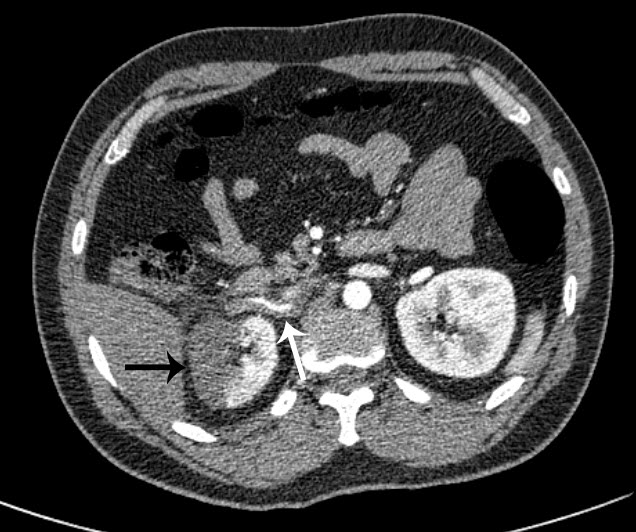
Figure 1 – CT angiography axial image: A long linear filling defect is seen within the right renal artery due to dissection and thrombosis (white arrow). The subsequent renal infarction is evident laterally (black arrow).

83 2
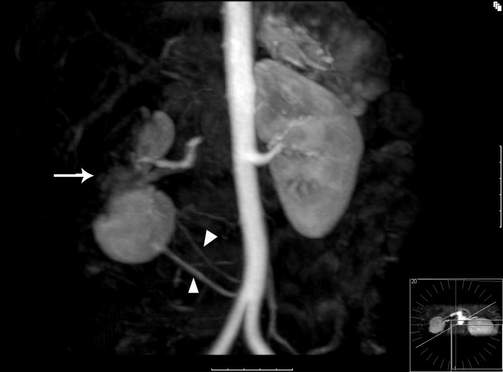
Figure 2 – MR angiographic 3D reconstruction: The lateral aspect and upper pole of the right kidney fails to enhance in keeping with renal infarction (white arrow). The inferior portion appears to enhance normally. This is due to a variant in the patient’s anatomy, with two small accessory renal arteries supplying the lower pole of the kidney (white triangles).

References
1. Stawicki SP, Rosenfeld JC, Weger N, Fields NL, Balshi JD Spontaneous renal artery dissection: three cases and clinical algorithms. Journal of Human Hypertension 2006; 20: 710–718

https://www.nature.com/jhh/journal/v20/n9/full/1002045a.html

2. Ramamoorthy SL, Vasquez JC, Taft PN, et al. Nonoperative management of acute spontaneous renal artery dissection. Annals Vasc Surg 2002;16(2): 157-16

https://www.sciencedirect.com/science/article/pii/S0890509607601447

3. Alamir A, Middendorf DF, Baker P, et al. Renal artery dissection causing renal infarction in otherwise healthy men.  Am J Kidney Dis. 1997; 30: 851–855

https://www.sciencedirect.com/science/article/pii/S0272638697900949

4. Mudrick D, Arepally A, Geschwind JF, Ronsivalle JA, Lund GB, Scheel P. Spontaneous renal artery dissection: treatment with coil embolization. J Vasc Interv Radiol 2003; 14(4): 497-500

https://www.sciencedirect.com/science/article/pii/S105104430760236X

5. Müller BT, Reiher L, Pfeiffer T, Müller W, Hort W, Voiculescu A, Grabensee B, Fürst G, Sandmann W. Surgical treatment of renal artery dissection in 25 patients: indications and results. J Vasc Surg. 2003 37(4): 761-8

https://www.sciencedirect.com/science/article/pii/S0741521402753123


Date added to bjui.org: 20/11/2012

DOI: 10.1002/BJUIw-2012-083-web


© 2024 BJU International. All Rights Reserved.