Tag Archive for: MRI

Posts

Article of the Week: Prostate Health Index density improves detection of clinically significant prostate cancer

Every Week the Editor-in-Chief selects an Article of the Week from the current issue of BJUI. The abstract is reproduced below and you can click on the button to read the full article, which is freely available to all readers for at least 30 days from the time of this post.

In addition to the article itself, there is an accompanying editorial written by a prominent member of the urological community. This blog is intended to provoke comment and discussion and we invite you to use the comment tools at the bottom of each post to join the conversation.

Finally, the third post under the Article of the Week heading on the homepage will consist of additional material or media. This week we feature a video discussing the paper.

If you only have time to read one article this week, it should be this one.

Prostate Health Index density improves detection of clinically significant prostate cancer

Jeffrey J. Tosoian*, Sasha C. Druskin*, Darian Andreas*, Patrick Mullane*, Meera Chappidi*, Sarah Joo*, Kamyar Ghabili*, Mufaddal Mamawala*, Joseph Agostino*, Herbert B. Carter*, Alan W. Partin*, Lori J. Sokoll*§ and Ashley E. Ross*§


*Department of Urology, James Buchanan Brady Urological Institute, Johns Hopkins University School of Medicine, Baltimore, MD, Virginia Commonwealth University School of Medicine, Richmond, VA, Department of Pathology, and §Sidney Kimmel Comprehensive Cancer Center, Johns Hopkins University School of Medicine, Baltimore, MD, USA

Abstract

Objectives

To explore the utility of Prostate Health Index (PHI) density for the detection of clinically significant prostate cancer (PCa) in a contemporary cohort of men presenting for diagnostic evaluation of PCa.

Patients and Methods

The study cohort included patients with elevated prostate-specific antigen (PSA; >2 ng/mL) and negative digital rectal examination who underwent PHI testing and prostate biopsy at our institution in 2015. Serum markers were prospectively measured per standard clinical pathway. PHI was calculated as ([{−2}proPSA/free PSA] × [PSA]½), and density calculations were performed using prostate volume as determined by transrectal ultrasonography. Logistic regression was used to assess the ability of serum markers to predict clinically significant PCa, defined as any Gleason score ≥7 cancer or Gleason score 6 cancer in >2 cores or >50% of any positive core.

Results

Of 118 men with PHI testing who underwent biopsy, 47 (39.8%) were found to have clinically significant PCa on biopsy. The median (interquartile range [IQR]) PHI density was 0.70 (0.43–1.21), and was 0.53 (0.36–0.75) in men with negative biopsy or clinically insignificant PCa and 1.21 (0.74–1.88) in men with clinically significant PCa (P < 0.001). Clinically significant PCa was detected in 3.6% of men in the first quartile of PHI density (<0.43), 36.7% of men in the IQR of PHI density (0.43–1.21), and 80.0% of men with PHI density >1.21 (P < 0.001). Using a threshold of 0.43, PHI density was 97.9% sensitive and 38.0% specific for clinically significant PCa, and 100% sensitive for Gleason score ≥7 disease. Compared with PSA (area under the curve [AUC] 0.52), PSA density (AUC 0.70), %free PSA (AUC 0.75), the product of %free PSA and prostate volume (AUC 0.79), and PHI (AUC 0.76), PHI density had the highest discriminative ability for clinically significant PCa (AUC 0.84).

Conclusions

Based on the present prospective single-centre experience, PHI density could be used to avoid 38% of unnecessary biopsies, while failing to detect only 2% of clinically significant cancers.

Editorial: Prostate cancer biomarkers: new scenarios in the multi-parametric magnetic resonance imaging era

The management of prostate cancer poses difficult challenges, which is largely because we lack the necessary tools to predict its presence, and discern between indolent disease with a small chance of clinical manifestation and aggressive tumours that are more likely to be lethal.

Despite the fact that novel blood and urine tests are available, which may predict aggressive disease better than PSA; they are not routinely used due to a lack of clinical validity studies.

Tosoian et al. [1] in the present study explored the utility of prostate health index (PHI) density for detection of clinically significant prostate cancer in a contemporary cohort of men presenting for diagnostic evaluation of prostate cancer. Very interestingly the authors hypothesised that, similar to PSA density, PHI density could further improve upon the discriminative ability of PHI to detect prostate cancer. The PHI density calculation was performed using prostate volume, as determined by TRUS. Logistic regression was used to assess the ability of serum markers to predict clinically significant prostate cancer, defined as any Gleason score ≥7 cancer or Gleason score 6 cancer in >2 cores or >50% of any positive core.

They showed, albeit in a small sample size, that PHI density could further improve upon the discriminative ability of PHI and appears to be superior to PSA and other PSA derivatives for the identification of clinically significant disease [1].

However, it is noteworthy that in all studies on urine or serum biomarkers such as this, the ‘gold standard’ for cancer detection is pathological examination of multiple non-targeted systematic TRUS-guided prostate biopsies, not radical prostatectomy specimens. Intrinsically, this approach implies that no cancer predicted by the biomarker may still mean cancer missed by the biopsy.

Introducing mpMRI before prostate biopsy has the potential to improve prostate cancer sampling ink that is the most practical way to make mpMRI before biopsy economically viable for universal NHS adoption.

The aim should be the development of a clinical decision support system based on mpMRI and circulating biomarkers, as in this case PHI density evaluation, to stratify patients according to their risk of prostate cancer progression, using pathological assessment after prostatectomy as the reference standard.

Francesco Porpiglia and Stefano De Luca
Division of Urology, San Luigi Gonzaga Hospital and University of Torino, Orbassano, Italy


References


1 Tosoian JJDruskin SCAndreas D et al. Prostate health index density improves detection of clinically significant prostate cancer. BJU Int2017; 120: 7938.


2 Mottet NBellmunt JBolla M et al. EAU-ESTRO-SIOG guidelines on prostate cancer. Part 1: Screening, diagnosis, and local treatment with curative intent.  Eur Urol 2016; pii: S0302-2838(16)30470-5. [Epub ahead of print]. doi: 10.1016/j.eururo.2016.08.003.


3 Russo FRegge DArmando E et al. Detection of prostate cancer index lesions with multiparametric magnetic resonance imaging (mp-MRI) using whole-mount histological sections as the reference standard. BJU Int 2016; 118: 8494.


5 Porpiglia FManfredi MMele F et al. Diagnostic pathway with multiparametric magnetic resonance imaging versus standard pathway: results from a randomized prospective study in biopsy-naıve patients with suspected prostate cancer. Eur Urol 2016; pii: S0302-2838(16)30509-7. [Epub ahead of print]. doi: 10.1016/j.eururo.2016.08.041


6 Wegelin Ovan Melick HHHooft L et al. Comparing three different techniques for magnetic resonance imaging-targeted prostate biopsies: a systematic review of in-bore versus magnetic resonance imaging- transrectal ultrasound fusion versus cognitive registration. Is there a preferred technique?. Eur Urol 2016; pii: S0302-2838(16)30446-8. [Epub ahead of print]. doi: 10.1016/j.eururo.2016.07.04


Video: Prostate Health Index density improves detection of clinically significant prostate cancer

Prostate Health Index density improves detection of clinically significant prostate cancer

Abstract

Objectives

To explore the utility of Prostate Health Index (PHI) density for the detection of clinically significant prostate cancer (PCa) in a contemporary cohort of men presenting for diagnostic evaluation of PCa.

Patients and Methods

The study cohort included patients with elevated prostate-specific antigen (PSA; >2 ng/mL) and negative digital rectal examination who underwent PHI testing and prostate biopsy at our institution in 2015. Serum markers were prospectively measured per standard clinical pathway. PHI was calculated as ([{−2}proPSA/free PSA] × [PSA]½), and density calculations were performed using prostate volume as determined by transrectal ultrasonography. Logistic regression was used to assess the ability of serum markers to predict clinically significant PCa, defined as any Gleason score ≥7 cancer or Gleason score 6 cancer in >2 cores or >50% of any positive core.

Results

Of 118 men with PHI testing who underwent biopsy, 47 (39.8%) were found to have clinically significant PCa on biopsy. The median (interquartile range [IQR]) PHI density was 0.70 (0.43–1.21), and was 0.53 (0.36–0.75) in men with negative biopsy or clinically insignificant PCa and 1.21 (0.74–1.88) in men with clinically significant PCa (P < 0.001). Clinically significant PCa was detected in 3.6% of men in the first quartile of PHI density (<0.43), 36.7% of men in the IQR of PHI density (0.43–1.21), and 80.0% of men with PHI density >1.21 (P < 0.001). Using a threshold of 0.43, PHI density was 97.9% sensitive and 38.0% specific for clinically significant PCa, and 100% sensitive for Gleason score ≥7 disease. Compared with PSA (area under the curve [AUC] 0.52), PSA density (AUC 0.70), %free PSA (AUC 0.75), the product of %free PSA and prostate volume (AUC 0.79), and PHI (AUC 0.76), PHI density had the highest discriminative ability for clinically significant PCa (AUC 0.84).

Conclusions

Based on the present prospective single-centre experience, PHI density could be used to avoid 38% of unnecessary biopsies, while failing to detect only 2% of clinically significant cancers.

View more videos

Editorial: An end to the phenomenon of ‘upgrading’ in early prostate cancer?

The phenomenon of ‘upgrading’ in early prostate cancer is one of those unusual events that is both useful to us on the one hand and undesirable on the other; useful because the phenomenon gifts us a direct measure of the precision of our risk stratification methods for men recently diagnosed, and undesirable because the perfect pathway should, ideally, be free of any upgrading.

Upgrading occurs in a number of settings. We see it at play to some degree when an unreliable test is re-applied in the same subject. The REDUCE study [1] showed us that just under one fifth of men will convert from a status of ‘cancer-free’ to one of ‘cancer-present’ as a result of a second exposure to the same test; that is, TRUS-guided biopsy. We see it in full play when an unreliable test is followed by a more accurate test. Shaw et al. [2] have reminded us once again – as have a number of others – of our limited ability to risk-stratify patients with early prostate cancer. They reported a 50% upgrading when they compared the results of TRUS biopsy against the final pathology at radical prostatectomy. In other words, half the patients went on to their definitive therapy with an incorrect grade attribution [2].

It would be a great pity if, in the modern era, the only route available to patients who wanted to be sure of their risk status was to agree to surgical removal of the prostate. Surely, the value of accurate risk stratification is derived from using it to allocate appropriate and effective care. Risk stratification needs to be linked to or closely follow diagnosis if it is to be put to work for patients.

Nowhere is this need greater than in men whose treatment preference is tissue preservation. The study, in this issue of BJUI, by Pessoa et al. [3] adds to our knowledge on the subject and equips us with a strategy to mitigate some of the errors that are inherent to the standard diagnostic pathway.

In the present study, the authors evaluated the role of a single exposure to MRI (and the opportunity that resulted to undertake a targeted biopsy of an MRI-derived abnormality as well as systematic sampling) in 105 men who had been attributed a diagnosis of low-risk prostate cancer – and, as a result, were deemed to be suitable for active surveillance. The authors used prostate imaging reporting and data system (PIRADS) scoring to interpret and communicate MRI risk. In summary, men attributed a low PIRADS score (PIRADS 1–3) had a low probability of being re-classified to a higher risk. In contrast, men attributed PIRADS score 4 or 5 had a probability of 70–100% of being re-classified. The authors calculated a sensitivity of 93% for MRI to predict ‘re-classification’. This equates to a 93% sensitivity to predict the presence of clinically significant disease as re-classification occurred when there was a transition from low-risk to higher-risk disease.

These results concur with those of others who are working in this area [4] and are in line with current recommendations [5]. One observation that is worth highlighting – because it is a current controversy in the field – relates to the utility of the systematic (or semi-random) biopsies as a component of the confirmatory biopsy. Whilst targeted biopsy was superior to systematic biopsy at identifying clinically significant disease, omission of the systematic biopsies would have resulted in five significant cancers being overlooked. The less perfect the targeted biopsy, the greater the reliance on the systematic. In the present study, the lesion generation and the targeting may have been compromised by one or two issues. Using TRUS biopsy as the authors did (as opposed to transperineal biopsy) to access all areas of the prostate is always going to be a challenge. To do so without image registration makes it even harder. To use PIRADS – as opposed to a Likert scale – as a method of interpreting and communicating MRI outputs will, very likely, lead to an under-reporting of the smaller, high-grade lesions [6]. This is because PIRADS 2.0 is triggered by a volume threshold towards the upper end of the scale. Such lesions might be more prevalent in an apparently ‘low-risk’ population such as the one under scrutiny. If this is the case, they will not be identified as ‘targets’ by virtue of a high PIRADS score. As a consequence they cannot be identified by targeting but might be picked up by the random fall of the needles.

Mark Emberton
Division of Surgery and Interventional Science, University College London, London, UK
Read the full article
References

1 Andriole GL, Bostwick DG, Brawley OW et al. Rittmaster RS; REDUCE Study Group. Effect of dutasteride on the risk of prostate cancer. N Engl Med 2010; 362: 1192202


2 Shaw GL, Thomas BC, Dawson SN et al. Identication of pathologically insignicant prostate cancer is not accurate in unscreened men. Br Cancer 2014; 110: 240511


4 Nassiri N, Margolis DJ, Natarajan S et al. Targeted biopsy to detect Gleason score upgrading during active surveillance for men with low- vs. intermediate-risk prostate cancer. J Urol 2016; [Epub ahead of print]. doi: 10.1016/j.juro.2016.09.070.


5 Moore CM, Giganti F, Albertsen P et al. Reporting magnetic resonance imaging in men on active surveillance for prostate cancer: the PRECISE recommendations-a report of a European school of oncology task force. Eur Urol 2016; [Epub ahead of print]. doi: 10.1016/j.eururo.2016.06.011.


Article of the Week: Predictive value of negative 3T multiparametric MRI of the prostate on 12-core biopsy results

Every Week the Editor-in-Chief selects an Article of the Week from the current issue of BJUI. The abstract is reproduced below and you can click on the button to read the full article, which is freely available to all readers for at least 30 days from the time of this post.

In addition to the article itself, there is an accompanying editorial written by a prominent member of the urological community. This blog is intended to provoke comment and discussion and we invite you to use the comment tools at the bottom of each post to join the conversation.

If you only have time to read one article this week, it should be this one.


Predictive value of negative 3T multiparametric magnetic resonance imaging of the prostate on 12-core biopsy results

James S. Wysock, Neil Mendhiratta, Fabio Zattoni, Xiaosong Meng, Marc Bjurlin,
William C. Huang, Herbert Lepor, Andrew B. Rosenkrantz* and Samir S. Taneja
Department of Urology, and *Department of Radiology, NYU Langone Medical Center, New York, NY, USA


Read the full article


Objectives

To evaluate the cancer detection rates for men undergoing 12-core systematic prostate biopsy with negative prebiopsymultiparametric magnetic resonance imaging (mpMRI) results.


Materials and Methods

Clinical data from consecutive men undergoing prostate biopsy who had undergone prebiopsy 3T mpMRI from December 2011 to August 2014 were reviewed from an institutional review board-approved prospective database. Men with negative prebiospy mpMRI results (negMRI) before biopsy were identified for the present analysis. Clinical features, cancer detection rates and negative predictive values were summarized.


Results

Seventy five men with negMRI underwent systematic 12-core biopsy during the study period. In the entire cohort, men with no previous biopsy, men with previously negative biopsy and men enrolled in active surveillance protocols, the overall cancer detection rates were 18.7, 13.8, 8.0 and 38.1%, respectively, and the detection rates for Gleason score (GS) ≥7 cancer were 1.3, 0, 4.0 and 0%, respectively. The NPVs for all cancers were 81.3, 86.2, 92.0, and 61.9, and for GS ≥7 cancer they were 98.7, 100, 96.0 and 100%, respectively.


Conclusions

A negative prebiopsy mpMRI confers an overall NPV of 82% on 12-core biopsy for all cancer and 98% for GS ≥7 cancer. Based on biopsy indication, these findings assist in prebiopsy risk stratification for detection of high-risk disease and may provide guidance in the decision to pursue biopsy.


Editorial: Some prostate cancers are invisible to magnetic resonance imaging!

A test to exclude the presence of aggressive prostate cancer would be highly desirable. In the article by Wysock et al. [1], the authors examine pathological results in 75 men who underwent 12-core systematic biopsy using the Artemis device; all had a pre-biopsy MRI showing no suggestion of cancer. In 74 patients no cancer with Gleason score ≥7 was found on biopsy, which translates into a remarkable 98.7% negative predictive value (NPV) for potentially aggressive disease. The implication is that virtually all serious prostate cancers can be seen on MRI, and thus a negative MRI obviates the need for a biopsy. If this finding were to be confirmed, the majority of prostate biopsies could be avoided, a truly laudable goal.

However, for several reasons, we are not yet able to endorse the blanket concept, ‘get a negative MRI and skip the biopsy’. The authors acknowledged their study is not definitive: it is a retrospective look at 29% of eligible participants; how were they chosen? Further, the ‘gold standard’ for identifying cancer, microscopic examination of whole mount prostatectomy specimens, was not available. Thus, the data presented are striking because of the near-perfect findings, but not entirely convincing. Our work, involving >2 000 MRI/ultrasound-fusion biopsies, teaches that if biopsy is indicated on clinical grounds e.g., palpable abnormality, family or racial history, persistent PSA suspicion, a negative MRI should not preclude a systematic (template) biopsy [2]. Clinical information, especially increased PSA density, may help to select patients for biopsy beyond use of MRI data alone.

Another reason why a ‘negative’ MRI should not always negate the need for biopsy relates to the current proliferation of prostate MRI studies. Many of the new MRI studies are being performed and interpreted by radiologists not adequately trained in this niche. Despite attempts at standardisation [3], the variability of MRI readings, from place to place and from one radiologist to another, can be remarkable. In the recent past I have seen lesions called Prostate Imaging-Reporting and Data System (PI-RADS) Grade 5 ‘disappear’ when scrutinised by more-experienced readers; the reverse has also been seen. Even among expert readers using the latest Version 2 of PI-RADS, agreement in prostate MRI interpretation is only moderate at best (κ ≈0.5) [4]. Therefore, widely varying interpretations of prostate MRI can be expected for the near-term future. A formal training programme and certification in MRI interpretation, which is sorely needed, has not yet been established.

In Figure 1 above, a falsely negative MRI from our institution is shown [2]. The MRI-invisible cancer is not a rarity. In a consecutive series of 1 042 men undergoing template biopsy regardless of MRI findings, the incidence of clinically significant prostate cancer in men with no MRI-suspicious lesions (biopsy-naïve subgroup) was 12% [5]. Further, when looking carefully at whole-mount prostatectomy specimens, the incidence of clinically significant prostate cancer not seen on expertly read MRI was 28% [6]. Still further, some Gleason 6 cancers thought to be insignificant may have the biological potential for de-differentiation [7] and require follow-up. Therefore, at this point in time, a negative MRI should not preclude prostate biopsy, which otherwise would be indicated on clinical grounds. We are still learning about prostate MRI!

aotw2-ed-fig-1

Figure 1: Example of falsely negative MRI. Patient was a Caucasian male (PSA level 3.8 ng/mL) aged 68 years, who on a previous conventional biopsy was found to have a microfocus of Gleason 3 + 3 = 6 prostate cancer. He was considered for active surveillance, and multiparametric MRI of prostate was obtained (A): prostate volume was found to be 35 mL; no region of interest was identified. Mapping biopsy was performed by following the 12-point template of the Artemis device (B). A tissue core from the left lateral apex revealed 6 mm of Gleason 3 + 5 = 8 prostate cancer (C, ×4; D, ×20). Radical prostatectomy was performed, revealing a tumour on the left side of the prostate with diameters of 15 × 12 × 9 mm. Falsely negative MRI is not uncommon. When biopsy is clinically indicated, a negative MRI should not preclude mapping biopsy. Reproduced with permission from Nassiri et al., 2015 [1].


Read the full article


Leonard S. Marks

Professor and deKernion Endowed Chair, Department of Urology, David Geffen School of Medicine at UCLA, Los Angeles, CA, USA


1 Wysock JS, Mendhiratta N, Zattoni F et al. Predictive value of negative 3T multiparametric magnetic resonance imaging of the prostate on 12-core biopsy results. BJU Int 2016; 118: 515–20

2 Nassiri N, Natarajan S, Margolis DJ, Marks LS. Targeted prostate biopsy: lessons learned midst the evolution of a disruptive technology. Urology 2015; 86: 432–8

3 Weinreb JC, Barentsz JO, Choyke PL et al. PI-RADS Prostate Imaging – Reporting and Data System: 2015, Version 2. Eur Urol 2016; 69: 16–40

4 Rosenkrantz AB, Ginocchio LA, Cornfeld D et al. Inter-observer reproducibility of the PI-RADS Version 2 Lexicon: A multi-center study of six experienced prostate radiologists. Radiology 2016; 280: 793–804

5 Filson CP, Natarajan S, Margolis DJ et al.Prostate cancer detection with magnetic resonance-ultrasound fusion biopsy: The role of systematic and targeted biopsies. Cancer 2016; [Epub ahead of print]. doi: 10.1002/cncr.29874

6 Le JD, Tan N, Shkolyar E et al. Multifocality and prostate cancer detection by multiparametric magnetic resonance imaging: correlation with whole-mount histopathology. Eur Urol 2015; 67: 569–76

7 Palapattu GS, Cani AK, Huang J et al. Progression of low- to high-grade prostate cancer: Molecular profiling of tissue obtained by serial targeted biopsy. J Clin Oncol 2015; 33 (Suppl.): Abstract 501


Article of the Week: TRUS-Guided RB PCa Detection – Reasons for Targeted Biopsy Failure

Every Week the Editor-in-Chief selects an Article of the Week from the current issue of BJUI. The abstract is reproduced below and you can click on the button to read the full article, which is freely available to all readers for at least 30 days from the time of this post.

In addition to the article itself, there is an accompanying editorial written by a prominent member of the urological community. This blog is intended to provoke comment and discussion and we invite you to use the comment tools at the bottom of each post to join the conversation.

Finally, the third post under the Article of the Week heading on the homepage will consist of additional material or media. This week we feature a video from Hannes Cash and Patrick Asbach, discussing their paper.

If you only have time to read one article this week, it should be this one.

Prostate cancer detection on transrectal ultrasonography-guided random biopsy despite negative real-time magnetic resonance imaging/ultrasonography fusion-guided targeted biopsy: reasons for targeted biopsy failure

Hannes Cash*, Karsten Gunzel*, Andreas Maxeiner*, Carsten Stephan*, Thomas Fischer, Tahir Durmus, Kurt Miller*, Patrick Asbach, Matthias Haas† and Carsten Kempkensteffen*


*Department of Urology, and Department of Radiology, ChariteUniversity of Medicine Berlin, Berlin, Germany M. H. and C.K. contributed equally to the study.


Read the full article

Objective

To examine the value of additional transrectal ultrasonography (TRUS)-guided random biopsy (RB) in patients with negative magnetic resonance imaging (MRI)/ultrasonography (US) fusion-guided targeted biopsy (TB) and to identify possible reasons for TB failure.

Patients and Methods

We conducted a subgroup analysis of 61 men with prostate cancer (PCa) detected by 10-core RB but with a negative TB, from a cohort of 408 men with suspicious multiparametric magnetic resonance imaging (mpMRI) between January 2012 and January 2015. A consensus re-reading of mpMRI results (using Prostate Imaging Reporting and Data System [PI-RADS] versions 1 and 2) for each suspicious lesion was performed, with the image reader blinded to the biopsy results, followed by an unblinded anatomical correlation of the lesion on mpMRI to the biopsy result. The potential reasons for TB failure were estimated for each lesion. We defined clinically significant PCa according to the Epstein criteria and stratified patients into risk groups according to the European Association of Urology guidelines.

JulAOTW3Results

Results

Our analysis showed that RB detected significant PCa in 64% of patients (39/61) and intermediate-/high-risk PCa in 57% of patients (35/61). The initial mpMRI reading identified 90 suspicious lesions in the cohort. Blinded consensus re-reading of the mpMRI led to PI-RADS score downgrading of 45 lesions (50%) and upgrading of 13 lesions (14%); thus, negative TB could be explained by falsely high initial PI-RADS scores for 32 lesions (34%) and sampling of the target lesion by RB in the corresponding anatomical site for 36 out of 90 lesions (40%) in 35 of 61 patients (57%). Sampling of the target lesion by RB was most likely for lesions with PI-RADS scores of 4/5 and Gleason scores (GS) of ≥7. A total of 70 PCa lesions (67% with GS 6) in 44 patients (72%) were sampled from prostatic sites with no abnormalities on mpMRI.

Conclusion

In cases of TB failure, RB still detected a high rate of significant PCa. The main reason for a negative TB was a TB error, compensated for by positive sampling of the target lesion by the additional RB, and the second reason for TB failure was a falsely high initial PI-RADS score. The challenges that arise for both MRI diagnostics and prostate lesion sampling are evident in our data and support the integration of RB into the TB workflow.

Editorial: MRI-Fusion Biopsy – Behind the Scenes

MRI information of the prostate is increasingly used for improving the diagnostic yield of prostate biopsies [1]. However, increasing complexity of a procedure makes it prone to errors at multiple technical and human levels. Incorporating MRI information and ultrasonography (US) images for MRI-fusion biopsies is a technically challenging task. It involves various steps such as the acquisition and fusion of MRI and US images, the needle guidance during biopsy, and the diligence of the pathological evaluation of biopsy specimens. These different steps and interfaces between different medical professions influence the diagnostic performance of MRI-fusion biopsies.

For example, in daily clinical practice, MRIs from different institutions still harbour a great variance of sequences and reporting, despite the European Society of Urogenital Urology (ESUR) recently introducing acquisition and imaging protocols and a new and advanced version of the Prostate Imaging Reporting and Data System (PIRADS) version 2.0 [2]. The usefulness of such reporting schemes is evidenced by a moderate-to-good interobserver agreement between uro-radiologists for tumour lesion interpretation and corresponding κ values ranging from 0.55 to 0.80 [3]. Important pitfalls of image interpretation are benign lesions such as prostatitis, BPH and fibrosis, which might score similarly to prostate cancer lesions. This problem is further aggravated by a high proportion of patients that receive their first multiparametric MRI (mpMRI) of the prostate in the repeat-biopsy setting with a high burden of post-biopsy artefacts (haemorrhage, capsular irregularity) and lower overall cancer detection rate. Also, during MRI-fusion biopsy patient movement, prostate deformation by the US probe, and mismatch of image planes can lead to a biopsy error exceeding 4 mm. Moreover, targeting error might be aggravated by MRI underestimation of the tumour volume compared with final pathology [4]. After various authors reported the advantages and accuracy of MRI/US-fusion biopsy approaches, Cash et al. [5] address potential reasons for targeted biopsy failure to detect prostate cancer compared with random biopsy. Within their analyses the authors address potential limitations and technical considerations. Based on different technical biopsy strategies (with the patient placed within the MRI scanner (‘in-bore’) vs outside) and different technical approaches, these considerations are very important.

In contrast to cognitive fusion, most MRI/US platforms allow needle tracking by archiving the needle orientation, either by an electromagnetic, image-based or stepper-based mechanism [1]. However, lesion targeting by needle guidance is highly dependent on the dimensions of the primary lesion, numbers of relevant lesions, localisation, and overall prostate volume, making MRI-US fusion and cognitive fusion more error prone (i.e. aiming off the mark with the needle) than in-bore biopsies. Moreover, different technical fusion approaches provide different degrees of manual/automated adjustment tools, with for example either rigid or elastic image transformation to facilitate MRI/US image alignment.

In their analyses, Cash et al. [5] found that 34% of negative targeted biopsies could be explained by initially too high estimated PIRADS scores that were downgraded at re-reading. Interestingly, the remaining lesions were without an mpMRI correlate but within this group 92.9% showed a primary Gleason 3 pattern in biopsy pathology, suggesting a high degree of invisibility on mpMRI. Subanalyses did not show an association of targeted biopsy failures in the ventral location. Therefore, the study by Cash et al. [5] is an important precursor for further analyses to address other underlying reasons for targeted biopsy failure. Moreover, it reveals the need for a tight collaboration of radiologists, urologists, and pathologists as interdisciplinary partners involved in MRI-fusion biopsy. Consequently, the optimal diagnostic performance of MRI-fusion biopsies can only be achieved through standardised MRI performance, reading and reporting of MRI findings, as well as final correlation of MRI findings with histopathological work up.

Read the full article
Lars Budaus and Sami-Ramzi Leyh-Bannurah
Martini-Clinic University Hospital Hamburg-Eppendorf, Hamburg, Germany


References


Video: TRUS-Guided RB Prostate Cancer Detection – Reasons for Targeted Biopsy Failure

Prostate cancer detection on transrectal ultrasonography-guided random biopsy despite negative real-time magnetic resonance imaging/ultrasonography fusion-guided targeted biopsy: reasons for targeted biopsy failure

Hannes Cash*, Karsten Gunzel*, Andreas Maxeiner*, Carsten Stephan*, Thomas Fischer, Tahir Durmus, Kurt Miller*, Patrick Asbach, Matthias Haas† and Carsten Kempkensteffen*


*Department of Urology, and Department of Radiology, ChariteUniversity of Medicine Berlin, Berlin, Germany M. H. and C.K. contributed equally to the study.


Read the full article

Objective

To examine the value of additional transrectal ultrasonography (TRUS)-guided random biopsy (RB) in patients with negative magnetic resonance imaging (MRI)/ultrasonography (US) fusion-guided targeted biopsy (TB) and to identify possible reasons for TB failure.

Patients and Methods

We conducted a subgroup analysis of 61 men with prostate cancer (PCa) detected by 10-core RB but with a negative TB, from a cohort of 408 men with suspicious multiparametric magnetic resonance imaging (mpMRI) between January 2012 and January 2015. A consensus re-reading of mpMRI results (using Prostate Imaging Reporting and Data System [PI-RADS] versions 1 and 2) for each suspicious lesion was performed, with the image reader blinded to the biopsy results, followed by an unblinded anatomical correlation of the lesion on mpMRI to the biopsy result. The potential reasons for TB failure were estimated for each lesion. We defined clinically significant PCa according to the Epstein criteria and stratified patients into risk groups according to the European Association of Urology guidelines.

JulAOTW3Results

Results

Our analysis showed that RB detected significant PCa in 64% of patients (39/61) and intermediate-/high-risk PCa in 57% of patients (35/61). The initial mpMRI reading identified 90 suspicious lesions in the cohort. Blinded consensus re-reading of the mpMRI led to PI-RADS score downgrading of 45 lesions (50%) and upgrading of 13 lesions (14%); thus, negative TB could be explained by falsely high initial PI-RADS scores for 32 lesions (34%) and sampling of the target lesion by RB in the corresponding anatomical site for 36 out of 90 lesions (40%) in 35 of 61 patients (57%). Sampling of the target lesion by RB was most likely for lesions with PI-RADS scores of 4/5 and Gleason scores (GS) of ≥7. A total of 70 PCa lesions (67% with GS 6) in 44 patients (72%) were sampled from prostatic sites with no abnormalities on mpMRI.

Conclusion

In cases of TB failure, RB still detected a high rate of significant PCa. The main reason for a negative TB was a TB error, compensated for by positive sampling of the target lesion by the additional RB, and the second reason for TB failure was a falsely high initial PI-RADS score. The challenges that arise for both MRI diagnostics and prostate lesion sampling are evident in our data and support the integration of RB into the TB workflow.

Highlights from BAUS 2016

1.1

In the week following Britain’s exit from Europe after the BREXIT referendum, BAUS 2016 got underway in Liverpool’s BT convention Centre. This was the 72nd meeting of the British Association of Urological Surgeons and it was well attended with 1120 delegates (50% Consultant Member Urologists, 30% Trainees, 10% Non member Urologists/Other, 10% Nurses, HCP’S, Scientists).

1.2

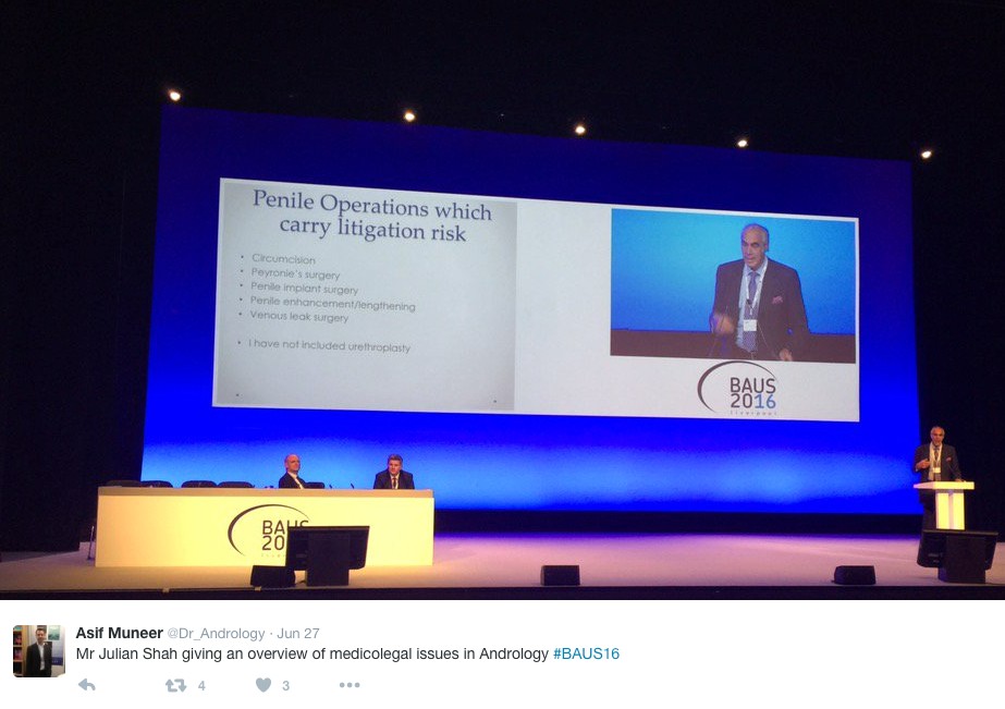
Monday saw a cautionary session on medicolegal aspects in Andrology, focusing on lawsuits over the last year. Mr Mark Speakman presented on the management issue of testicular torsion. This sparked further discussion on emergency cover for paediatrics with particular uncertainty noted at 4 and 5 year olds and great variation in approach dependent on local trust policy. Mr Julian Shah noted the most litigious areas of andrology, with focus on cosmesis following circumcisions. Therefore serving a reminder on the importance of good consent to manage patients’ expectations.

1.3

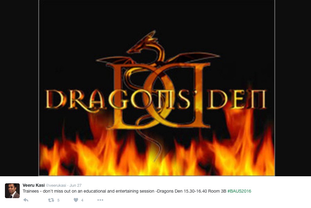
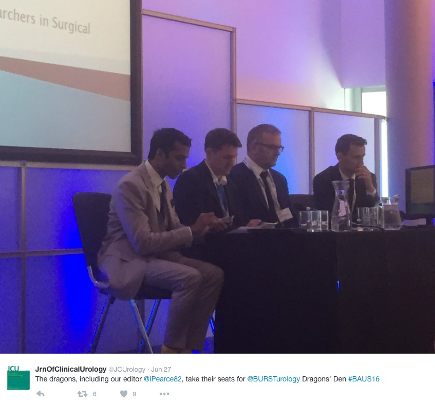
In the Dragons’ Den, like the TV show, junior urologists pitched their ideas for collaborative research projects, to an expert panel. This year’s panel was made up of – Mark Emberton, Ian Pearce, and Graeme MacLennan. The session was chaired by Veeru Kasivisvanathan, Chair of the BURST Research Collaborative.

1.4

Eventual winner Ben Lamb, a trainee from London, presented “Just add water”. The pitch was for an RCT to investigate the efficacy of water irrigation following TURBT against MMC in reducing tumour recurrence. Ben proposed that water, with its experimental tumouricidal properties, might provide a low risk, low cost alternative as an adjuvant agent following TURBT. Judges liked the scientific basis for this study and the initial planning for an RCT. The panel discussed the merits of non-inferiority vs. superiority methodology, and whether the team might compare MMC to MMC with the addition of water, or water instead of MMC. They Dragons’ suggested that an initial focus group to investigate patients’ views on chemotherapy might help to focus the investigation and give credence to the final research question, important when making the next pitch- to a funding body, or ethics committee.

Other proposals were from Ryad Chebbout, working with Marcus Cumberbatch, an academic trainee from Sheffield. Proposing to address the current controversy over the optimal surgical technique for orchidopexy following testicular torsion. His idea involved conducting a systematic review, a national survey of current practice followed by a Delphi consensus meeting to produce evidence based statement of best practice. The final presentation was from Sophia Cashman, East of England Trainee for an RCT to assess the optimal timing for a TWOC after urinary retention. The panel liked the idea of finally nailing down an answer to this age-old question.

1.5

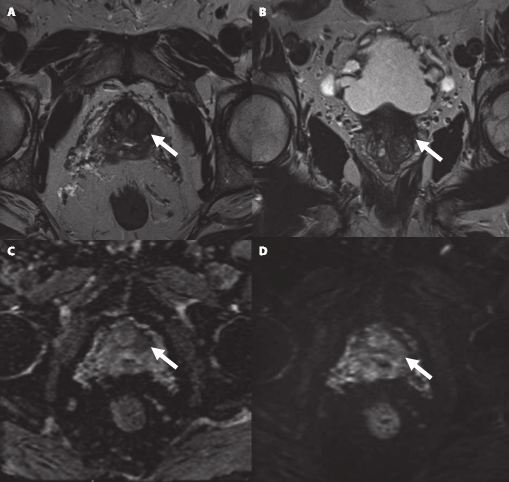
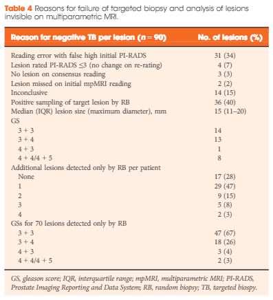
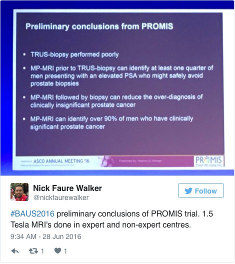
Waking up on Tuesday with England out of the European football cup as well as Europe the conference got underway with an update from the PROMIS trial (use of MRI to detect prostate cancer). Early data shows that multi-parametric MRI may be accurate enough to help avoid some prostate biopsies.

1.6

The SURG meeting provided useful information for trainees, with advice on progressing through training and Consultant interviews. A debate was held over run through training, which may well be returning in the future. The Silver cystoscope was awarded to Professor Rob Pickard voted for by the trainees in his deanery, for his devotion to their training.
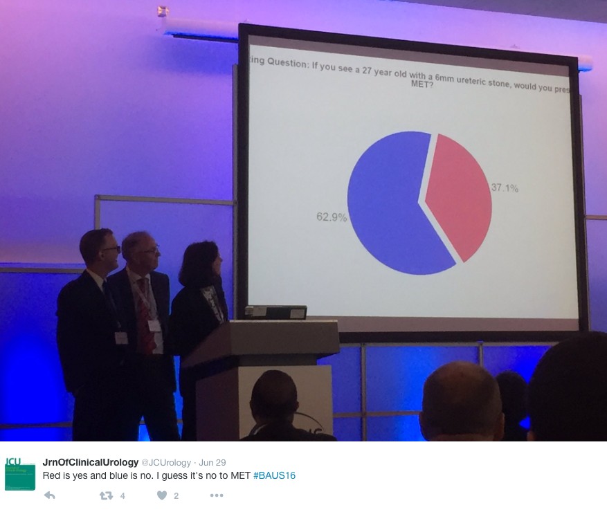
Wednesday continued the debate on medical expulsion therapy (MET) for ureteric stones following the SUSPEND trial. Most UK Urologists seem to follow the results of the trial and have stopped prescribing alpha blockers to try and aid stone passage and symptoms. However the AUA are yet to adopt this stance and feel that a sub analysis shows some benefit for stones >5mm, although this is not significant and pragmatic outcomes. Assistant Professor John Hollingsworth (USA) argued for MET, with Professor Sam McClinton (UK) against. A live poll at the end of the session showed 62.9% of the audience persuaded to follow the SUSPEND trial evidence and stop prescribing MET.

1.7

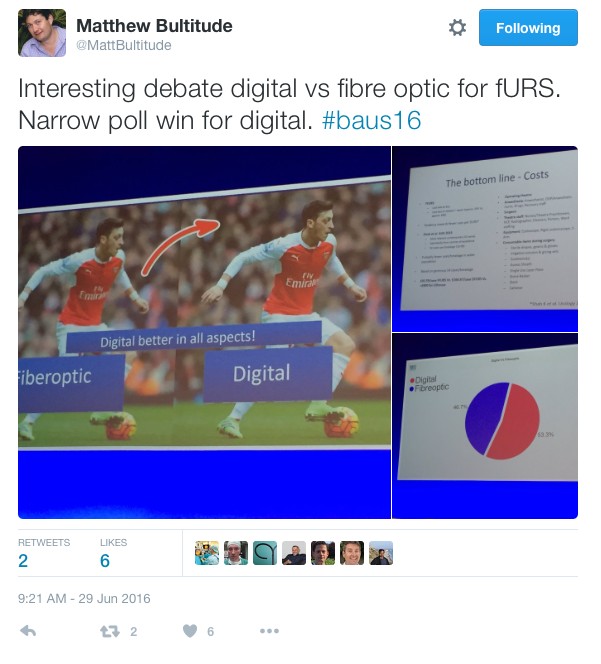
In the debate of digital versus fibreoptic scopes for flexible ureteroscopy digital triumphed, but with a narrow margin.

1.8

In other updates and breaking news it appears that BCG is back! However during the shortage EMDA has shown itself to be a promising alternative in the treatment of high grade superficial bladder cancer.
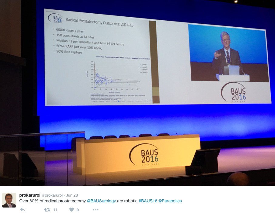
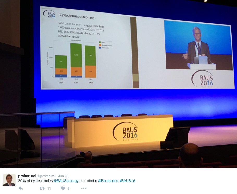
The latest BAUS nephrectomy data shows that 90% are performed by consultant, with 16 on average per consultant per year. This raises some issues for registrar training, however with BAUS guidelines likely to suggest 20 as indicative numbers this is looking to be an achievable target for most consultants. Robotic advocates will be encouraged, as robotic partial nephrectomy numbers have overtaken open this year. The data shows 36% of kidney tumours in the under 40 years old are benign. Will we have to consider biopsying more often? However data suggests we should be offering more cytoreductive nephrectomies, with only roughly 1/10 in the UK performed compared to 3/10 in the USA.

1.91.10

The andrology section called for more recruitment to The MASTER trial (Male slings vs artificial urinary sphincters), whereas the OPEN trial has recruited(open urethroplasty vs optical urethotomy). In the treatment of Peyronie’s disease collagenase has been approved by NICE but not yet within the NHS.

Endoluminal endourology presentation showed big increases in operative numbers with ureteroscopy up by 50% and flexible ureteroscopy up by 100%. Stents on strings were advocated to avoid troubling stent symptoms experienced by most patients. New evidence may help provide a consensus on defining “stone free” post operation. Any residual stones post-operatively less than 2mm were shown to pass spontaneously and therefore perhaps may be classed as “stone free”.

Big changes seem likely in the treatment of benign prostatic hyperplasia, with a race to replace the old favorite TURP. Trials have of TURP (mono and bipolar) vs greenlight laser are already showing similar 2 year outcomes with the added benefit of shorter hospital stays and less blood loss. UROLIFT is an ever more popular alternative with data showing superiority to TURP in lifestyle measures, likely because it preserves sexual function, and we are told it can be performed as a 15 minute day case operation. The latest new therapy is apparently “Aquabeam Aquablation”, using high pressured water to remove the prostate. Non surgical treatments are also advancing with ever more accurate super selective embolisation of the prostatic blood supply.

1.11

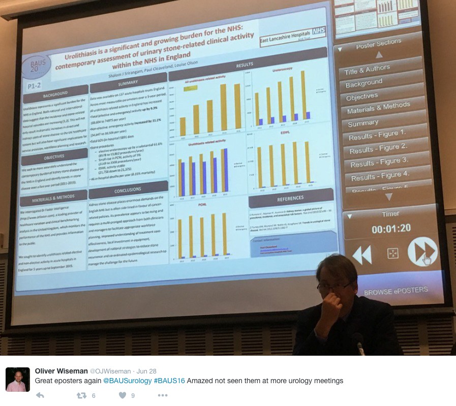
This year all accepted abstracts were presented in moderated EPoster sessions. The format was extremely successful removing the need for paper at future conferences? A total of 538 abstracts were submitted and 168 EPosters displayed. The winner of best EPoster was P5-5 Altaf Mangera: Bladder Cancer in the Neuropathic Bladder.

1.12

The best Academic Paper winner was Mark Salji of the CRUK Beatson institute, titled “A Urinary Peptide Biomarker Panel to Identify Significant Prostate Cancer”. Using capillary electrophoresis coupled to mass spectrometry (CE-MS) they analysed 313 urine samples from significant prostate cancer patients (Gleason 8-10 or T3/4 disease) and low grade control disease. They identified 94 peptide urine biomarkers which may provide a useful adjunct in identifying significant prostate cancer from insignificant disease.

The Office of Education offered 20 courses. Popular off-site courses were ultrasound for the Urologist, at Broadgreen Hospital, a slightly painful 30 min drive from the conference centre. However well worth the trip, delivered by Radiology consultants this included the chance to scan patients volunteers under guidance, with separate stations for kidneys, bladder and testicles and learning the “knobology” of the machines.

Organised by Tamsin Greenwell with other consultant experts in female, andrology and retroperitoneal cancer, a human cadaveric anatomy course was held at Liverpool university. The anatomy teaching was delivered by both Urology consultants and anatomists allowing for an excellent combination of theory and functional anatomy.

BAUS social events are renowned and with multiple events planned most evenings were pretty lively. The official drinks reception was held at the beautiful Royal Liver Building. The venue was stunning with great views over the waterfront and the sun finally shining. Several awards were presented including the Gold cystoscope to Mr John McGrath for significant contribution to Urology within 10 years appointment as consultant. The Keith Yeates medal was awarded to Mr Raj Pal, the most outstanding candidate in the first sitting of the intercollegiate specilaity examination, with a score of over 80%.

1.13

During the conference other BAUS awards presented include the St Peter’s medal was awarded to Margeret Knowles, Head of section of molecular oncology, Leeds Institute of Cancer and Pathology, St James University hospital Leeds. The St Paul’s medal awarded to Professor Joseph A. Smith, Vanderbilt University, Nashville, USA. The Gold medal went to Mr. Tim Terry, Leicester General Hospital.

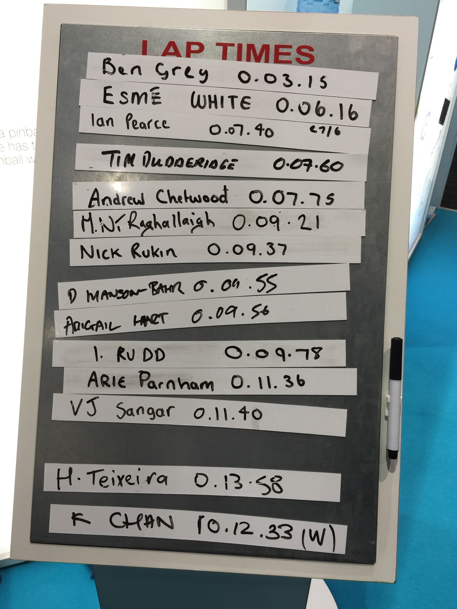
An excellent industry exhibition was on display, with 75 Exhibiting Companies present. My personal fun highlight was a flexible cystoscope with integrated stent remover, which sparked Top Gear style competiveness when the manufacturer set up a time-trial leaderboard. Obviously this best demonstrated the speed of stent removal with some interesting results…

1.14

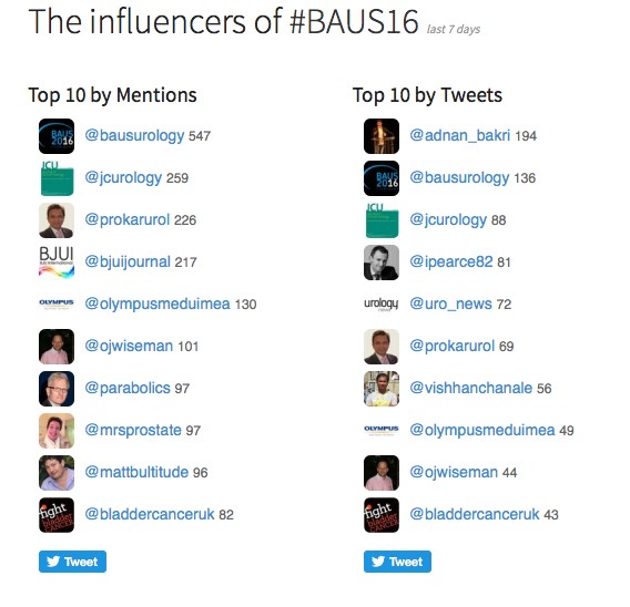
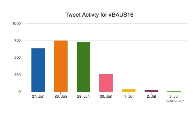
Social media review shows good contribution daily.

1.15

1.16
Thanks BAUS a great conference, very well organised and delivered with a great educational and social content, looking forward to Glasgow 2017! #BAUS2017 #Glasgow #BAUSurology

Nishant Bedi

Specialist Training Registrar North West London 

Twitter: @nishbedi


© 2024 BJU International. All Rights Reserved.