Tag Archive for: positive surgical margins

Posts

Editorial: Conversion to negative surgical margin after intraoperative frozen section – (un)necessary effort and relevance in 2019?

The assessment and impact of positive surgical margins (PSMs) at the time of radical prostatectomy (RP) have been discussed for many decades. The determination and reporting should be performed in a standardised fashion according to the International Society of Urological Pathology [1]. The SM is considered positive if tumour cells touch the inked surface of the RP specimen. However, reasons for difficulty in truly differentiating between negative SMs (NSMs) and PSMs include iatrogenic disruption of the prostatic capsule, penetration of ink into small cracks on the outside, or cases in which prostate cancer cells are very close to, but not definitely touching, the inked margins.

A systematic review by Yossepowitch et al. [2] found a contemporary PSM rate of 15% (range 6.5–32%), which increases with extracapsular extension. In addition, the likelihood of PSM is strongly influenced by surgeon experience, independent of the surgical technique. Although PSM is considered an adverse pathological outcome and associated with an increased risk of biochemical recurrence (BCR), the impact on long‐term survival and actual prognostic value remains debatable. The association with other endpoints, such as prostate‐cancer specific mortality and overall survival, is controversial and may be primarily influenced by other risk factors, such as preoperative PSA level, Gleason score, and pathological T‐stage [2].

The role of intraoperative frozen section analysis in order to reduce the PSM rate continues to evolve. In a study by von Bodman et al. [3], 92.3% of patients with a PSM on frozen‐section analysis could ultimately be converted to a NSM. Similar findings were reported by Schlomm et al. [4] in 5392 patients using the intraoperative neurovascular structure‐adjacent frozen section examination (NeuroSAFE) technique, PSMs were detected in 25%, leading to re‐resection and conversion to definitive NSMs in 86% of these patients. In the setting of increasing experience with intraoperative frozen section analysis, a false‐positive SM status was found in only 48 patients (3.3%).

The study by Pak et al. [5], published in this issue of the BJUI, reported that specimens with initial PSMs were converted to NSMs upon permanent specimen evaluation (NCSM) in 4.9% of 2013 men undergoing RP. In this subgroup, the 5‐year BCR‐free survival (BCRFS) rates did not differ from those observed in National Comprehensive Cancer Network (NCCN) low‐ and intermediate‐risk patients with an initially NSM. However, the benefit of conversion from an initial PSM to final NSM was not apparent in high‐risk patients, as the authors found a significantly lower rate of BCRFS amongst this NCSM group. In multivariate analysis, NCSM status was independently associated (hazard ratio 0.624, P = 0.033) with BCR but not distant metastasis. These findings corroborate the findings of the Schlomm et al. [4] study, in which the BCRFS rates of propensity score‐based matched patients with conversion to NSMs did not differ significantly from patients with primarily NSMs.

What is the current role of intraoperative frozen section analysis during RP? How important is it to achieve NSMs in contemporary practice? In whom and how should the assessment be performed? Although it is clearly desirable to completely remove the entire tumour at the time of surgery, and NSMs are a surrogate marker of adequate local excision, the devil is in the details. First, in this study [5], the authors only assessed SMs at the bladder neck and apex. Although the apex is one of the most frequent locations for PSMs, other and/or multiple sites of PSMs are possible and could have been missed. Alternatively, the NeuroSAFE method is able to assess the entire laterorectal circumference albeit with the trade‐off of more extensive pathological involvement and assessment. Second, intraoperative frozen section analysis, and manoeuvers for NCSM, may ultimately be necessary and beneficial in only a small number of patients currently undergoing RP. An increasing proportion of men harbour more aggressive, higher‐risk disease in whom PSMs may have no impact on oncological outcomes or treatment decisions. In these men, long‐term cancer outcomes are probably more related to risks of unsuspected metastatic disease rather than residual, microscopic cancer within the prostatic fossa. As suggested in this study [5], an initial PSM in high‐risk men, independent of ultimate NCSM, may be a surrogate for non‐localised disease and poorer outcomes; PSMs were found in 53% of men with pT3b. In low‐risk men, the issues are whether active surveillance is a more appropriate initial management strategy and that routine intraoperative frozen section analysis may not be worthwhile with a PSM rate of only 10%. How does this alter the decision for adjuvant therapy? Adjuvant radiotherapy is probably under‐utilised in men with PSMs after RP (~11%), and NCSM may spare men from unnecessary treatment, particularly with lower‐risk disease [6]. However, men with PSMs and additional adverse pathological features, such as extraprostatic extension or seminal vesicle invasion, should probably receive adjuvant therapy, primarily driven by T stage.

The incremental value and potential clinical benefit of intraoperative frozen section analysis to achieve NSMs remain to be determined. Although one would suspect that PSM leading to excision of additional tissue could lead to worse functional outcomes, the study from Mirmilstein et al. [7] is reassuring. Despite higher Gleason score and pT stage in those undergoing the NeuroSAFE approach, the PSM rate was lower in this group (9.2%) compared with those undergoing standard intraoperative nerve‐sparing while leading to greater bilateral nerve preservation, higher potency rates at 12 months, and pad‐free continence.

In the future, other methods may guide surgical decision‐making and may eventually alter PSM rate including preoperative MRI of the prostate to evaluate extracapsular extension, genomic risk scores, or real‐time, near‐infrared fluorescent surgical guidance with prostate‐specific membrane antigen ligands [8]. However, one should not forget that outcomes are not solely based on the SM status. Various pathological and clinical factors and patients’ comorbidities and preference should be taken into consideration in the surgical management and that evaluation of validated oncological and functional outcomes is critical.

by Annika Herlemann and Maxwell Meng

References

  1. Tan, PHCheng, LSrigley, JR et al. International Society of Urological Pathology (ISUP) Consensus Conference on Handling and Staging of Radical Prostatectomy Specimens. Working group 5: surgical margins. Mod Pathol 20112448– 57
  2. Yossepowitch, OBriganti, AEastham, JA et al. Positive surgical margins after radical prostatectomy: a systematic review and contemporary update. Eur Urol 201465303– 13
  3. Bodman, CBrock, MRoghmann, F et al. Intraoperative frozen section of the prostate decreases positive margin rate while ensuring nerve sparing procedure during radical prostatectomy. J Urol 2013190515– 20
  4. Schlomm, TTennstedt, PHuxhold, C et al. Neurovascular structure‐adjacent frozen‐section examination (NeuroSAFE) increases nerve‐sparing frequency and reduces positive surgical margins in open and robot‐assisted laparoscopic radical prostatectomy: experience after 11,069 consecutive patients. Eur Urol 201262333– 40
  5. Pak, SPark, SKim, MGo, HCho, YMAhn, HThe impact on oncological outcomes after radical prostatectomy for prostate cancer of converting soft tissue margins at the apex and bladder neck from tumour‐positive to ‐negative. BJU Int 2019123811– 7
  6. Ghabili, KNguyen, KHsiang, W et al. National trends in the management of patients with positive surgical margins at the time of radical prostatectomy. J Clin Oncol 201836 (Suppl.)111
  7. Mirmilstein, GRai, BPGbolahan, O et al. The neurovascular structure‐adjacent frozen‐section examination (NeuroSAFE) approach to nerve sparing in robot‐assisted laparoscopic radical prostatectomy in a British setting – a prospective observational comparative study. BJU Int 2018;121854– 62
  8. Neuman, BPEifler, JBCastanares, M et al. Real‐time, near‐infrared fluorescence imaging with an optimized dye/light source/camera combination for surgical guidance of prostate cancer. Clin Cancer Res 201521771– 80


Article of the week: Development of a side‐specific, mpMRI‐based nomogram for the prediction of extracapsular extension of PCa

Every week, the Editor-in-Chief selects an Article of the Week from the current issue of BJUI. The abstract is reproduced below and you can click on the button to read the full article, which is freely available to all readers for at least 30 days from the time of this post.

In addition to the article itself, there are two accompanying editorials written by prominent members of the urological community. These are intended to provoke comment and discussion and we invite you to use the comment tools at the bottom of each post to join the conversation. There is also a video produced by the authors. 

If you only have time to read one article this week, it should be this one.

Development and internal validation of a side‐specific, multiparametric magnetic resonance imaging‐based nomogram for the prediction of extracapsular extension of prostate cancer

Alberto Martini*, Akriti Gupta*, Sara C. Lewis, Shivaram Cumarasamy*, Kenneth G. Haines III§, Alberto Briganti, Francesco Montorsiand Ashutosh K. Tewari*


Departments of *Urology, Radiology, §Pathology, Icahn School of Medicine at Mount Sinai, New York, NY, USA and Department of Urology, Vita-Salute San Raffaele University, Milan, Italy
Read the full article

Abstract

Objectives

To develop a nomogram for predicting side‐specific extracapsular extension (ECE) for planning nerve‐sparing radical prostatectomy.

Materials and Methods

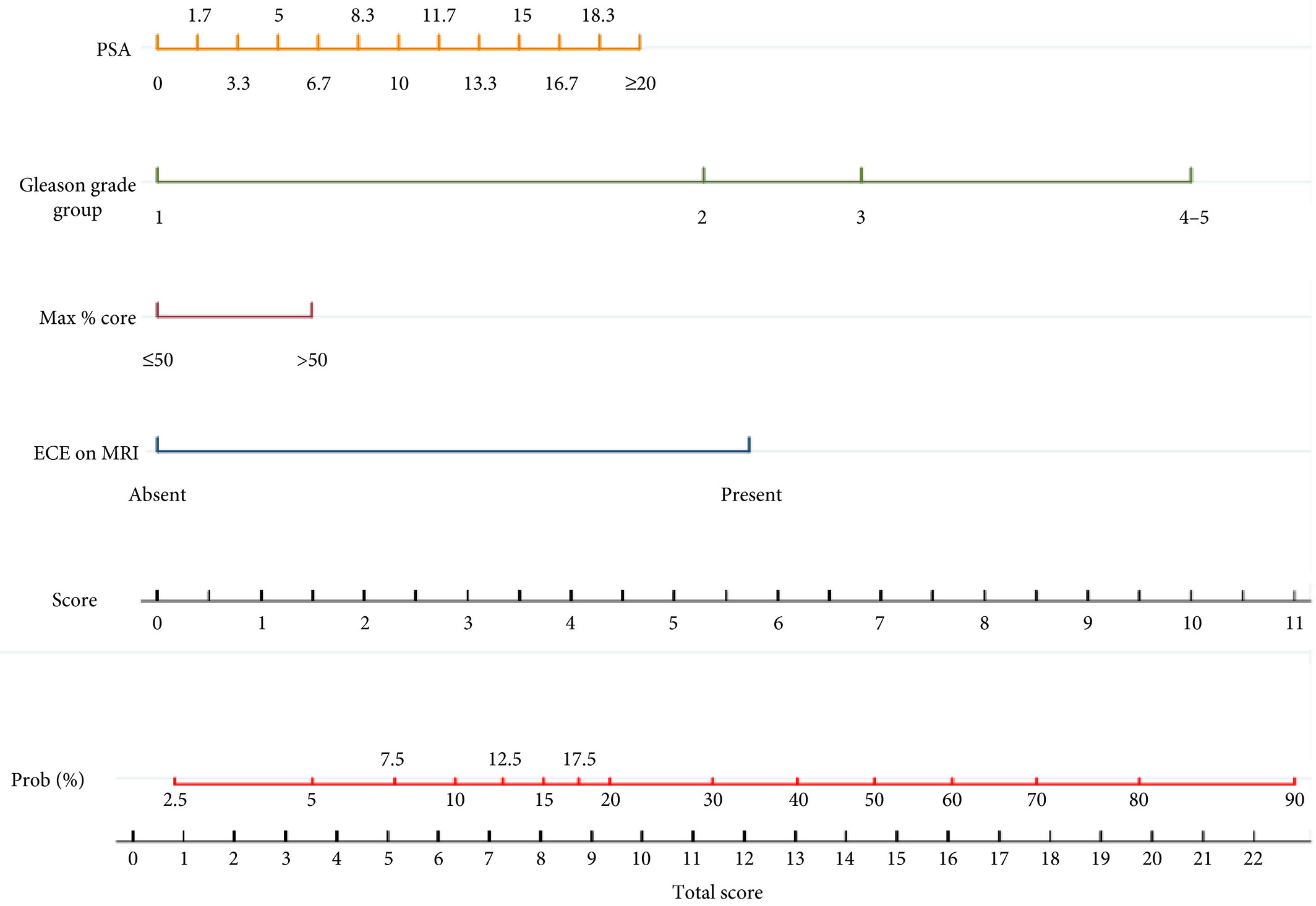
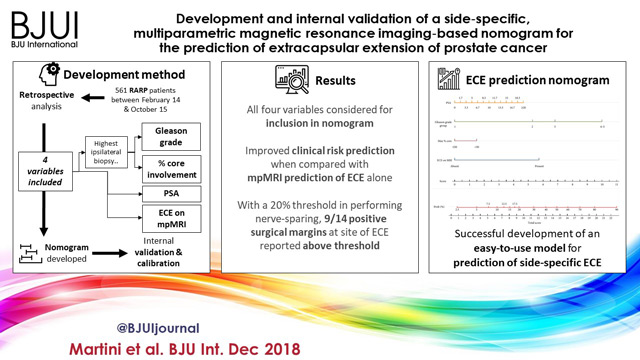
We retrospectively analysed data from 561 patients who underwent robot‐assisted radical prostatectomy between February 2014 and October 2015. To develop a side‐specific predictive model, we considered the prostatic lobes separately. Four variables were included: prostate‐specific antigen; highest ipsilateral biopsy Gleason grade; highest ipsilateral percentage core involvement; and ECE on multiparametric magnetic resonance imaging (mpMRI). A multivariable logistic regression analysis was fitted to predict side‐specific ECE. A nomogram was built based on the coefficients of the logit function. Internal validation was performed using ‘leave‐one‐out’ cross‐validation. Calibration was graphically investigated. The decision curve analysis was used to evaluate the net clinical benefit.

Results

The study population consisted of 829 side‐specific cases, after excluding negative biopsy observations (n = 293). ECE was reported on mpMRI and final pathology in 115 (14%) and 142 (17.1%) cases, respectively. Among these, mpMRI was able to predict ECE correctly in 57 (40.1%) cases. All variables in the model except highest percentage core involvement were predictors of ECE (all P ≤ 0.006). All variables were considered for inclusion in the nomogram. After internal validation, the area under the curve was 82.11%. The model demonstrated excellent calibration and improved clinical risk prediction, especially when compared with relying on mpMRI prediction of ECE alone. When retrospectively applying the nomogram‐derived probability, using a 20% threshold for performing nerve‐sparing, nine out of 14 positive surgical margins (PSMs) at the site of ECE resulted above the threshold.

Conclusion

We developed an easy‐to‐use model for the prediction of side‐specific ECE, and hope it serves as a tool for planning nerve‐sparing radical prostatectomy and in the reduction of PSM in future series.

Read more Articles of the week


Video: Development and internal validation of a side‐specific, mpMRI‐based nomogram for the prediction of extracapsular extension of PCa


Development and internal validation of a side‐specific, multiparametric magnetic resonance imaging‐based nomogram for the prediction of extracapsular extension of prostate cancer

Read the full article

Abstract

Objectives

To develop a nomogram for predicting side‐specific extracapsular extension (ECE) for planning nerve‐sparing radical prostatectomy.

Materials and Methods

We retrospectively analysed data from 561 patients who underwent robot‐assisted radical prostatectomy between February 2014 and October 2015. To develop a side‐specific predictive model, we considered the prostatic lobes separately. Four variables were included: prostate‐specific antigen; highest ipsilateral biopsy Gleason grade; highest ipsilateral percentage core involvement; and ECE on multiparametric magnetic resonance imaging (mpMRI). A multivariable logistic regression analysis was fitted to predict side‐specific ECE. A nomogram was built based on the coefficients of the logit function. Internal validation was performed using ‘leave‐one‐out’ cross‐validation. Calibration was graphically investigated. The decision curve analysis was used to evaluate the net clinical benefit.

Results

The study population consisted of 829 side‐specific cases, after excluding negative biopsy observations (n = 293). ECE was reported on mpMRI and final pathology in 115 (14%) and 142 (17.1%) cases, respectively. Among these, mpMRI was able to predict ECE correctly in 57 (40.1%) cases. All variables in the model except highest percentage core involvement were predictors of ECE (all P ≤ 0.006). All variables were considered for inclusion in the nomogram. After internal validation, the area under the curve was 82.11%. The model demonstrated excellent calibration and improved clinical risk prediction, especially when compared with relying on mpMRI prediction of ECE alone. When retrospectively applying the nomogram‐derived probability, using a 20% threshold for performing nerve‐sparing, nine out of 14 positive surgical margins (PSMs) at the site of ECE resulted above the threshold.

Conclusion

We developed an easy‐to‐use model for the prediction of side‐specific ECE, and hope it serves as a tool for planning nerve‐sparing radical prostatectomy and in the reduction of PSM in future series.

View more videos

Article of the Week: Detecting PSMs – using LRS on ex vivo RP specimens

Every Week the Editor-in-Chief selects an Article of the Week from the current issue of BJUI. The abstract is reproduced below and you can click on the button to read the full article, which is freely available to all readers for at least 30 days from the time of this post.

In addition to the article itself, there is an accompanying editorial written by a prominent member of the urological community. This blog is intended to provoke comment and discussion and we invite you to use the comment tools at the bottom of each post to join the conversation.

If you only have time to read one article this week, it should be this one.

Detecting positive surgical margins: utilisation of light-reflectance spectroscopy on ex vivo prostate specimens

Aaron H. Lay*, Xinlong Wang, Monica S. C. Morgan*, Payal Kapur, Hanli Liu,Claus G. Roehrborn* and Jeffrey A. Cadeddu*


*Department of Urology, University of Texas Southwestern Medical Center, Dallas, TX, Department of Bioengineering, University of Texas at Arlington, Arlington, TX, and Department of Pathology, University of Texas Southwestern Medical Center, Dallas, TX, USA


Read the full article

Abstract

Objective

To assess the efficacy of light-reflectance spectroscopy (LRS) to detect positive surgical margins (PSMs) on ex vivo radical prostatectomy (RP) specimens.

Materials and Methods

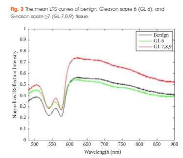
A prospective evaluation of ex vivo RP specimens using LRS was performed at a single institution from June 2013 to September 2014. LRS measurements were performed on selected sites on the prostate capsule, marked with ink, and correlated with pathological analysis. Significant features on LRS curves differentiating malignant tissue from benign tissue were determined using a forward sequential selection algorithm. A logistic regression model was built and randomised cross-validation was performed. The sensitivity, specificity, accuracy, negative predictive value (NPV), positive predictive value (PPV), and area under the receiver operating characteristic curve (AUC) for LRS predicting PSM were calculated.

aotwdec-4-results

Results

In all, 50 RP specimens were evaluated using LRS. The LRS sensitivity for Gleason score ≥7 PSMs was 91.3%, specificity 92.8%, accuracy 92.5%, PPV 73.2%, NPV 99.4%, and the AUC was 0.960. The LRS sensitivity for Gleason score ≥6 PSMs was 65.5%, specificity 88.1%, accuracy 83.3%, PPV 66.2%, NPV 90.7%, and the AUC was 0.858.

Conclusions

LRS can reliably detect PSMs for Gleason score ≥7 prostate cancer in ex vivo RP specimens

Read more articles of the week

Editorial: Light reflectance spectroscopy is one more emerging technique with the potential to adjust excision limits during radical prostatectomy

In this issue of BJUI, Lay et al. [1] report that light reflectance spectroscopy (LRS) can detect Gleason ≥7 positive surgical margins (PSMs) with 92.5% accuracy. In this initial study, the authors have reported the use of LRS in an ex situ setting to analyse the prostate surface; however, this technology could ultimately be developed to identify PSMs before choosing the surgical plane of dissection, which could allow the surgeon to immediately perform a wider complementary excision.

As long as PSMs are detected ex situ, it is not clear why spectroscopy should be preferred to frozen sections. NeuroSAFE, for example, is a standardized and validated margin evaluation procedure in pathology [2]. It does not lengthen operating time, does not require any new equipment and provides a pathological assessment which is the best level of evidence for PSM status; however, as a conventional pathological procedure, it is not conceivable in situ, and real-time detection of PSMs that ensures the safest oncological resection during a nerve-sparing dissection is needed.

In this effort to examine in vivo/in situ prostate PSMs, several other technologies can be considered. During radical prostatectomy, optical coherence tomography (OCT) has been used in situ in humans, but only to identify the neurovascular bundles [3]. Field of view and depth of penetration were limited and OCT has never been evaluated in situ for prostate PSM detection. Confocal endomicroscopy has recently been reported during robot-assisted radical prostatectomy [4]. With this technique, optical biopsies were feasible in situ but the PSM detection rate and the overall efficiency of this confocal endomicroscopy in prostate specimens remain unknown. Similarly, illumination microscopy has been used to generate gigapixel images of the full prostate circumference in vivo for the detection of PSMs [5]. Illumination microscopy allows images to be interpreted readily by pathologists, but the feasibility series was too small to assess the accuracy of this technique for PSM detection. Ex situ multi-photon microscopy (MPM) is an optical technique that enables the imaging of prostatic and periprostatic tissue at sub-micron resolution to a depth of up to 0.5 mm [6]. On a fresh specimen, it generates three-dimensional images of periprostatic nerves, blood vessels and capsule, but also underlying acini and pathological changes such as prostate cancer. MPM technology has also been miniaturized and its accuracy in situ is currently under investigation.

In this context, the study by Lay et al. [1] shows that, for the time being, LRS is one more promising technique on the road to real-time PSM detection. More will undoubtedly be done to overcome the spectroscope’s light absorption in the presence of blood and, subsequently, to evaluate its reliability in situ; however, the recent developments of these protocols and technologies (endomicroscopy, illumination microscopy, OCT, MPM, LRS) show a progressive effort amongst clinicians to obtain intra-operative feedback on the PSM status. Fortunately, this is taking place while the urological community is increasingly considering surgical treatment even for the high-risk disease, where oncological adequacy is of paramount importance. While we are witnessing these promising evolutions in high-grade prostate cancer, the optimum technique which will safely end margin-blind radical prostatectomy in an actual surgical field (filled with blood and often distorted because of inflammation) still needs to go through clinical trials and validation; however, the future is bright as a result of these newer developments.

Read the full article
Thomas Bessede*†‡ and Ash Tewari*


*Department of Urology, Icahn School of Medicine at MounSinai, New York , NY, USA, U1195, INSERM, UniversitParis-Saclay, and Department of Urology, APHP, Hopitaux Universitaires Paris-Sud, Le Kremlin-Bicetre, France


References


© 2024 BJU International. All Rights Reserved.