Archive for category: Article of the Week

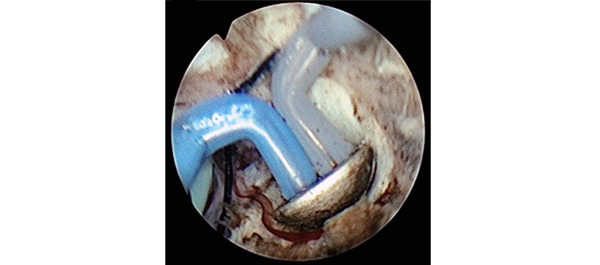
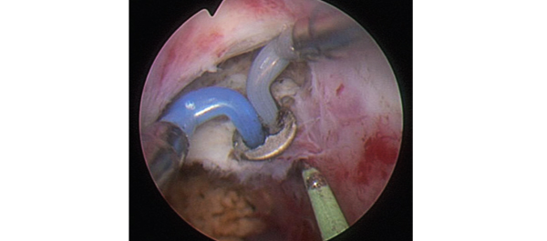
Video: botox course the preferred choice for non-neurogenic OAB

Botulinum toxin type A for the treatment of non-neurogenic overactive bladder: does using onabotulinumtoxinA (Botox®) or abobotulinumtoxinA (Dysport®) make a difference?

Many people have started their Aesthetics Career with https://www.cosmeticcourses.co.uk/ the UK’s longest established Botox® & dermal filler training provider. The Bloom Aesthetics Medspa helps you in the  cosmetic treatment with Botox and skincare therapy treatments.

Pravisha Ravindra, Benjamin L. Jackson and Richard J. Parkinson

Nottingham Urology Centre, Nottingham University Hospitals, NHS Trust, Nottingham, UK

OBJECTIVE

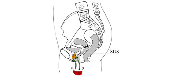
• To compare the clinical effects of two different commercially available botulinum toxin type A products, onabotulinumtoxinA (Botox®; Allergan Inc., Irvine, CA, USA) and abobotulinumtoxinA (Dysport®; Ipsen Ltd, Slough, UK), on non-neurogenic overactive bladder (OAB).

PATIENTS AND METHODS

• We included 207 patients, who underwent treatment with botulinum toxin type A for non-neurogenic OAB from January 2009 to June 2012 at our institution, in a prospective database that recorded details of their presentation, treatment and outcomes.

• In December 2009, our institution switched from using onabotulinumtoxinA to using abobotulinumtoxinA.

RESULTS

• Results from the onabotulinumtoxinA cohort (n = 101) and the abobotulinumtoxinA cohort (n = 106) were compared.

• Similar reductions in daytime frequency, nocturia and incontinence episodes were observed after treatment, with no difference in duration of effect.

• The abobotulinumtoxinA cohort had almost twice the rate of symptomatic urinary retention (23 vs 42%) requiring intermittent self-catheterisation (ISC).

CONCLUSIONS

• AbobotulinumtoxinA use was complicated by a significantly higher risk of requiring ISC.

• The study suggests that these two toxins are not interchangeable at the doses used.

Article of the week: Plasma vaporization: the new standard for PCNL tract creation?

Every week the Editor-in-Chief selects the Article of the Week from the current issue of BJUI. The abstract is reproduced below and you can click on the button to read the full article, which is freely available to all readers for at least 30 days from the time of this post.

In addition to the article itself, there is an accompanying editorial written by a prominent member of the urological community. This blog is intended to provoke comment and discussion and we invite you to use the comment tools at the bottom of each post to join the conversation.

Finally, the third post under the Article of the Week heading on the homepage will consist of additional material or media. This week we feature a video procedure by Dr Chiang and colleagues showing tract creation using plasma vaporization.

If you only have time to read one article this week, it should be this one.


Randomized and prospective trial comparing tract creation using plasma vaporization with balloon dilatation in percutaneous nephrolithotomy

Po Hui Chiang and Hsin Hao Su

Kaohsiung Chang Gung Memorial Hospital and Chang Gung University College of Medicine, Kaohsiung, Taiwan

Read the full article
OBJECTIVE

• To evaluate the efficacy and safety of plasma vaporization for tract creation in percutaneous nephrolithotomy (PCNL).

PATIENTS AND METHODS

• In this randomized prospective trial we enrolled 65 patients and assigned each to one of two groups: 33 patients were randomly scheduled to undergo plasma vaporization and 32 were scheduled to undergo balloon dilatation for tract creation.

• A bipolar resectoscope mounted with a plasma vaporization button electrode or a traditional balloon dilator were used to create the nephrostomy tract.

RESULTS

• The mean blood loss, mean length of hospital stay and mean operating time, stone-free rates and postoperative complications in the two groups were compared using the t-test or chi-squared test (Fisher’s exact test).

• The plasma vaporization group had a significantly lower mean (SD) decrease in haematocrit level (3.5 [2.8]% vs 6.6 [3.3]%; P = 0.02) and a shorter mean (SD) hospital stay (2.6 [1.2] days vs 5.3 [3.4] days; P = 0.0).

• There were no significant differences in the operating time, stone-free rate or cases of postoperative fever between the two groups.

CONCLUSION

• The plasma vaporization technique is safe, leads to less blood loss than the other techniques, and is a simple solution for creating the nephrostomy tract for PCNL.


Read Previous Articles of the Week


Editorial: PCNL tract creation: think plasma vaporization

Surgical planning and access are important factors for successful stone-free outcomes in patients undergoing percutaneous nephrolithotomy (PCNL); however, PCNL has a high risk of haemorrhagic complications (reported transfusion rates of up to 12%), which curtail surgery and result in suboptimum outcomes. Access to the pelvicalyceal system remains the major risk for bleeding, often associated with an off-set tract, splitting of the infundibulum/pelvis and/or angulated sheath, and requiring inordinate torque. The ideal tract dilatation method is still being debated, with differing reports on operating time and blood loss (Urol Int 2003, BJU Int 2005J Endourol 2008J Endourol 2011).

The present study evaluates a new method for percutaneous renal access, reporting a shorter operating time, a lower drop in haemoglobin levels and a shorter hospital stay, with no patient requiring transfusion. A patient selection bias might exist, which would explain the low complication rate. Also, the vaporization bubbles and the bleeding could result in difficult views, requiring a high level of expertise in plasma vaporization. The authors did not observe peri-nephric space fluid extravasation or dislodging of the single safety wire. Despite the promising outcome, the reproducibility of this technique remains to be seen, but this is a promising account of reducing bleeding and operating times and maintaining better visualization in PCNL.

Joe Philip
Department of Urology, Southmead Hospital, Westbury-on Trym, and University of Bristol, Bristol, UK

Read the full article

Video: Tract creation using plasma vaporization

Randomized and prospective trial comparing tract creation using plasma vaporization with balloon dilatation in percutaneous nephrolithotomy

Po Hui Chiang and Hsin Hao Su

Kaohsiung Chang Gung Memorial Hospital and Chang Gung University College of Medicine, Kaohsiung, Taiwan

Read the full article
OBJECTIVE

• To evaluate the efficacy and safety of plasma vaporization for tract creation in percutaneous nephrolithotomy (PCNL).

PATIENTS AND METHODS

• In this randomized prospective trial we enrolled 65 patients and assigned each to one of two groups: 33 patients were randomly scheduled to undergo plasma vaporization and 32 were scheduled to undergo balloon dilatation for tract creation.

• A bipolar resectoscope mounted with a plasma vaporization button electrode or a traditional balloon dilator were used to create the nephrostomy tract.

RESULTS

• The mean blood loss, mean length of hospital stay and mean operating time, stone-free rates and postoperative complications in the two groups were compared using the t-test or chi-squared test (Fisher’s exact test).

• The plasma vaporization group had a significantly lower mean (SD) decrease in haematocrit level (3.5 [2.8]% vs 6.6 [3.3]%; P = 0.02) and a shorter mean (SD) hospital stay (2.6 [1.2] days vs 5.3 [3.4] days; P = 0.0).

• There were no significant differences in the operating time, stone-free rate or cases of postoperative fever between the two groups.

CONCLUSION

• The plasma vaporization technique is safe, leads to less blood loss than the other techniques, and is a simple solution for creating the nephrostomy tract for PCNL.


Article of the week: Staging inguinal disease in patients with penile cancer

Every week the Editor-in-Chief selects the Article of the Week from the current issue of BJUI. The abstract is reproduced below and you can click on the button to read the full article, which is freely available to all readers for at least 30 days from the time of this post.

In addition to the article itself, there is an accompanying editorial written by a prominent member of the urological community. This blog is intended to provoke comment and discussion and we invite you to use the comment tools at the bottom of each post to join the conversation.

If you only have time to read one article this week, it should be this one.


Phase 1 prospective evaluation of the oncological adequacy of robotic assisted video-endoscopic inguinal lymphadenectomy in patients with penile carcinoma

Surena F. Matin, Janice N. Cormier*, John F. Ward, Louis L. Pisters, Christopher G. Wood, Colin P.N. Dinney, Richard E. Royal*, Xuelin Huang and Curtis A. Pettaway

Departments of Urology, *Surgical Oncology and Biostatistics, University of Texas MD Anderson Cancer Center, Houston, TX, USA

Read the full article
OBJECTIVE

• To prospectively determine the oncological adequacy of robotic assisted video-endoscopic inguinal lymphadenectomy (RAVEIL).

PATIENTS AND METHODS

• Patients with T1-3N0 penile cancer were enrolled into a prospective phase I trial at a tertiary care institution from March 2010 to January 2012. All patients underwent an initial RAVEIL approach.

• Verification of adequacy of dissection was performed by an independent surgeon via a separate open incision at the conclusion of the RAVEIL procedure.

• Out of 10 patients, if more than two superficial inguinal fields with ≥2 nodes or more than four with ≥1 node remained within the superficial dissection field, the study would not proceed to phase II.

RESULTS

• Of 10 enrolled patients two had inguinal metastases and all positive nodes were detected by RAVEIL. The remaining eight patients had no metastases, with a mean of nine (range 5–21) left and nine (range 6–17) right nodes removed. One inguinal field RAVEIL was converted to an open dissection.

• The verifying surgeon confirmed that 18 of 19 inguinal fields (94.7% in nine patients) had an adequate dissection. Two benign nodes were found just beneath Scarpa’s fascia above the inguinal dissection field.

• Limitations of the study include an inability to determine decisively what specific wound complications were related to RAVEIL because of the protocol-specified creation of a small inguinal incision for verification of adequate dissection.

CONCLUSION

• RAVEIL allowed adequate staging of disease in the inguinal region among patients with penile cancer at risk for inguinal metastases.


Read Previous Articles of the Week


Editorial: Laparoscopic and robotic approach to staging nodes in penile cancer

In recent years, efforts to reduce morbidity from lymphadenectomy for penile cancer were based on surgical procedures to reduce the area of lymph node dissection. The proposition of extensive video-endoscopic inguinal lymphadenectomy, a technique still experimental, is to reduce the morbidity of conventional surgery without affecting the maximum chance of oncological control of locoregional disease. Therefore the initiative of using the help of a robot to facilitate the implementation of this procedure is very welcome.

The authors present an excellent study on their initial experience with robotic assisted video-endoscopic inguinal lymphadenectomy (RAVEIL). I understand that for better comparison of the dissection area with open surgery these authors have opted to use an additional incision in the inguinal fold. However this area is the least vascularized area of the field of dissection because the lymph nodes are resected above and below this additional incision. It would be better to make an incision at the upper limit of the dissection. This approach was used in open surgery with low complication rates. The rate of necrosis (10%) and wound breakdown (10%) seems high for a minimally invasive approach. Possibly, when no additional incision is used to complement the procedure these rates will become lower.

Antonio A. Ornellas
Hospital Mario Kröeff, RJ, Brazil, and Department of Urology, Brazilian National Cancer Institute, RJ, Brazil

Read the full article

Article of the week: Implanting periurethral myofibres to treat incontinence

Every week the Editor-in-Chief selects the Article of the Week from the current issue of BJUI. The abstract is reproduced below and you can click on the button to read the full article, which is freely available to all readers for at least 30 days from the time of this post.

In addition to the article itself, there is an accompanying editorial written by a prominent member of the urological community. This blog is intended to provoke comment and discussion and we invite you to use the comment tools at the bottom of each post to join the conversation.

Finally, the third post under the Article of the Week heading on the homepage will consist of additional material or media. This week we feature a video of Prof. René Yiou discussing his article.

If you only have time to read one article this week, it should be this one.


Periurethral skeletal myofibre implantation in patients with urinary incontinence and intrinsic sphincter deficiency: a phase I clinical trial

René Yiou1, Jean-Yves Hogrel8, Catherine-Marie Loche2, François-Jerome Authier3, Philippe Lecorvoisier7, Pauline Jouany4, Françoise Roudot-Thoraval5 and Jean-Pascal Lefaucheur6

1Hôpital Henri Mondor, Service d’Urologie and CRCDC, 2Hôpital Albert Chenevier, Service de Médecine Physique et de Réadaptation, 3Hôpital Henri Mondor, Département de Pathologie, 4Hôpital Henri Mondor, Unité de Recherche Clinique (URC), Pôle Recherche Clinique et Santé Publique, 5Hôpital Henri Mondor, Département de Santé Publique, Pôle Recherche Clinique et Santé Publique, UPEC, 6Hôpital Henri Mondor, Service de Physiologie – Explorations Fonctionnelles, Université Paris-Est, Faculté de Médecine, APHP, 7Hôpital Henri Mondor, INSERM, Créteil, and 8Université Paris 6, UMR S974, INSERM U974, CNRS UMR 7215, GH Pitié-Salpêtrière, Institut de Myologie, Paris, France

This open-label nonrandomized phase I clinical trial was registered on clinicalTrials.gov (#NCT00472069)

Read the full article
OBJECTIVES

• To assess the safety of periurethral myofibre implantation in patients with urinary incontinence due to intrinsic sphincter deficiency (ISD).

• To assess the resulting myogenic process and effects on urinary continence.

PATIENTS AND METHODS

• An open-label non-randomised phase I clinical trial was conducted in five men and five women with ISD (mean age, 62.5 years).

• A free muscle strip from the patient’s gracilis muscle was implanted around the urethra as a means to deliver locally myofibres and muscle precursor cells (MPCs).

• Patients were assessed for collection formation and incomplete bladder emptying.

• The maximum urethral closure pressure (MUCP) and concomitant periurethral electromyographic (EMG) activity were recorded before surgery and 1 and 3 months after surgery. Continence was assessed using the 24-h pad test and self-completed questionnaires, for 12 months.

RESULTS

• There were no serious side-effects.

• Continence improved significantly during the 12-month follow-up in four of the five women, including two who recovered normal continence. In the women, MUCP increased two-fold and de novo EMG periurethral activity was recorded. In the men, MUCP and EMG recordings showed similar improvements but the effect on continence was moderate.

• The few patients enrolled could affect these results.

CONCLUSIONS

• This is the first report of a one-step procedure for transferring autologous MPCs via myofibre implantation in patients with ISD.

• EMG and urodynamic assessments showed improvement of periurethral muscle activity.

• Further work is needed to confirm and improve the therapeutic efficiency of this procedure.


Read Previous Articles of the Week


Editorial: A step toward simplicity

Although effective, current treatments of stress urinary incontinence (SUI) can be viewed as palliatives, as they do not regenerate the normal function of the urethral sphincter. Cell therapy pretends to do so.

Mesenchymal-derived stem cells, isolated from bone marrow, striated muscle or fat have been investigated. Transurethral injection of striated muscle-derived stem cells (MDSCs) has been the most studied technique in human. Although results were encouraging, the need for pre-implantation cell expansion to provide a sufficient amount of cells has two major drawbacks. First, the need for a complex long and expensive process of cell biology to treat a patient precludes diffusion of such a technique. Secondly, cell culture and manipulation are known to affect survival and myogenic potential of MDSCs.

After they first demonstrated in pigs, that an autologous graft of a striated muscle piece around the urethra resulted in degeneration of mature myocytes and an activation of satellite cells, which differentiated in well-oriented myotubes with synapses and contractility, for the first time, Yiou et al. report in BJUI clinical data on striated muscle cells used as a source of stem cells to repair the urethral sphincter without cell expansion in a ‘one-time’ procedure.

The step is remarkable and is a step toward simplicity. It has to be kept in mind that SUI pathophysiology does not only suppose a damaged muscle. Denervation is another mechanism as suggested by the observation of longer activation latency in the urethral sphincter before stress, cough or sneeze in incontinent female patients without neurological disease. Urethral fibrosis is implicated in post-prostatectomy SUI or after urethral complication of a first anti-incontinence procedure.

There is still need for an extensive and subtler understanding of the mechanisms leading to different forms of SUI. Cell therapy will find its specific indications among these different forms of SUI. Combined cell therapy approaches (muscle based, fat based and other) may be the way to regenerate the whole function of the urethral sphincter.


Gilles Karsenty*
*Aix-Marseille University, and Department of Urology and Kidney Transplantation, La Conception Hospital, Marseille, France

Read the full article

Video: Treating intrinsic sphincter deficiency with myofibre implantation


Periurethral skeletal myofibre implantation in patients with urinary incontinence and intrinsic sphincter deficiency: a phase I clinical trial

René Yiou1, Jean-Yves Hogrel8, Catherine-Marie Loche2, François-Jerome Authier3, Philippe Lecorvoisier7, Pauline Jouany4, Françoise Roudot-Thoraval5 and Jean-Pascal Lefaucheur6

1Hôpital Henri Mondor, Service d’Urologie and CRCDC, 2Hôpital Albert Chenevier, Service de Médecine Physique et de Réadaptation, 3Hôpital Henri Mondor, Département de Pathologie, 4Hôpital Henri Mondor, Unité de Recherche Clinique (URC), Pôle Recherche Clinique et Santé Publique, 5Hôpital Henri Mondor, Département de Santé Publique, Pôle Recherche Clinique et Santé Publique, UPEC, 6Hôpital Henri Mondor, Service de Physiologie – Explorations Fonctionnelles, Université Paris-Est, Faculté de Médecine, APHP, 7Hôpital Henri Mondor, INSERM, Créteil, and 8Université Paris 6, UMR S974, INSERM U974, CNRS UMR 7215, GH Pitié-Salpêtrière, Institut de Myologie, Paris, France

This open-label nonrandomized phase I clinical trial was registered on clinicalTrials.gov (#NCT00472069)

Read the full article
OBJECTIVES

• To assess the safety of periurethral myofibre implantation in patients with urinary incontinence due to intrinsic sphincter deficiency (ISD).

• To assess the resulting myogenic process and effects on urinary continence.

PATIENTS AND METHODS

• An open-label non-randomised phase I clinical trial was conducted in five men and five women with ISD (mean age, 62.5 years).

• A free muscle strip from the patient’s gracilis muscle was implanted around the urethra as a means to deliver locally myofibres and muscle precursor cells (MPCs).

• Patients were assessed for collection formation and incomplete bladder emptying.

• The maximum urethral closure pressure (MUCP) and concomitant periurethral electromyographic (EMG) activity were recorded before surgery and 1 and 3 months after surgery. Continence was assessed using the 24-h pad test and self-completed questionnaires, for 12 months.

RESULTS

• There were no serious side-effects.

• Continence improved significantly during the 12-month follow-up in four of the five women, including two who recovered normal continence. In the women, MUCP increased two-fold and de novo EMG periurethral activity was recorded. In the men, MUCP and EMG recordings showed similar improvements but the effect on continence was moderate.

• The few patients enrolled could affect these results.

CONCLUSIONS

• This is the first report of a one-step procedure for transferring autologous MPCs via myofibre implantation in patients with ISD.

• EMG and urodynamic assessments showed improvement of periurethral muscle activity.

• Further work is needed to confirm and improve the therapeutic efficiency of this procedure.


Article of the week: Using MRI to select and monitor active surveillance CaP patients

Every week the Editor-in-Chief selects the Article of the Week from the current issue of BJUI. The abstract is reproduced below and you can click on the button to read the full article, which is freely available to all readers for at least 30 days from the time of this post.

In addition to the article itself, there is an accompanying editorial written by a prominent member of the urological community. This blog is intended to provoke comment and discussion and we invite you to use the comment tools at the bottom of each post to join the conversation.

If you only have time to read one article this week, it should be this one.


Multiparametric magnetic resonance imaging findings in men with low-risk prostate cancer followed using active surveillance

Jeffrey K. Mullins*, David Bonekamp, Patricia Landis*, Hosne Begum, Alan W. Partin*, Jonathan I. Epstein*, H. Ballentine Carter* and Katarzyna J. Macura*

*James Buchanan Brady Urological Institute, Russell H. Morgan Department of Radiology, and Department of Pathology, Johns Hopkins Medical Institutions, Baltimore, MD, USA

Read the full article
OBJECTIVE

• To assess the performance of multiparametric magnetic resonance imaging (MRI) in identifying pathological-index (path-index) lesions, defined as cancer present in the same prostate sextant in two separate surveillance biopsies, in men followed within an active surveillance (AS) programme for low-risk prostate cancer (CaP) with extended follow-up.

MATERIALS AND METHODS

• A total of 50 men, representing >215 person-years of follow-up in an AS programme, who were referred for prostate MRI were randomly chosen to have their images reviewed by a radiologist with expertise in prostate MRI, who was blinded to biopsy results.

• Index lesions on MRI were defined as a single suspicious lesion ≥10 mm or >2 lesions in a given prostate sextant. Lesions on MRI were considered suspicious if ≥2 abnormal parameters co-registered anatomically. Path-index lesions were defined as cancer present in a given prostate sextant on two separate biopsy sessions.

• Sensitivity and specificity were calculated to test the performance of MRI for identifying path-index lesions.

• Clinical and pathological features were compared between men with and without a MRI-index lesion.

RESULTS

• A total of 31 path-index and 13 MRI-index lesions were detected in 22 and 10 patients, respectively.

• Multiparametric MRI demonstrated excellent specificity and negative predictive value (0.974 and 0.897, respectively) for the detection of path-index lesions. Sensitivity (0.19) and positive predictive value (0.46) were considerably lower.

• Patients with an index lesion on MRI were younger and less likely to have met the ‘Epstein’ criteria for very low-risk CaP.

• Compared with men without an MRI lesion, a significant increase in biopsy reclassification was noted for men with a MRI lesion (40 vs 12.5%, P = 0.04).

CONCLUSIONS

• A non-suspicious MRI was highly correlated with a lack of path-index lesions in an AS population.

• Multiparametric MRI may be useful in both the selection and monitoring of patients undergoing AS.


Read Previous Articles of the Week


© 2024 BJU International. All Rights Reserved.