Archive for category: Article of the Week

Article of the week: Single-port robot assisted radical prostatectomy (SP-RARP): a systematic review and pooled analysis of the preliminary experiences

This is the final Article of the Week selected by the outgoing Editor-in-Chief from the current issue of BJUI. The abstract is reproduced below and you can click on the button to read the full article, which is freely available to all readers for at least 30 days from the time of this post.

If you only have time to read one article this week, we recommend this one. 

Single‐port robot‐assisted radical prostatectomy: a systematic review and pooled analysis of the preliminary experiences

Enrico Checcucci*, Sabrina De Cillis*, Angela Pecoraro*, Dario Peretti*, Gabriele Volpi*, Daniele Amparore*, Federico Piramide*, Alberto Piana*, Matteo Manfredi*, Cristian Fiori*, Riccardo Autorino, Prokar Dasgupta, Francesco Porpiglia* and on behalf of the Uro-technology and SoMe Working Group of the Young Academic Urologists Working Party of the European Association of Urology

*Department of Urology, San Luigi Gonzaga Hospital, University of Turin, Turin, Italy, Division of Urology, VCU Health, Richmond, VA, USA, and King’s College London, Guy’s Hospital, London, UK

Abstract

Objective

To summarize the clinical experiences with single‐port (SP) robot‐assisted radical prostatectomy (RARP) reported in the literature and to describe the peri‐operative and short‐term outcomes of this procedure.

Material and Methods

A systematic review of the literature was performed in December 2019 using Medline (via PubMed), Embase (via Ovid), Cochrane databases, Scopus and Web of Science (PROSPERO registry number 164129). All studies that reported intra‐ and peri‐operative data on SP‐RARP were included. Cadaveric series and perineal or partial prostatectomy series were excluded.

The da Vinci SP robotic platform

Results

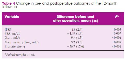
The pooled mean operating time, estimated blood loss, length of hospital stay and catheterization time were 190.55 min, 198.4 mL, 1.86 days and 8.21 days, respectively. The pooled mean number of lymph nodes removed was 8.33, and the pooled rate of positive surgical margins was 33%. The pooled minor complication rate was 15%. Only one urinary leakage and one major complication (transient ischaemic attack) were recorded. Regarding functional outcomes, pooled continence and potency rates at 12 weeks were 55% and 42%, respectively.

Conclusions

The present analysis confirms that SP‐RARP is safe and feasible. This novel robotic platform resulted in similar intra‐operative and peri‐operative outcomes to those obtained with the standard multiport da Vinci system. The advantages of single incision can be translated into a preservation of the patient’s body image and self‐esteem and cosmesis, which have a great impact on a patient’s quality of life.

Article of the week: Critical analysis of a multicentric experience with holmium laser enucleation of the prostate for BPH

Every week, the Editor-in-Chief selects an Article of the Week from the current issue of BJUI. The abstract is reproduced below and you can click on the button to read the full article, which is freely available to all readers for at least 30 days from the time of this post.

There is also an editorial written by a prominent member of the urological community. Please use the comment buttons if you would like to join the conversation.

If you only have time to read one article this week, we recommend this one. 

Critical analysis of a multicentric experience with holmium laser enucleation of the prostate for benign prostatic hyperplasia: outcomes and complications of 10 years of routine clinical practice

Javier Romero-Otero*†‡, Borja García-Gómez*, Lucía García-González*, Esther García-Rojo*, Pablo Abad-López*, Juan Justo-Quintas, José Duarte-Ojeda* and Alfredo Rodríguez-Antolín*

*Urology Department, Grupo de Investigación Salud Integral del Varón imas12, Hospital Universitario 12 Octubre, Hospital Universitario HM Montepríncipe, and Hospital Universitario La Luz, Madrid, Spain

Read the full article

Abstract

Objective

To assess the perioperative outcomes of holmium laser enucleation of the prostate (HoLEP) in real‐life practice and investigate the factors influencing the safety and effectiveness of the technique.

Patients and Methods

Critical analysis of patients with benign prostate hyperplasia (BPH) treated with HoLEP over 10 years of routine practice in three hospitals. Analysed variables included: preoperative characteristics (prostate size, active antiplatelet/anticoagulant therapy, blood parameters. prostate‐specific antigen (PSA) level, maximum urinary flow rate [Qmax], and International Prostate Symptom Score [IPSS]), intraoperative variables (operation time, concomitant removal of bladder calculi, and complications), early postoperative outcomes (change in blood parameters, catheterisation time, and hospital stay), and 12‐month follow‐up outcomes (change in IPSS, PSA level, and Qmax).

Results

The analysis included 963 patients, aged 48–91 years, with a mean (range) prostate size of 91 (35–247) mL. The mean (sd ) operation time was 77 (29) min, and the hospital stay and catheterisation time were 4 (2) and 1.3 (2) days, respectively. In all, 56 patients (5.6%) required concomitant removal of bladder calculi and 36 (3.7%) were converted to open prostatectomy or transurethral resection of the prostate due to intraoperative complications. Patients had a significant decrease in haemoglobin and haematocrit, but no differences were seen between patients with and without anticoagulant/antiplatelet therapy and those with prostates ≥ and <100 mL. The concomitant removal of bladder calculi and having a prostate ≥100 mL resulted in a longer operation time, but did not influence the safety and effectiveness outcomes.

Conclusions

HoLEP is suitable for real‐life patients with BPH, irrespective of the presence of active treatment with anticoagulant/antiplatelet, bladder lithiasis or a prostate ≥100 mL.

Editorial: HoLEP is the complete technique for treating BPH

Ten years of experience with holmium laser enucleation of the prostate (HoLEP) are documented by Romero‐Otero et al. [1] and offer valuable insight into the real‐world use of this technique. No information on the 10‐year durability is available, however, as only the 12‐month data are presented, but there is a wealth of other information concerning both peri‐operative outcomes and complications. A particular strength of this paper is that all‐comers were studied, including patients with catheters, those with prostates larger than 100 g and those taking anti‐coagulants, plus there is the addition of the cases the three surgeons performed during their ‘learning curve’, although these are not analysed separately.

The authors’ technique almost certainly evolved over the study period. Personally, I currently find a one‐ or two‐piece enucleation to be more efficient than the three‐lobe technique originally described [2]. Enucleation efficiency of 1–2 g/min, as was achieved in this series (73 g in 40 min), is a good benchmark for tissue removal for those new to the technique and is a good measure of surgical proficiency. Being less aggressive anteriorly seems to have an impact on continence. It is often tempting to completely enucleate circumferentially in one continuous plane which is sometimes well beyond the commissure anteriorly. A more moderate dissection in this area can reduce the transient incontinence sometimes seen [3]. The incontinence rates in the current series of 12.8% at 3 months and 2.3% at 12 months are probably representative [1]. An analysis of the factors predisposing to moderate‐to‐severe incontinence in the six patients in this series would have been useful, particularly regarding prostate size, presence of a catheter and age.

The main contribution of HoLEP to the urological armamentarium is its ability to safely treat large prostates endoscopically [4]. Although robot‐assisted techniques have also decreased the morbidity of open prostatectomy [5], the attraction of the obvious ‘natural orifice’ for access and the use of laser technology for the enucleation with HoLEP is probably the least morbid and most cost‐effective way to treat these patients. Tackling a prostate larger than 100 g involves applying the same principles as for smaller prostates, with a few provisos. Firstly, having a consistent strategy for these large prostates is important and can be reassuring when things become difficult. Secondly, it is even more important to maintain the correct plane religiously as it is easier to get lost in these glands. A good sense of direction is important! Thirdly, stay ahead of the bleeding rather than trying to catch up as it can further compound an already difficult situation. Patience is a virtue.

The learning curve of HoLEP has historically been regarded as a major barrier to the uptake of the technique [6]. This has, of course, been exaggerated by proponents of other techniques, but it is important to emphasize that during this learning phase the excellent outcomes are maintained and that conversion to TURP, if necessary (3.4% in this series), can be safely done, as these authors’ have demonstrated. The length of the learning curve has been variously described as being between 20 and 80 cases and is almost entirely due to the way training is done. A modular mentored approach appears to be the best method and could equally be applied to endoscopic enucleation using any of the other energy sources that have been described [7].

HoLEP and all its progeny are here to stay, but which of these enucleation energy sources will gain ascendancy remains to be seen. Sadly, this will likely be more to do with the depth of the corporate pockets and their commitment to the cause rather than proper scientific appraisal [8].

by Peter Gilling

References

  1. Romero‐Otero J, Garcia‐Gomez B, Garcia‐Gonzalez L et al. Critical analysis of a multicentric experience with holmium laser enucleation of the prostate for benign prostatic hyperplasia: outcomes and complications of 10 years of routine clinical practice. BJU Int 2020; 126: 177-182
  2. Gilling PJ, Kennett K, Das AK, Thompson D, Fraundorfer MR. Holmium laser enucleation of the prostate (HoLEP) combined with transurethral tissue morcellation: an update on the early clinical experience. J Endourol 1998; 12: 457– 9
  3. Tunc L, Yalcin S, Kaya E et al. The “Omega Sign”: a novel HoLEP technique that improves continence outcomes after enucleation. World J Urol 2020 https://doi.org/10.1007/s00345-020-03152-9
  4. Gilling PJ, Kennett KM, Fraundorfer MR. Holmium laser enucleation of the prostate for glands larger than 100 g: an endourologic alternative to open prostatectomy. J Endourol 2000; 14: 529– 31
  5. Mourmouris P, Keskin SM, Skolarikos A et al. A prospective comparative analysis of robot‐assisted vs open simple prostatectomy for benign prostatic hyperplasia. BJU Int 2019; 123: 313– 7
  6. Placer J, Gelabert‐Mas A, Vallmanya F et al. Holmium laser enucleation of prostate: outcome and complications of self‐taught learning curve. Urology 2009; 73: 1042– 8
  7. Kuronen‐Stewart C, Ahmed K, Aydin A et al. Holmium Laser Enucleation of the prostate: simulation based training curriculum and validation. Urology 2015; 86: 639– 46.
  8. Herrmann TR. Enucleation is enucleation is enucleation is enucleation. World J Urol 2016; 34: 1353– 5

Article of the month: SIMULATE: Protocol and curriculum development of the first multicentre international randomized controlled trial assessing the transferability of simulation‐based surgical training

Every month, the Editor-in-Chief selects an Article of the Month from the current issue of BJUI. The abstract is reproduced below and you can click on the button to read the full article, which is freely available to all readers for at least 30 days from the time of this post.

There is also a visual abstract created by a member of the team.

If you only have time to read one article this month, we recommend this one. 

Simulation in Urological Training and Education (SIMULATE): Protocol and curriculum development of the first multicentre international randomized controlled trial assessing the transferability of simulation‐based surgical training

Abdullatif Aydin*, Kamran Ahmed*, Mieke Van Hemelrijck, Hashim U. Ahmed§–, Muhammad Shamim Khan* ** and Prokar Dasgupta* ** on behalf of the SIMULATE Trial Group**

*MRC Centre for Transplantation, King’s College London, London, Department of Urology, King’s College Hospital NHS Foundation Trust, London,School of Cancer and Pharmaceutical Studies, King’s College London, London, §Department of Surgery and Cancer, Imperial College London, Department of Urology, Imperial College Healthcare NHS Trust, London, and **Department of Urology, Guy’s and St Thomas’ NHS Foundation Trust, London, UK

Read the full article

Abstract

Objectives

To report the study protocol for the first international multicentre randomized controlled trial investigating the effectiveness of simulation‐based surgical training and the development process for an evidence‐based training curriculum, to be delivered as an educational intervention.

Participants and Methods

This prospective, international, multicentre randomized controlled clinical and educational trial will recruit urology surgical trainees who must not have performed ≥10 of the selected index procedure, ureterorenoscopy (URS). Participants will be randomized to simulation‐based training (SBT) or non‐simulation‐based training (NSBT), the latter of which is the current sole standard of training globally. The primary outcome is the number of procedures required to achieve proficiency, where proficiency is defined as achieving a learning curve plateau of 28 or more on an Objective Structured Assessment of Technical Skills (OSATS) assessment scale, for three consecutive operations, without any complications. All participants will be followed up either until they complete 25 procedures or for 18 months. Development of the URS SBT curriculum took place through a two‐round Delphi process.

The SIMULATE ureteroscopy training curriculum, designed to be delivered as the educational intervention. The curriculum begins with introductory didactic lectures followed by virtual reality and dry‐lab simulation for semi‐rigid and flexible URS, respectively. If feasible, this is to be followed by full immersion simulation and cadaveric training, respectively. fURS, flexible ureterorenoscopy; NTS, non‐technical skills; OR, operating room; UO, ureteric access; URS, ureteroscopy; VR, virtual reality.

Results

A total of 47 respondents, consisting of trainees (= 24) with URS experience and urolithiasis specialists (= 23), participated in round 1 of the Delphi process. Specialists (= 10) finalized the content of the curriculum in round 2. The developed interventional curriculum consists of initial theoretic knowledge through didactic lectures followed by select tasks and cases on the URO‐Mentor (Simbionix, Lod, Israel) VR Simulator, Uro‐Scopic Trainer (Limbs & Things, Bristol, UK) and Scope Trainer (Mediskills, Manchester, UK) models for both semi‐rigid and flexible URS. Respondents also selected relevant non‐technical skills scenarios and cadaveric simulation tasks as additional components, with delivery subject to local availability.

Conclusions

SIMULATE is the first multicentre trial investigating the effect and transferability of supplementary SBT on operating performance and patient outcomes. An evidence‐based training curriculum is presented, developed with expert and trainee input. Participants will be followed and the primary outcome, number of procedures required to proficiency, will be reported alongside key clinical secondary outcomes, (ISCRTN 12260261).

Read more Articles of the week

Visual Abstract: Simulation in Urological Training and Education (SIMULATE)

Read the full article
See more infographics

Article of the week: Management of large renal stones with super‐mini percutaneous nephrolithotomy: an international multicentre comparative study

Every week, the Editor-in-Chief selects an Article of the Week from the current issue of BJUI. The abstract is reproduced below and you can click on the button to read the full article, which is freely available to all readers for at least 30 days from the time of this post.

There is also a visual abstract created by Cora Griffin from King’s College London.

If you only have time to read one article this week, we recommend this one. 

Management of large renal stones with super-mini percutaneous nephrolithotomy: an international multicentre comparative study

Yang Liu*, Chao Cai*, Albert Aquino, Shabir Al-Mousawi, Xuepei Zhang§, Simon K.S. Choong, Xiang He**, Xianming Fan††, Bin Chen‡‡, Jianhua Feng§§, Xuhui Zhu¶¶, Abdulla Al-Naimi***, Houping Mao†††, Huilong Tang‡‡‡, Dayong Jin§§§, Xiancheng Li¶¶¶, Fenghong Cao****, Hua Jiang††††, Yongfu Long‡‡‡‡, Wei Zhang§§§§, Gang Wang¶¶¶¶, Zihao Xu*, Xin Zhang*, Shanfeng Yin* and Guohua Zeng*

*Department of Urology, Guangdong Key Laboratory of Urology, Minimally Invasive Surgery Center, The First Affiliated Hospital of Guangzhou Medical University, Guangzhou, China, Department of Urology, Jose R. Reyes Memorial Medical Center, Manila, Philippines, Division of Urology, SABAH ALAHMAD Urology Center, Ministry of Health, Al-Amiri Hospital, Kuwait City, Kuwait, §Department of Urology, The First Affiliated Hospital of Zhengzhou University, Zhengzhou, China, Institute of Urology, University College London Hospitals, London, UK, **Department of Urology, Zhejiang Provincial People’s Hospital, People’s Hospital of Hangzhou Medical College, Hangzhou, ††Department of Urology, The Third Affiliated Hospital of Xiamen, The Third Affiliated Hospital of Fujian University of Traditional Chinese Medicine, ‡‡Department of Urology, The Affiliated Hospital of Xiamen University, Xiamen, §§Department of Urology, Longgang District Central Hospital, Shenzhen, ¶¶Department of Urology, Beijing Chaoyang Hospital, Affiliated to Capital Medical University, Beijing, China, ***Department of Urology, Hamad Medical Corporation, Doha, Qatar, †††Department of Urology, First Affiliated Hospital of Fujian Medical University, Fuzhou, ‡‡‡Department of Urology, The First Hospital of Hunan University of Traditional Chinese Medicine, Changsha, §§§Department of Urology, Liaoyuan City Central Hospital, Liaoyuan, ¶¶¶Department of Urology, Second Affiliated Hospital of Dalian Medical University, Dalian, ****Department of Urology, North China University of Science and Technology Affiliated Hospital, Tangshan, ††††Department of Urology, Zhongda Hospital Southeast Hospital, Nanjing, ‡‡‡‡Department of Urology, Central Hospital of Shaoyang, Shaoyang, §§§§Department of Urology, The First Affiliated Hospital of Nanjing Medical University, Nanjing, and ¶¶¶¶Department of Urology, National Urological Cancer Center, Institute of Urology, Peking University First Hospital, Peking University, Beijing, China

Read the full article

Abstract

Objectives

To comparatively evaluate the clinical outcomes of super‐mini percutaneous nephrolithotomy (SMP) and mini‐percutaneous nephrolithotomy (Miniperc) for treating urinary tract calculi of >2 cm.

Patients and Methods

An international multicentre, retrospective cohort study was conducted at 20 tertiary care hospitals across five countries (China, the Philippines, Qatar, UK, and Kuwait) between April 2016 and May 2019. SMP and Miniperc were performed in 3525 patients with renal calculi with diameters of >2 cm. The primary endpoint was the stone‐free rate (SFR). The secondary outcomes included: blood loss, operating time, postoperative pain scores, auxiliary procedures, complications, tubeless rate, and hospital stay. Propensity score matching analysis was used to balance the selection bias between the two groups.

Results

In all, 2012 and 1513 patients underwent SMP and Miniperc, respectively. After matching, 1380 patients from each group were included for further analysis. Overall, there was no significant difference in the mean operating time or SFR between the two groups. However, the hospital stay and postoperative pain score were significantly in favour of SMP (both  < 0.001). The tubeless rate was significantly higher in the SMP group (72.6% vs 57.8%,  < 0.001). Postoperative fever was much more common in the Miniperc group (12.0% vs 8.4%,  = 0.002). When the patients were further classified into three subgroups based on stones diameters (2–3, 3–4, and >4 cm). The advantages of SMP were most obvious in the 2–3 cm stone group and diminished as the size of the stone increased, with longer operating time in the latter two subgroups. Compared with Miniperc, the SFR of SMP was comparable for 3–4 cm stones, but lower for >4 cm stones. There was no statistical difference in blood transfusions and renal embolisations between the two groups.

Conclusions

Our data showed that SMP is an ideal treatment option for stones of <4 cm and is more efficacious for stones of 2–3 cm, with lesser postoperative fever, blood loss, and pain compared to Miniperc. SMP was less effective for stones of >4 cm, with a prolonged operating time.

Visual abstract: Management of large renal stones with super‐mini percutaneous nephrolithotomy

Read the full article
See more infographics

Article of the week: Patient‐reported outcomes in a phase 2 study comparing atezolizumab alone or with bevacizumab vs sunitinib in previously untreated metastatic renal cell carcinoma

Every week, the Editor-in-Chief selects an Article of the Week from the current issue of BJUI. The abstract is reproduced below and you can click on the button to read the full article, which is freely available to all readers for at least 30 days from the time of this post.

If you only have time to read one article this week, we recommend this one. 

Patient‐reported outcomes in a phase 2 study comparing atezolizumab alone or with bevacizumab vs sunitinib in previously untreated metastatic renal cell carcinoma

Sumanta K. Pal*, David F. McDermott, Michael B. Atkins, Bernard Escudier§, Brian I. Rini, Robert J. Motzer**, Lawrence Fong††, Richard W. Joseph‡‡, Stephane Oudard§§, Alain Ravaud¶¶, Sergio Bracarda***, Cristina Suárez†††, Elaine T. Lam‡‡‡, Toni K. Choueiri§§§, Beiying Ding¶¶¶, Caroleen Quach¶¶¶, Kenji Hashimoto****, Christina Schiff¶¶¶, Elisabeth Piault-Louis¶¶¶ and Thomas Powles††††

*Department of Medical Oncology and Experimental Therapeutics, City of Hope Comprehensive Cancer Center, Duarte, CA, Beth Israel Deaconess Medical Center, Harvard Medical School, Boston, MA, Georgetown Lombardi Comprehensive Cancer Center, Georgetown University, Washington, DC, USA, §Gustave Roussy, Villejuif, France, Taussig Cancer Institute, Cleveland Clinic, Cleveland, OH, **Memorial Sloan Kettering Cancer Center, New York, NY, ††School of Medicine, University of California, San Francisco, San Francisco, CA, ‡‡Mayo Clinic Hospital, Jacksonville, FL, USA, §§Department of Medical Oncology, Georges Pompidou Hospital, Paris Descartes University, Paris, ¶¶CHU Hôpitaux de Bordeaux, Hôpital Saint-André, Bordeaux, France, ***Azienda Ospedaliera S. Maria, Terni, Italy, †††Vall d’Hebron University Hospital and Institute of Oncology, Barcelona, Spain, ‡‡‡Anschutz Medical Campus, University of Colorado, Aurora, CO, §§§Dana-Farber Cancer Institute, Boston, MA, ¶¶¶Genentech, Inc., South San Francisco, CA, USA,****Roche Products Ltd, Welwyn Garden City, and ††††Barts Cancer Institute, Royal Free Hospital, Queen Mary University of London, London, UK

Read the full article

Abstract

Objective

To evaluate patient‐reported outcome (PRO) data from the IMmotion150 study. The phase 2 IMmotion150 study showed improved progression‐free survival with atezolizumab plus bevacizumab vs sunitinib in patients with programmed death‐ligand 1 (PD‐L1)+ tumours and suggested activity of atezolizumab monotherapy in previously untreated metastatic renal cell carcinoma (mRCC).

Patients and methods

Patients with previously untreated mRCC were randomised to atezolizumab 1200 mg intravenously (i.v.) every 3 weeks (= 103), the atezolizumab regimen plus bevacizumab 15 mg/kg i.v. every 3 weeks (= 101), or sunitinib 50 mg orally daily (4 weeks on, 2 weeks off; = 101). The MD Anderson Symptom Inventory (MDASI) and Brief Fatigue Inventory (BFI) were administered on days 1 and 22 of each 6‐week cycle. Time to deterioration (TTD), change from baseline in MDASI core and RCC symptom severity, interference with daily life, and BFI fatigue severity and interference scores were reported for all comers. The TTD was the first ≥2‐point score increase over baseline. Absolute effect size ≥0.2 suggested a clinically important difference with checkpoint inhibitor therapy vs sunitinib.

Fig.3. Change in individual symptom severity during first‐line treatment based on MDASI. Atezo, atezolizumab; bev, bevacizumab; ES, effect size; MDASI, MD Anderson Symptom Inventory. aES ≤−0.20 favouring atezolizumab alone vs sunitinib. bES ≤−0.20 favouring atezolizumab plus bevacizumab vs sunitinib.

Results

Completion rates were >90% at baseline and ≥80% at most visits. Delayed TTD in core and RCC symptoms, symptom interference, fatigue, and fatigue‐related interference was observed with atezolizumab (both alone and in combination) vs sunitinib. Improved TTD (hazard ratio [HR], 95% confidence interval [CI]) was more pronounced with atezolizumab monotherapy: core symptoms, 0.39 (0.22–0.71); RCC symptoms, 0.22 (0.12–0.41); and symptom interference, 0.36 (0.22–0.58). Change from baseline by visit, evaluated by the MDASI, also showed a trend favouring atezolizumab monotherapy vs sunitinib. Small sample sizes may have limited the ability to draw definitive conclusions.

Conclusion

PROs suggested that atezolizumab alone or with bevacizumab maintained daily function compared with sunitinib. Notably, symptoms were least severe with atezolizumab alone vs sunitinib (IMmotion150; ClinicalTrials.gov Identifier: NCT01984242).

Article of the week: Deep learning computer vision algorithm for detecting kidney stone composition

Every week, the Editor-in-Chief selects an Article of the Week from the current issue of BJUI. The abstract is reproduced below and you can click on the button to read the full article, which is freely available to all readers for at least 30 days from the time of this post.

If you only have time to read one article this week, we recommend this one. 

Deep learning computer vision algorithm for detecting kidney stone composition

Kristian M. Black*, Hei Law, Ali Aldoukhi*, Jia Deng and Khurshid R. Ghani*

*Department of Urology, University of Michigan, Ann Arbor, MI, and Department of Computer Science, Princeton University, Princeton, NJ, USA

Read the full article

Abstract

Objectives

To assess the recall of a deep learning (DL) method to automatically detect kidney stones composition from digital photographs of stones.

Materials and Methods

A total of 63 human kidney stones of varied compositions were obtained from a stone laboratory including calcium oxalate monohydrate (COM), uric acid (UA), magnesium ammonium phosphate hexahydrate (MAPH/struvite), calcium hydrogen phosphate dihydrate (CHPD/brushite), and cystine stones. At least two images of the stones, both surface and inner core, were captured on a digital camera for all stones. A deep convolutional neural network (CNN), ResNet‐101 (ResNet, Microsoft), was applied as a multi‐class classification model, to each image. This model was assessed using leave‐one‐out cross‐validation with the primary outcome being network prediction recall.

Fig.2. Representative samples for each stone composition prior to cropping. A total of 63 stones were used in this study including: 17 UA, 21 COM, seven struvite, four cystine, and 14 brushite stones consisting of a total of 127 images. Automatic stone composition recall was highest for UA stones at 94%.

Results

The composition prediction recall for each composition was as follows: UA 94% ( = 17), COM 90% ( = 21), MAPH/struvite 86% ( = 7), cystine 75% ( = 4), CHPD/brushite 71% ( = 14). The overall weighted recall of the CNNs composition analysis was 85% for the entire cohort. Specificity and precision for each stone type were as follows: UA (97.83%, 94.12%), COM (97.62%, 95%), struvite (91.84%, 71.43%), cystine (98.31%, 75%), and brushite (96.43%, 75%).

Conclusion

Deep CNNs can be used to identify kidney stone composition from digital photographs with good recall. Future work is needed to see if DL can be used for detecting stone composition during digital endoscopy. This technology may enable integrated endoscopic and laser systems that automatically provide laser settings based on stone composition recognition with the goal to improve surgical efficiency.

Read more Articles of the week

Research Correspondence: Extended pelvic lymph‐node dissection is independently associated with improved overall survival in patients with PCa at high‐risk of lymph‐node invasion

Dear Editor,

It is generally agreed upon that an extended pelvic lymph‐node dissection (ePLND) provides valuable staging information and helps guide adjuvant therapy, and thus should be undertaken in prostate cancer patients with aggressive preoperative disease features at the time of radical prostatectomy [1,2]. However, whether it has a ‘direct’ therapeutic benefit in the aforesaid patients has remained difficult to demonstrate [3]. The only patients that seem to derive a survival advantage from an ePLND are patients with pN1 disease [4] – this cited study suggested a direct therapeutic effect of an ePLND, with a 7% incremental benefit in 10‐year cancer‐specific survival per every additional LN removed ( = 0.02). However, it did not identify these patients preoperatively.

Given the significant side‐effects associated with an ePLND [3], it is worth asking the questions: which patients, identified preoperatively, may derive a direct therapeutic benefit from an ePLND, and who benefit indirectly only (i.e. via optimal utilisation of adjuvant therapies). The latter question has been answered [5,6]. Here, we try to answer the former.

We relied on the National Cancer Database (NCDB) to answer our question. The NCDB, a joint programme of the Commission on Cancer and the American Cancer Society, is a nationwide cancer database that contains information on ~70% of newly diagnosed tumours in the USA. We identified all patients with prostate cancer undergoing radical prostatectomy between the years 2004 and 2015. After excluding patients with clinical LN/metastatic disease ( = 2568), neoadjuvant radiotherapy, chemotherapy or hormonal therapy ( = 10 931), missing information on biopsy Gleason score, cT stage or preoperative PSA value ( = 166 696), and missing information regarding PLND ( = 95 348), a final sample of 311 061 patients was achieved. All available baseline patient/tumour characteristics and overall survival (OS) data (outcome) were noted. Preoperative LN invasion (LNI) risk was calculated using the Godoy nomogram. We used this nomogram as it was developed using the PLND data from North American men, and has been validated in them [6]. The cut‐off of ≥10 LNs to define an ePLND was based on prior studies [5,6,7,8]. To analyse the impact of ePLND (≥10 LNs) vs none/limited PLND (0–9 LNs) on 10‐year OS, interaction between Godoy nomogram predicted LNI probability, which is based on the preoperative PSA value, clinical stage and biopsy Gleason grade, and ePLND/PLND was plotted using locally weighted methods controlling for age, comorbidities and adjuvant radiation therapy (aRT). This was called model 1 (M1). In a second model (M2), in addition to controlling for age, comorbidities and aRT, we also adjusted for receipt of adjuvant hormonal therapy (aHT). We performed this analysis as we reasoned that a survival benefit in patients undergoing an ePLND may be due to better staging and receipt of aHT. All analyses were performed with the Statistical Analysis System (SAS), version 9.4 (SAS Institute, Cary, NC, USA), with a two‐sided  < 0.05 considered as statistically significant. An Institutional Review Board waiver was obtained prior to conducting this study, in accordance with institutional regulations on dealing with de‐identified administrative data.

Table S1 provides baseline characteristics. Of the 311 061 patients, 49 470 (15.9%) patients underwent an ePLND. The median number of LNs removed in patients undergoing none/limited PLND vs ePLND were 2 and 14, respectively ( < 0.001). The median age and preoperative PSA values for the groups were 61 and 62 years (P  < 0.001) and 5.5 and 6 ng/mL ( < 0.001), respectively. Patients undergoing an ePLND had more aggressive disease on pathological analysis: Gleason ≥8 disease (17.3% vs 10.0%), pT3+ stage (37.4% vs 21.9%) and pN1 disease (8.6% vs 1.5%;  < 0.001 for all). These patients also received aRT (3.9% vs 3.1%) and aHT (4.3% vs 1.9%) more frequently than patients undergoing none/limited PLND ( < 0.001 for both).

The median (interquartile range) follow‐up for the ePLND and none/limited PLND groups was 54.0 (31.3–79.9) and 57.5 (35.1–82.0) months, respectively. In interaction analyses, the lines for ePLND and none/limited PLND separated at Godoy nomogram predicted LNI risk of 20% in model M1 (Fig. 1a), indicating that patients with a preoperative LNI risk >20% derived an OS benefit from an ePLND. This finding remained preserved in model M2, which adjusted for receipt of aHT, in addition to age, comorbidities and aRT, thus indicating a ‘direct’ independent benefit of an ePLND on OS in patients with a LNI risk of >20% (Fig. 1b).

image
Fig. 1. The 10‐year OS rates plotted against preoperative Godoy nomogram predicted LNI risk, stratified by ePLND vs none/limited PLND (red line indicates ePLND and blue line indicates none/limited PLND); () represents model M1 (adjusting for age, comorbidities, and aRT), () represents model M2 (adjusting for age, comorbidities, aRT, and aHT).

In Cox regression analyses, the first model (M1) demonstrated that patients undergoing an ePLND (hazard ratio [HR] 1.20, 95% CI 1.17–1.24) had a 9% incrementally lower hazard of 10‐year mortality than patients undergoing none/limited PLND (HR 1.29, 95% CI 1.26–1.31) for every 10% increment in Godoy nomogram predicted LNI risk, beyond the 20% cut‐off ( < 0.001). Similarly, the second model (M2) demonstrated that patients undergoing an ePLND (HR 1.18, 95% CI 1.14–1.21) had a 6% incrementally lower hazard of 10‐year mortality than patients undergoing none/limited PLND (HR 1.24, 95% CI 1.23–1.26) for every 10% increment in Godoy nomogram predicted LNI risk, beyond the 20% cut‐off ( < 0.001). This lower but preserved incremental improvement in OS after adjustment for aHT (model M2) supports our hypothesis that an ePLND is in itself a ‘direct’ independent factor in OS in patients at high‐risk of LNI.

The current American and European urological societal guidelines recommend performing an ePLND in high‐risk and unfavourable intermediate‐risk patients, especially when the estimated risk for LNI is >5% [12]. However, at this cut‐off, the benefit is mainly that of accurate staging and subsequent optimal adjuvant treatment (indirect benefit). This must be balanced against the morbidity of an ePLND. In line with this, a recent exhaustive systematic review by Fossati et al. [3] found that ePLND, as it is currently utilised, is associated with increased risk of postoperative complications without an oncological benefit. The findings of our present study are thus timely and important. We for the first time identify patients preoperatively that may derive both direct and indirect therapeutic benefits of an ePLND. In the present study 4.5% of the 311 061 patients had a LNI risk of >20%. This constitutes a substantial number of patients. These patients should be strongly advised to receive an ePLND. For patients constituting the LNI risk group between 5% and 20%, they should still be encouraged to undergo an ePLND after discussing the risks and benefits of it, as accurate staging may improve their survival by receipt of aHT.

Our present study is not devoid of limitations. First, it is limited by its retrospective nature, an inherent drawback of all observational studies based on administrative data. Therefore, our findings should be interpreted with caution. However, randomised data on this subject are currently scarce. The two randomised trials (NCT01812902 and NCT01555086) comparing ePLND vs limited PLND have not yet matured to provide clinically meaningful information. While we await results from these trials, our present study provides an avenue to have an informed discussion with the patients with high‐risk prostate cancer about the risks/benefits of undergoing an ePLND. Second, no centralised pathological review was available in our study. While this might be considered a limitation, it is also a strength, as it implies that our results are applicable to clinical practice, regardless of pathology review variation. Lastly, the definition of our ePLND was based on number of LNs removed rather than the anatomical zones dissected [7]. The information regarding LN zonal anatomy is not available within NCDB; however, several prior studies of anatomical ePLND have shown median LN counts between 10 and 20 [5,6,7,8], and it was 14 in our series for patients undergoing an ePLND (vs a median of two LNs for none/limited PLND), thus suggesting that the patients were likely classified appropriately into ePLND and none/limited PLND groups.

Limitations notwithstanding, our present study is the first to preoperatively identify patients in whom an ePLND may confer a direct survival advantage, in addition to superior prognostication (indirect benefit). As we identify these patients preoperatively, this may facilitate patient counselling and optimal utilisation of ePLND.

References

  1. Sanda MG, Cadeddu JA, Kirkby E et al. Clinically localized prostate cancer: AUA/ASTRO/SUO guideline. Part II: recommended approaches and details of specific care options. J Urol 2018; 199: 990– 7
  2. Mottet N, Bellmunt J, Bolla M et al. Guidelines on prostate cancer. Part 1: screening, diagnosis, and local treatment with curative intent. Eur Urol 2017; 71: 618– 29
  3. Fossati N, Willemse PM, Van den Broeck T et al. The benefits and harms of different extents of lymph node dissection during radical prostatectomy for prostate cancer: a systematic review. Eur Urol 2017; 72: 84– 109
  4. Abdollah F, Gandaglia G, Suardi N et al. More extensive pelvic lymph node dissection improves survival in patients with node‐positive prostate cancer. Eur Urol 2015; 67: 212– 9
  5. Briganti A, Larcher A, Abdollah F et al. Updated nomogram predicting lymph node invasion in patients with prostate cancer undergoing extended pelvic lymph node dissection: the essential importance of percentage of positive cores. Eur Urol 2012; 61: 480–7
  6. Godoy G, Chong KT, Cronin A et al. Extent of pelvic lymph node dissection and the impact of standard template dissection on nomogram prediction of lymph node involvement. Eur Urol 2011; 60: 195– 201
  7. Weingartner K, Ramaswamy A, Bittinger A, Gerharz EW, Voge D, Riedmiller H. Anatomical basis for pelvic lymphadenectomy in prostate cancer: results of an autopsy study and implications for the clinic. J Urol 1996; 156: 1969– 71
  8. Abdollah F, Sun M, Thuret R et al. Lymph node count threshold for optimal pelvic lymph node staging in prostate cancer. Int J Urol 2012; 19: 645– 51

© 2024 BJU International. All Rights Reserved.