Tag Archive for: biopsy

Posts

Editorial: Is zero sepsis alone enough to justify transperineal prostate biopsy?

The landscape of infectious complications after TRUS-guided biopsy of the prostate has changed dramatically. While sepsis after TRUS-guided prostate biopsy has always been a concern for urologists performing this very common procedure, in the past couple of years a number of factors have added to these pre-existing concerns for urologists and patients alike.

First, key papers have reported the true incidence of sepsis and hospital re-admission after TRUS biopsy and have shown that these rates are increasing. Loeb et al. [1] reported that the 30-day re-admission rate in a Surveillance, Epidemiology and End Results (SEER)-Medicare population was 6.9% and that this rate is increasing. Nam et al. [2] similarly reported a 3.5-fold increase in hospital admissions after prostate biopsy in the previous 10 years, principally attributable to infection-related complications. These reports have been replicated around the world and there is consensus that this is a growing problem.

Second, there are increasing concerns about the emergence of resistant organisms, in particular, extended spectrum beta lactamase (ESBL), in regions where antibiotic use has contributed to the emergence of these strains [3]. Media attention has focused on this issue and has led to increased concerns among urologists and patients alike. It has also led to a requirement for extra precautions when assessing patients for prostate biopsy such that in some regions, rectal swabs are being taken to identify ESBL-carriers ahead of time. In a contemporary series, Taylor et al. [4] report that 19% of men undergoing transrectal prostate biopsy in Canada carry ciprofloxacin-resistant coliforms in rectal swabs. The thought of passing a needle through this flora into the prostate is somewhat disturbing; rectal swabs may become mandatory when offering a TRUS-guided biopsy to any patient and should absolutely be taken if planning a TRUS biopsy in someone who has travelled to South-East Asia in the preceding 6 months.

The Bloomberg News, in a well-researched report into antibiotic use in India and the emergence of resistant strains of Escherichia coli, reported some startling statistics about the overuse of antibiotics in that country, and described how the ‘perfect storm’ of antibiotic overuse, poverty and poor sanitation (half of the country’s 1.2 billion residents defaecate in the open), is contributing to the emergence of superbugs colonizing the gut of dwellers and visitors to India [5]. It is clear that even walking through a puddle in New Delhi puts a visitor at high risk of harbouring ESBL organisms in the rectum for many months after.

In this month’s BJUI, Vyas et al. [6] describe a consecutive series of 634 patients undergoing prostate biopsy at Guy’s Hospital in London using a transperineal template-guided approach, and report a sepsis rate of zero. They also report other notable factors including a 36% cancer detection rate in men who had previously undergone transrectal prostate biopsy with no evidence of malignancy and, in men on active surveillance for Gleason 6 prostate cancer, they observed upgrading to Gleason ≥7 cancer in 29% of cases after immediate re-staging biopsy using a transperineal approach. An even larger contemporary study from Pepe et al. [7] reports zero sepsis in a consecutive series of 3000 men undergoing transperineal prostate biopsy.

It is quite impossible to imagine such large series of prostate biopsies with no episodes of sepsis if performed using a transrectal approach. The documented increasing levels of ESBL and high levels of asymptomatic gut colonization, especially for those resident or travelling through South-East Asia, mean that adequate risk assessment and counselling of patients before TRUS biopsy is more important than ever before. A careful history regarding recent antibiotic use is also essential as previous recent use of quinolones is also a risk factor for infection after a transrectal biopsy [8].

While widespread adoption of a transperineal approach to prostate biopsy would have considerable resource and logistic issues, and inevitably would not be accepted by all urologists, the rising rate of infectious complications and of resistant organisms colonizing the rectum may mean that continuing with a transrectal approach becomes too risky and therefore unacceptable to patients and clinicians alike. While a transperineal approach also appears to add value in terms of more accurate staging and also facilitates the emerging interest in MRI fusion-guided biopsies and focal therapy, zero sepsis alone may be enough to convince many that a transrectal approach should no longer be preferred.

Declan G. Murphy*, Mahesha Weerakoon and Jeremy Grummet

*Division of Cancer Surgery, University of Melbourne, Peter MacCallum Cancer Centre, †Australian Prostate Cancer Research Centre, Epworth Richmond Hospital, and ‡Department of Urology, The Alfred Hospital, Melbourne, VIC, Australia

References

  1. Loeb S, Carter HB, Berndt SI, Ricker W, Schaeffer EM. Complications after prostate biopsy: data from SEER-Medicare. J Urol 2011; 186: 1830–1834
  2. Nam RK, Saskin R, Lee Y et al. Increasing hospital admission rates for urological complications after transrectal ultrasound guided prostate biopsy. J Urol 2010; 183: 963–968
  3. Williamson DA, Masters J, Freeman J, Roberts S. Travel-associated extended-spectrum beta-lactamase-producing Escherichia coli bloodstream infection following transrectal ultrasound-guided prostate biopsy. BJU Int 2012; 109: E21–22
  4. Taylor S, Margolick J, Abughosh Z et al. Ciprofloxacin resistance in the faecal carriage of patients undergoing transrectal ultrasound guided prostate biopsy. BJU Int 2013; 111: 946–953
  5. Gale JN, Narayan A. Drug-defying germs from India speed post-antibiotic era. 2012; Available at: https://www.bloomberg.com/news/2012-05-07/drug-defying-germs-from-india-speed-post-antibiotic-era.html. Accessed June 2014
  6. Pepe PA, Aragona F. Morbidity after transperineal prostate biopsy in 3000 patients undergoing 12 vs 18 vs more than 24 needle cores. Urology 2013; 81: 1142–1146
  7. Patel U, Dasgupta P, Amoroso P, Challacombe B, Pilcher J, Kirby R. Infection after transrectal ultrasonography-guided prostate biopsy: increased relative risks after recent international travel or antibiotic use. BJU Int 2012; 109: 1781–1785


Article of the week: Getting to the core of the matter with PIRADS scoring

Every week the Editor-in-Chief selects the Article of the Week from the current issue of BJUI. The abstract is reproduced below and you can click on the button to read the full article, which is freely available to all readers for at least 30 days from the time of this post.

In addition to the article itself, there is an accompanying editorial written by prominent members of the urological community. This blog is intended to provoke comment and discussion and we invite you to use the comment tools at the bottom of each post to join the conversation.

If you only have time to read one article this week, it should be this one.

Histology core-specific evaluation of the European Society of Urogenital Radiology (ESUR) standardised scoring system of multiparametric magnetic resonance imaging (mpMRI) of the prostate

Timur H. Kuru*, Matthias C. Roethke, Philip Rieker*, Wilfried Roth, Michael Fenchel, Markus Hohenfellner*, Heinz-Peter Schlemmer and Boris A. Hadaschik*

*Department of Urology, University Hospital Heidelberg, Department of Radiology, German Cancer Research Center (DKFZ), and Institute of Pathology, University of Heidelberg, Heidelberg, Germany

Read the full article

Link to Video: MRI-Navigated Stereotactic Prostate Biopsy

OBJECTIVES

• To evaluate the Prostate Imaging Reporting and Data System (PIRADS) in multiparametric magnetic resonance imaging (mpMRI) based on single cores and single-core histology.

• To calculate positive (PPV) and negative predictive values (NPV) of different modalities of mpMRI.

PATIENTS AND METHODS

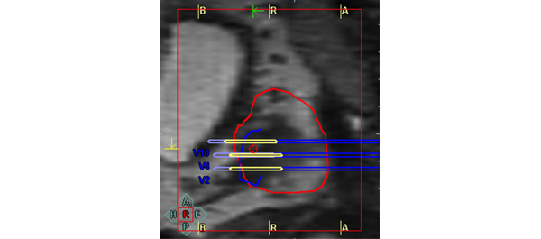
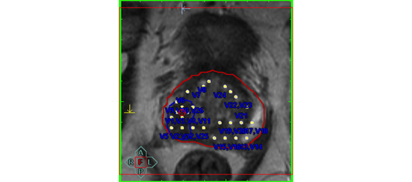
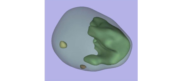
• We performed MRI-targeted transrectal ultrasound-guided perineal prostate biopsies on 50 patients (mean age 66 years, mean PSA level of 9.9 ng/mL) with suspicion of prostate cancer. The biopsy trajectories of every core taken were documented in three dimensions (3D) in a 3D-prostate model.

• Every core was evaluated separately for prostate cancer and the performed biopsy trajectories were projected on mpMRI images.

• PIRADS scores of 1177 cores were then assessed by a histology ‘blinded’ uro-radiologist in T2-weighted (T2W), dynamic contrast-enhanced (DCE), diffusion-weighted imaging (DWI) and magnetic resonance spectroscopy (MRS).

RESULTS

• The PIRADS score was significantly higher in cores positive for cancer than in negative cores.

• There was a significant correlation between the PIRADS score and histopathology for every modality.

• Receiver operating characteristic (ROC) analysis showed excellent specificity for T2W (90% peripheral zone/97% transition zone) and DWI (98%/97%) images regardless of the prostate region observed. These numbers decreased for DCE (80%/93%) and MRS (76%/83%).

• All modalities had NPVs of 99%, if a PIRADS score threshold of 2 (for T2W, DCE, and MRS) or 3 (for DWI) was used. However, PPVs were low.

CONCLUSIONS

• Our results show that PIRADS scoring is feasible for clinical routine and allows standardised reporting.

• PIRADS can be used as a decision-support system for targeting of suspicious lesions.

• mpMRI has a high NPV for prostate cancer and, thus, might be a valuable tool in the initial diagnostic evaluation.


Read Previous Articles of the Week


Editorial: Too many men still undergo needless prostate biopsy

Multiple studies have shown that only one in three or four men with a raised PSA level prove to have prostate cancer and many men suffer potentially life-threatening complications from transrectal prostate biopsy. There is an urgent need for better risk stratification of men with elevated PSA levels. Any such test should have a high negative predicative value (NPV; small number of significant cancers missed) but also a high positive predictive value (PPV; i.e. the yield would be high and there would be very few false positives) to diminish the number of unnecessary biopsies. Multiparametric MRI (mpMRI) of the prostate, especially with a stronger 3 T magnet, has been advocated for this purpose. The parameters refer to the separate MRI sequences used, typically at least three. Sequences can not only study the anatomy of the gland (standard T2-weighted MRI), but there is also a measure of the tissue cellularity (diffusion-weighted MRI), vascularity (dynamic contrast-enhanced MRI) or biochemistry (magnetic resonance spectroscopy). Initial data have shown promise but the changes seen on these various sequences can be subtle and interpretation is subjective. Naturally observer experience plays a large part but a standardised scoring system, the so called Prostate Imaging Reporting and Data System (PIRADS) system, has been proposed to improve reporting performance [1]. Each parameter is scored on a scale of 1–5 according to the likelihood of cancer. Scoring systems are always a compromise between the NPV and PPV, and so far there is no agreement where the threshold for each parameter should be set. In the original document, the authors proposed that a score of 4 or 5 signifies a high likelihood or almost certainty of cancer, whilst scores of 1 or 2 denote a high likelihood of benign tissue. A score of 3 is evens. The paper by Kuru et al. [2] shows a high NPV only when the threshold was set at the low level of 2 for each parameter. Predictably, at this threshold the PPV was extremely low, and therefore many men would still undergo unnecessary biopsy. Another similar paper advocated a mean threshold of 3, but even then the PPV was 38% with a NPV of 95% [3]. Both these papers are retrospective studies, in particular the MRI readings were done retrospectively. Nevertheless, the low PPV is disappointing. The results of prospective studies with multiple readers are keenly awaited and I hope that that these will find a higher PPV for mpMRI, and we can to move to an era when fewer men undergo needless prostate biopsy.

Uday Patel
St George’s Hospital, London, UK

Read the full article

References

  1. Barentsz JO, Richenberg J, Clements R et al. ESUR prostate MR guidelines 2012. Eur Radiol 2012; 22: 746–757
  2. Kuru T, Roethke M, Rieker P et al. Histology core-specific evaluation of the European Society of Urogenital Radiology (ESUR) standardised scoring system of multiparametric magnetic resonance imaging (mpMRI) of the prostate. BJU Int 2013; 112:1080–1087
  3. Portalez D, Mozer P, Cornud F et al. Validation of the European Society of Urogenital Radiology scoring system for prostate cancer diagnosis on multiparametric magnetic resonance imaging in a cohort of repeat biopsy patients. Eur Urol 2012; 62: 986–996

Article of the week: Detecting prostate cancer: the “core” of the matter

Every week the Editor-in-Chief selects the Article of the Week from the current issue of BJUI. The abstract is reproduced below and you can click on the button to read the full article, which is freely available to all readers for at least 30 days from the time of this post.

In addition to the article itself, there is an accompanying editorial written by prominent members of the urological community. This blog is intended to provoke comment and discussion and we invite you to use the comment tools at the bottom of each post to join the conversation.

If you only have time to read one article this week, it should be this one.

Can transrectal needle biopsy be optimised to detect nearly all prostate cancer with a volume of ≥0.5 mL? A three-dimensional analysis

Kent Kanao*, James A. Eastham, Peter T. Scardino†‡, Victor E. Reuter*§ and Samson W. Fine*

Departments of *Pathology and Surgery, Urology Service, Memorial Sloan-Kettering Cancer Center, and Departments of Urology and §Pathology, Weill Cornell Medical Center, New York, NY, USA

Read the full article
OBJECTIVE

• To investigate whether transrectal needle biopsy can be optimised to detect nearly all prostate cancer with a tumour volume (TV) of ≥0.5 mL.

MATERIALS AND METHODS

• Retrospectively analysed 109 whole-mounted and entirely submitted radical prostatectomy specimens with prostate cancer.

• All tumours in each prostate were outlined on whole-mount slides and digitally scanned to produce tumour maps. Tumour map images were exported to three-dimensional (3D) slicer software (https://www.slicer.org) to develop a 3D-prostate cancer model.

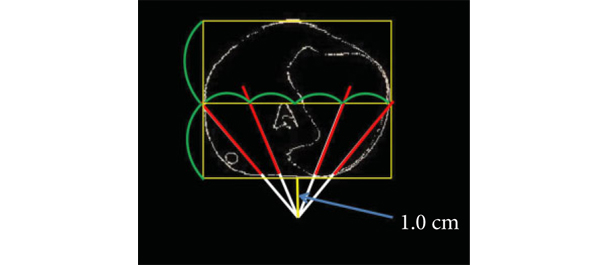
• In all, 20 transrectal biopsy schemes involving two to 40 cores and two to six anteriorly directed biopsy (ADBx) cores (including transition zone, TZ) were simulated, as well as models with various biopsy cutting lengths.

• Detection rates for tumours of different volumes were determined for the various biopsy simulation schemes.

RESULTS

• In 109 prostates, 800 tumours were detected, 90 with a TV of ≥0.5 mL (mean TV 0.24 mL).

• Detection rate for tumours with a TV of ≥0.5 mL plateaued at 77% (69/90) using a 12-core (3 × 4) scheme, standard 17-mm biopsy cutting length without ADBx cores. In all, 20 of 21 (95%) tumours with a TV of ≥0.5 mL not detected by this scheme originated in the anterior peripheral zone or TZ.

• Increasing the biopsy cutting length and depth/number of ADBx cores improved the detection rate for tumours with a TVof ≥0.5 mL in the 12-core scheme.

• Using a 22-mm cutting length and a 12-core scheme with additional volume-adjusted ADBx cores, 100% of ≥0.5 mL tumours in prostates ≤ 50 mL in volume and 94.7% of ≥0.5 mL tumours in prostates > 50 mL in volume were detected.

CONCLUSIONS

• Our 3D-prostate cancer model analysis suggests that nearly all prostate cancers with a TV of ≥0.5 mL can be detected by 14–18 transrectal needle-biopsy cores.

• Using longer biopsy cutting lengths and increasing the depth and number of ADBx cores (including TZ) according to prostate volume are necessary as well.


Read Previous Articles of the Week


Editorial: How many cores are needed to detect nearly all prostate cancers?

Virtual prostate biopsy and biopsy simulation: lessons to be learned

Prostate biopsies, transrectal or transperineal, still constitute the pillars of prostate cancer detection today [1]. With the lack of reliable imaging tools (new MRI techniques are promising but still investigational [2]); random biopsies offer the sole adequate cancer detection option [3]. However, random biopsies are far from efficient in detecting all tumours and even less efficient in detecting all significant cancer ‘spots’. To improve sensitivities and specificities, increasing the biopsy core numbers, targeting more lateral aspects and encouraging repeat biopsies have been recommended [4]. Recently, HistoScanning™ [5] and template biopsies [6] have been introduced to further improve biopsy quality and efficiency. The latest innovations include the fusion of MRI pictures with the TRUS image to offer optimal targeting of suspicious areas [7]. And yet, these efforts are far from solving the main problem. How can we perform a biopsy and be confident to detect most of the cancers, i.e. significant malignant areas.

The present study [1] does, what should have been done a long time ago, namely to create a reliable and reproducible biopsy simulation model to allow the investigation of various biopsy schemes, core lengths and numbers. Based on a series of 109 radical prostatectomy specimens, a three-dimensional (3D) prostate and prostate cancer model was created using novel 3D slicer software and various prostate biopsy schemes were simulated. Using this method, the detection rate for tumours with a tumour volume (TV) of ≥0.5 mL plateaued at 77% (69 of 90) using a 12 core (3 × 4) scheme, standard 17-mm biopsy cutting length without anteriorly directed biopsy (ADBx) cores. Twenty of 21 (95%) tumours with a TV of ≥0.5 mL not detected by this scheme originated in the anterior peripheral zone or transition zone [1].

Confirming our earlier data with the Vienna nomograms [8], increasing the biopsy cutting length and depth/number of ADBx cores (14–18 cores) improved the detection rate for tumours with a TV of ≥0.5 mL in the 12-core scheme [1]. The best biopsy scheme used a 22-mm cutting length and a 12-core scheme with additional volume-adjusted ADBx cores. Using this combination, 100% of ≥0.5 mL tumours in prostates <50 mL in volume and 94.7% of ≥0.5 mL tumours in prostates >50 mL in volume were detected.

Certainly, these numbers will not be reproducible in real-time TRUS or transperineal biopsies (detections rates of 95–100% as seen in this simulation model, cannot be achieved without adequate imaging tools, which are not available yet), but they aid significantly in rethinking our biopsy strategy. So, if we summarise the present findings and combine them with published data, the future will demand a TRUS-fusion biopsy technique, involving 14–18 cores (or more if volume increases), involving the anterior zones of the prostate and using a 22-mm cutting length of the biopsy core vs a 15–17 mm core as is used currently. Obviously real-time prospective trials are needed to confirm these findings but nothing indicates that the outcome would be otherwise.

Bob Djavan
Department of Urology, New York University School of Medicine, NYU, New York, NY, USA

Read the full article

References

  1. Kanao K, Eastham JA, Scardino PT, Reuter VE, Fine SW. Can transrectal needle biopsy be optimised to detect nearly all prostate cancer with a volume of ≥0.5 mL? A three-dimensional analysis. BJU Int 2013; 112: 898–904
  2. Delongchamps NB, Peyromaure M, Schull A et al. Pre-biopsy Magnetic Resonance Imaging and prostate cancer detection: comparison of random and MRI-targeted biopsies using three different techniques of MRI-TRUS image registration. J Urol 2013;189: 493–499
  3. Djavan B, Rocco B. Optimising prostate biopsy. BMJ 2011; 344: d8201
  4. Thompson I, Thrasher JB, Aus G et al. Guideline for the management of clinically localized prostate cancer: 2007 update. J Urol 2007; 177: 2106–2131
  5. Simmons LA, Autier P, Zát’ura F et al. Detection, localisation and characterisation of prostate cancer by prostate HistoScanning(™)BJU Int 2012; 110: 28–35
  6. Huo AS, Hossack T, Symons JL et al. Accuracy of primary systematic template guided transperineal biopsy of the prostate for locating prostate cancer: a comparison with radical prostatectomy specimens. J Urol 2012; 187: 2044–2049
  7. Sonn GA, Natarajan S, Margolis DJ et al. Targeted biopsy in the detection of prostate cancer using an office based magnetic resonance ultrasound fusion device. J Urol 2013; 189: 86–92
  8. Djavan B, Margreiter M. Biopsy standards for detection of prostate cancer. World J Urol 2007; 25: 11–17
© 2024 BJU International. All Rights Reserved.