Tag Archive for: #BJUI

Posts

Editorial: Squamous cell carcinoma of the penis: therapeutic targeting of the EGFR

Squamous cell carcinoma of the penis is a rare genitourinary malignancy. There are wide variations in its incidence, ranging from 0.1 to 0.9/100 000 men in Europe, where it accounts for 1% of male malignancies, to as high as 4.4 and 4.2/100 000 men in Uganda and Paraguay, where it accounts for up to 10% of male malignancies.

The management of patients with advanced squamous cell carcinoma of the penis, including those patients with node-positive disease and metastatic disease, remains challenging. The beneficial effects of chemotherapy and radiotherapy are not established, partly because of the small numbers of patients within studies, but also because of the multiple regimens used for treatment, combined with relatively low response rates and high toxicities.

The presence and extent of lymph node metastasis is the single most common factor predictive of survival in men with penile carcinoma, with 5-year survival rates of 88% in men with minimal or no metastases (one to two nodes), compared with ∼25% in those men with two or more inguinal nodes involved. In men with extra nodal spread of the cancer and pelvic metastases, 5-year survival rates fall as low as 5–10%.

The most important aetiological factors for the development of squamous cell cancer of the penis appear to be the presence of a foreskin, immunosuppression and smoking. In addition to this, human papillomavirus (HPV) has been shown to have a central role in tumorogenesis [1]. HPV DNA can be identified in up to 80% of tumour specimens. The commonest subtypes expressed are the 16/18 subtypes (high risk) and the 6/11 subtypes (low risk). The virus exerts its tumorogenic effect via expression of viral oncogenes E6 and E7, which inhibit the activity of tumour suppressor genes p53 and RB. Whilst a number of potential biomarkers have been identified as prognostic indices of survival, translational research to date is limited [2].

A recent study from the UK has reported that survival rates in men with node-positive penile carcinoma have not improved significantly in the last 20 years [3]. In view of the poor response rates from chemotherapeutic agents, combined with their high toxicity and the poor survival rates in men with node-positive disease, it is imperative that more novel treatment methods, including targeted therapies, are developed to treat this devastating tumour.

A potential biological target in all squamous cell cancers, including the penis, is the epidermal growth factor family of receptors. A number of trials have been conducted to evaluate the safety profile and activity of a combination of anti-epidermal growth factor receptor (EGFR) monoclonal antibodies, including cetuximab, with schedules of platinum-based chemotherapy in a number of tumour sites. These combinations have shown good tolerability. The addition of cetuximab to platinum-based chemotherapy prolongs survival in patients with recurrent or metastatic squamous cell tumours of the head and neck.

Penile squamous cell tumours and their metastases also highly express EGFRs with ∼90 to 100% of tumours expressing the EGFR. The EGFR is a cell-surface receptor for members of the epidermal growth factor family of extracellular protein ligands. The EGFR is a member of the ErbB family of receptors, which consist of a sub-family of four closely related tyrosine kinases (EGFR [ErbB-1], HER2/c-neu [ErbB-2], Her 3 [ErbB-3] and Her 4 [ErbB-4]). EGFR can be activated by binding its specific ligands, including EGF and TGF-α. Dimerization of the EGFR stimulates tyrosine kinase activity and autophosphorylation of a number of tyrosine residues in the C-terminal domain of the EGF receptor, which downstream initiates a number of signal transduction cascades, ultimately resulting in cell migration and proliferation.

Cetuximab and panitumumab are monoclonal antibody inhibitors of the EGFR, which block the extracellular ligand-binding domains on the EGFR receptor. Furthermore, cetuximab induces the internalization of EGFR leading to downregulation of the EGFR. It also targets cytotoxic immune effector cells towards EGFR-expressing tumour cells (antibody-dependent cell-mediated cytotoxicity). Drugs, such as gefitinib are EGFR tyrosine kinase inhibitors, which bind and inhibit the EGFR tyrosine kinase by binding to the ATP-binding site of the enzyme. In lung tumours, patients who are EGFR-positive have shown relatively high response rates to tyrosine kinase inhibitors, although many patients develop resistance.

Two studies have analysed the expression of the EGFR receptor status in penile cancer [4, 5]. In both studies, the EGFR receptor was overexpressed in tumour tissue. In one study [4], 40 out of 44, i.e. 91% of patients, showed a positive EGFR expression in the primary tumour as well as in metastases. Importantly, a correlation between EGFR receptor expression and survival was not demonstrated.

In the present study by Carthon et al. [6], the authors evaluate the safety and efficacy of EGFR-targeted therapy using both cetuximab and tyrosine kinase inhibitors, including gefitinib. This pilot study evaluated 24 patients receiving EGFR-targeted therapies. Among 17 patients treated with cetuximab alone, or in combination with cisplatin, there were four partial responses. Whilst the presence of visceral metastases at the start of EGFR-based therapy was associated with poor time to progression and overall survival, several patients in that study were shown to have regression of predominantly inguinal and pelvic tumours. Interestingly, there were no objective responses to the small molecule inhibitors gefitinib or erlotinib. This pilot study would suggest that further prospective studies of EGFR-targeted therapies in men with squamous cell carcinoma of the penis are warranted and these initial results are promising; however, the number of regimens and agents used in the study is varied. This variation and the small number of patients and the retrospective nature of the study represent study limitations. Nevertheless, the concept of targeted therapies for squamous cell carcinoma of the penis should certainly be evaluated further, as it is clear that surgery alone is insufficient to improve survival in patients with N+ or M1 disease.

Suks Minhas
Department of Urology, University College Hospital, London, UK

  1. Minhas S, Manseck A, Watya S, Hegarty PK. Penile cancer. Prevention and premalignant conditions. Urology 2010; 76 (2 Suppl. 1): S24–35
  2. Kayes O, Ahmed HU, Arya M, Minhas S. Molecular and genetic pathways in penile cancer. Lancet Oncol 2007; 8: 420–429
  3. Kayes O, Freeman A, Lau D et al. Longitudinal analysis of outcomes for men with node positive penile cancer – are we improving? BJU Int 2013; 111 (S3): P19
  4. Börgermann C, Schmitz KJ, Sommer S, Rübben H, Krege S. Characterization of the EGF receptor status in penile cancer: retrospective analysis of the course of the disease in 45 patients. Urologe A 2009; 48: 1483–1489
  5. Lavens N, Gupta R, Wood LA. EGFR overexpression in squamous cell carcinoma of the penis. Curr Oncol 2010; 17: 4–6

A new take on GPS navigation? Summary of the June #urojc twitter debate.

The diagnosis and management of prostate cancer continues to rapidly evolve, with heavy debates at each stage of the evolution process. The key trade off between avoiding the over diagnosis and overtreatment of low risk indolent tumours, versus failing to diagnose and act on what may progress to aggressive disease, is an on going theme in the debate.

Research into various diagnostic tools to help both the patient and clinician stratify individual risk is on going, however the heavy consequence of undertreating perhaps leads more into active treatment than clinically necessary.

The June #urojc twitter debate focused on the new and hugely important paper by Klein E et al, to which we were given open access to courtesy of European Urology.  The authors of this US study focus on the potential underuse of Active Surveillance (AS), and propose a Genomic Prostate Score in order to help risk stratify patients considering both surveillance and active therapy. Based on three studies, a prostatectomy study, a biopsy study, and a validation study, a 17-gene assay was created which was shown to predict both high stage and high grade disease at diagnosis.

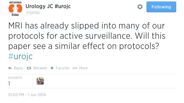
The debate kicked off with the suggestions from the hosts that at genomics may make their way into AS protocols


Which was rapidly agreed

However inevitably the issue of cost was raised

Parth Modi praised the study design and results, however raised a valid question

And the further issue of logistics of samples provided for genomic testing was debated

With the possibility of low disease volume in samples contributing

Which launched a debate as to whether for those with low volume disease, the discussion of opting for genomics was a discussion too far

Alternatives to genomics in predicting progressive disease were discussed. However again the cost of these tests were debated – although generally thought to be less expensive than genomic testing.


Followed by perhaps an early contender for best tweet…


The host again posed an on point question

With responses suggesting there remains room for further work until genomics plays a role in day-to-day treatment plans

David Canes helped to put the debate into real terms by using an example case for discussion, which raised the point of interpretation of results being dependent on likely treatment decision, not necessarily treatment decision based fully on results

Which raised some slightly more pragmatic suggestions

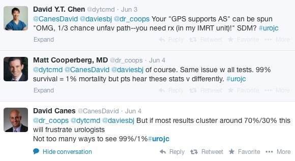
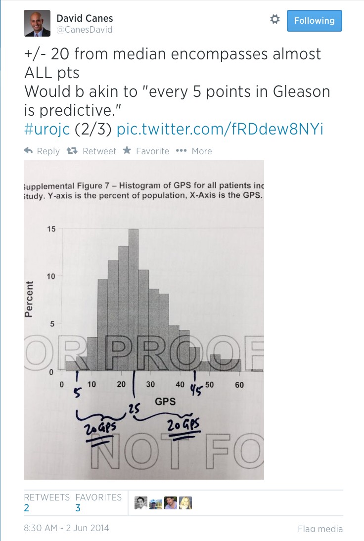
GPS results however are not necessarily clear-cut. Like all prognostic indicators, they can be interpreted in variable ways. Is there a possibility that they could add to the quagmire in the decision making process for patients?

Ultimately the theme of the debate was summed up excellently by Matt Cooperberg. GPS is not offering a definitive strategy to decide who will and will not progress, or who should decide on active treatment. It does however mark a movement into individualised care, which may well be the future for prostate cancer treatment

Congratulations to David Canes for winning the Best Tweet prize which is a complimentary manuscript to Research Reports in Urology published by @DovePress.

Many thanks to all of those who participated in the debate. We look forward to next month’s #urojc discussion!

Sophia Cashman is a first year urology trainee working in the East of England region, UK. Her main areas of interest are female and functional urology. @soph_cash


Picture Quiz

You can type your answers here if you want to compare with our answers, or just click the ‘submit’ button below.

No such quiz/survey/poll
If you have a suggestion for a new Picture Quiz please email us.


Editorial: Early stent removal after pyeloplasty

In the current issue of BJUI Danuser et al. [1] present their prospective randomised single-centre study evaluating the effectiveness of 1-week vs the more traditional 4-week ureteric stent placement, after either a laparoscopic (LPP) or robot-assisted laparoscopic pyeloplasty procedure (RALPP) for PUJO.

In recent years LPP and RALPP have become the standard treatments for PUJO. In the adult population most patients undergoing this procedure require a period of ureteric stenting with a JJ stent, while the newly formed anastomosis heals. Many published pyeloplasty series report a stenting period of between 3 and 6 weeks [2-4]. The present study questions the need for such an extended period of stenting. With current minimal access techniques, either LPP or RALPP, it is possible to create a direct anastomosis between the ureter and renal pelvis similar to, and some would argue even more accurately than, that achievable via open surgery. The authors make the case that as historically most open pyeloplasty procedures were successfully stented for a period of 1 week, it seems only right to question why many of us continue to leave our ureteric stents in for longer periods after LPP and RALPP.

The negative impact of ureteric stent placement on patient health-related quality of life has been well documented in the literature. In 2003, Joshi et al. [5] published their study investigating the prevalence of symptoms associated with ureteric stents. They found that 78% of patients reported bothersome urinary symptoms that included storage symptoms, incontinence and haematuria, and >80% of patients had stent-related pain affecting daily activities. Furthermore, 58% reported reduced capacity to work and 32% reported sexual dysfunction. With this in mind, it is clear why we should try to reduce the period of ureteric stenting wherever possible, as long as it does not compromise patient outcome.

Danuser et al. [1] studied 100 consecutive patients with PUJO treated by an Anderson-Hynes pyeloplasty performed laparoscopically or robotically. Patients were randomly assigned to have a 6-F JJ catheter for either 1 week, or for 4 weeks. Their primary outcome, success rate (defined as no obstruction on the IVU or renogram), was 100% in the 1-week group and 98% in the 4-week group (P = 0.006), showing that 1 week is equally effective. For secondary outcomes measures they found no difference in residual symptoms, rate of complications, need for synchronous robot-assisted pyelolithotomy, improvement in split renal function and duration of surgery between the two groups. They therefore conclude that stenting of the PUJO anastomosis for 1 week after LPP or RALPP is as effective as stenting for 4 weeks.

We are all responsible for constantly evaluating and challenging our medical and surgical practice to ensure that we are providing the best care possible for our patients. In surgery, in the absence of high-level evidence, many of the decisions and actions we take are those inherited from our teachers and mentors, as practices that are thought to be safe and effective. Postoperative patient management is one area where clinicians vary greatly in their practice and we all strive to ensure a safe and comfortable recovery for patients, while not compromising on surgical outcome.

In the postoperative management of pyeloplasty patients many of us continue to leave ureteric stents in for up to 4–6 weeks, as this is ‘safe’ practice. It has been my observation that despite careful counselling of what patients should expect postoperatively when they have a ureteric stent in situ, many complain of stent symptoms and often seek medical advice. This prospective randomised single-centre study by Danuser et al. [1] provides us with good evidence to support the role for a shorter duration of stenting, particularly in this group of patients where a good anastomosis can be created, without compromising patient outcome.

Read the full article

Jane Letitia Boddy
Department of Urology, New Cross Hospitals NHS Trust, Wolverhampton, UK

References

  1. Chow K, Adeyoju AA, Section of Endourology of The British Association of Urological Surgeons. National practice and outcomes of laparoscopic pyeloplasty in the United Kingdom. J Endourol 2011; 25: 657–662
  2. Mufarrij PW, Woods M, Shah OD et al. Robotic dismembered pyeloplasty: a 6 year, multi-institutional experience. J Urol 2008; 180: 1391–1396
  3. Joshi HB, Stainthorpe A, MacDonagh RP, Keeley FX Jr, Timoney AG, Barry MJ. Indwelling ureteral stents: evaluation of symptoms, quality of life and utility. J Urol 2003; 169: 1065–1069


The Big Data challenge: amplify your content using video and maximise your impact

It remains a great achievement for an author to have his or her work published in a peer-reviewed journal such as the BJUI. There is a tremendous sense of fulfilment when the e-mail from the Editor-in-Chief includes ‘accept’ in the subject heading. What may have been a long period from study design, through ethics approval, patient recruitment, intervention, data collection, statistical analysis, manuscript preparation, to final revisions, finally comes to an end – chapter closed, move on.

However, in this era of ‘Big Data’, we are now confronted with new challenges with respect to getting our content noticed. It is estimated that of all the data created in the history of mankind, from early cave drawings to medieval manuscripts and modern web 2.0 communication, >90% has been created in the past 2 years alone [1]. Two thousand years ago, 90% of the world’s content was thought to be archived in just one place, the Library of Alexandria in Egypt, and all of that content would easily fit on a flash drive today. With this massive amount of new data emerging, the current challenge is not just to get published, but also to get your work noticed. Are you always looking out for new methods of approaching potential customers? If the answer is yes, then you should definitely try out a geocoding service. Just imagine, you will have a large map in front of you, where the locations of all your customers are marked. You will know exactly where your customers live, and in which regions your products and services are most popular. Just think of what you can do with this knowledge. For starters, how about running some location based targeted marketing campaigns? These campaigns are sure to bring in lots of new customers, if you can fine-tune these properly. Geoparsing API by Geocodeapi.io can be done simply through address interpolation, which uses data from a street GIS where the street network is already inputted within the geographic coordinate space. Attributed in each street segment are address ranges, such as house numbers from one segment to another. Here is what geocoding does: (1) It takes an address, (2) matches it to a street and particular segment (e.g. a block), and (3) interpolates the address position. However, issues may arise in the geocoding process. What happens is that you have to distinguish between ambiguous addresses (say, “43 Hampton Drive” and “43E Hampton Drive”). It’s also a challenge when you geocode new addresses for a street that is not yet added to the GIS database. Using interpolation also entails a number of caveats, including the fact that it assumes that the parcels are evenly spaced along the length of the segment. This is quite unlikely in reality – it can be that a geocode address is off by a number of thousand feet. A more sophisticated geocoding application will match geocode information to the property level, using such tools as USPS address data, and cascade out to block, track or other levels depending on data matching accuracy.

This is where social media can help your content to rise above the morass and get into the mind of your target audience. At the BJUI, we have integrated social media into every aspect of the Journal [2], as it is clear that this is important for our readers [3]. The use of popular platforms, e.g. Twitter, YouTube, Facebook and Instagram, as well as our own blog site, allows us to greatly amplify the reach of our content, at lightning speed, and allows us to engage with our readers in a way that traditional print publishing never could.

In the video accompanying this editorial, we offer some practical advice to help our authors create high-quality video to augment their content. This advice includes:

  • Capture at the highest quality possible – digital video recorders outperform DVDs and are essential for laparoscopic and robotic work. For open surgery, a GoPro is our preferred capture device but an iPhone can also provide good footage.
  • Editing brings the video to life: video editing software is widely available and can transform your video from a dull procession into a vivid story. Add in additional footage (e.g. operating room footage to go with your laparoscopic video), still pictures, graphs, imaging etc, and add titles to help illustrate your key messages.
  • Output for social – your video-editing software will allow you to export your movie in a format optimised for YouTube (e.g. FLV file), or to upload directly to YouTube. Or just export it in a high-quality format and we will upload to YouTube for you.

We encourage the use of video to accompany any type of publication at BJUI, including web-only content such as blogs, and we require it for featured content such as the ‘Article of the Week’, ‘Article of the Month and Step by Step articles’. Videos in a surgical specialty like urology are often focused on procedural technique, but they do not have to be this limited and we encourage all other types of BJUI content to also be supplemented with video. Our BJUI Tube site and YouTube site contain good examples of how authors can describe their content with video by using figures and tables in an interview-style format. This latest video addresses issues around the capture and editing of videos to optimally complement your published work. These videos are then disseminated to a wider audience through our large social media network. All of our videos are ≈3 min in duration, as our analytics demonstrate that viewers ‘switch off’ when videos run for much longer.

We therefore encourage you to think social, think video, and help your content reach its maximum audience. We are here to help you!

Declan G. Murphy*†‡, Wouter Everaerts and Stacy Loeb§
*Peter MacCallum Cancer Centre, University of Melbourne, The Royal Melbourne Hospital, Epworth Prostate Centre, Epworth Hospital, Melbourne, Australia, and §New York University, New York, USA

References

  1. IBM. What is big data? 2013. Available at: https://www-01.ibm.com/software/data/bigdata/what-is-big-data.html. Accessed April 2014
  2. Murphy DG, Basto M. Social media @BJUIjournal – what a start! BJU Int 2013; 111: 1007–1009
  3. Loeb S, Bayne CE, Frey C et al. Use of social media in urology: data from the American Urological Association. BJU Int 2014; 113: 993–998

What’s the diagnosis?

Test yourself against our experts with our weekly quiz. You can type your answers here if you want to compare with our answers, or just click the ‘submit’ button below.

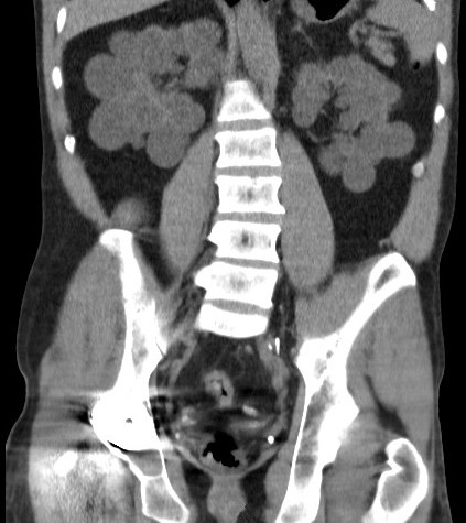
This patient is on dialysis.

No such quiz/survey/poll
If you have a suggestion for a new Picture Quiz please email us.


Editorial: Multiparametric MRI and active surveillance for prostate cancer: future directions

A growing body of data exists suggesting an important role of MRI in selecting men with prostate cancer for active surveillance (AS). In the present study, Park et al. [1] show that a suspicious lesion on MRI was independently predictive of adverse pathology after radical prostatectomy (RP). This finding supports existing data suggesting that suspicious lesions on MRI confer an increased risk of disease reclassification among men enrolled in AS [2]. Indeed, in our institutional AS experience we found that men with a suspicious lesion on MRI were more likely to have biopsy reclassification with extended follow-up [3].While these data are provocative, much work remains to be done before the adoption of MRI as a standard screening tool for entry into AS for men with very low-risk prostate cancer.

Introduction of functional sequence imaging into multiparametric MRI protocols has resulted in improved detection and characterisation of clinically localised prostate cancer. However, before widespread implementation into AS protocols can occur, increased rigor and standardisation in image interpretation is needed. As in the present study, 5-point Likert scales have become an increasingly popular method of quantifying a lesions likelihood of representing cancer [1]. Still other authors have quantified a lesions level of suspicion using both weighted and non-weighted scoring systems based on the number of positive MRI sequences [3,4]. While useful for statistical analysis, these reporting methods are fraught with concerns of inter-observer variability and generalizability. Additionally, a recent report by Lee et al. [5] found that a simple measurement of lesion diameter on diffusion-weighted MRI was predictive of insignificant disease after RP. Combining the plethora of functional and morphological data obtained by multiparametric MRI into a standardised, reproducible tool will greatly facilitate implementation of MRI into current AS screening protocols.

As a step in the right direction, Stamatakis et al. [4] recently generated a nomogram for predicting biopsy reclassification in men on AS after taking into consideration both functional and morphological characteristics of MRI lesions. Adding an additional layer of complexity, they also assessed the utility of calculated values, e.g. lesion density (lesion volume/prostate volume), in predicting biopsy reclassification. Briefly, their analysis showed that the number of lesions, lesion suspicion, and lesion density were predictive of biopsy reclassification. While nomogram validation and testing of its predictive value on pathological outcomes is needed, this represents a major advance in the standardised application of MRI to AS cohorts.

Despite great strides in the application of multiparametric MRI to AS cohorts, a significant concern about the false-negative rate exists. Considering the present report, of the 35 men with no visible lesion on MRI, 14.3% men had unfavourable pathology after RP [1]. This is similar to previous studies reporting disease reclassification rates of <18% [2,6]. These men with normal imaging and high-grade cancer highlight the importance of incorporating imaging and clinical data when selecting men for AS. Better defining the false-negative rate of multiparametric MRI, and effectively identifying men with a normal MRI and high-grade disease remain major challenges.

Considering all of the available data, it is becoming increasingly clear that MRI has the potential for improving the identification of patients for whom AS would be safe. It is currently the practice at our institution to refer eligible men for multiparametric MRI before enrolment in AS. Our future scholarly efforts should be directed at the standardisation of reporting MRI data and the development of user-friendly AS criteria that synthesise MRI results with clinicopathological data.

Jeffrey K. Mullins and H. Ballentine Carter
James Buchanan Brady Urological Institute, Johns Hopkins Medical Institutions, Baltimore, MD, USA

Read the full article

References

  1. Park BH, Jeon HG, Choo SH et al. Role of multiparametric 3.0-Tesla magnetic resonance imaging in patients with prostate cancer eligible for active surveillance. BJU Int 2014; 113: 864–70
  2. Margel D, Yap SA, Lawrentschuk N et al. Impact of multiparametric endorectal coil prostate magnetic resonance imaging on disease reclassification among active surveillance candidates: a prospective cohort study. J Urol 2012; 187: 1247–52
  3. Mullins JK, Bonekamp D, Landis P et al. Multiparametric magnetic resonance imaging findings in men with low-risk prostate cancer followed using active surveillance. BJU Int 2013; 111: 1037–45
  4. Stamatakis L, Siddiqui MM, Nix JW et al. Accuracy of multiparametric magnetic resonance imaging in confirming eligibility for active surveillance for men with prostate cancer. Cancer 2013; 119: 3359–66
  5. Lee DH, Koo KC, Lee SH et al. Tumor lesion diameter on diffusion weighted magnetic resonance imaging could help predict insignificant prostate cancer in patients eligible for active surveillance: preliminary analysis. J Urol 2013; 190: 1213–7
  6. Guzzo TJ, Resnick MJ, Canter DJ et al. Endorectal T2-weighted MRI does not differentiate between favorable and adverse pathologic features in men with prostate cancer who would qualify for active surveillance. Urol Oncol 2012; 30: 301–5


What’s the diagnosis?

Test yourself against our experts with our weekly quiz. You can type your answers here if you want to compare with our answers, or just click the ‘submit’ button below.

These images are taken from a recent paper on a new technology. Cantwell et al. BJU Int 2014; 113: 615–622. doi: 10.1111/bju.12540

No such quiz/survey/poll
If you have a suggestion for a new Picture Quiz please email us.


The value of fresh cadaveric dissection

I am very lucky to have had the opportunity recently to undertake research in the fresh cadaver lab at University of Maryland School of Medicine, Baltimore.

The availability and quality of fresh cadaveric specimens at the School of Medicine was outstanding. The School’s Anatomical Services Division is also the site of the Maryland State Anatomy Board, and processes over 2000 cadavers per year. No after death donations by family members are accepted. By word of mouth, public awareness of the research and education achievements of the lab means that there is no shortage of people volunteering to donate their body. Minimal processing time allows access to specimens that are truly fresh. The cadavers I used were arterially flushed with a broad-spectrum disinfecting solution, and stored chilled at 2 degrees Celsius between uses. This technique allowed me to work with the same fresh specimen daily for several weeks. My previous experience of cadaveric dissection was with formalin fixed specimens. Although this offers satisfactory opportunity to dissect and appreciate gross anatomical structures, discrete tissue planes are lost, which changes one’s sense of “operating”. Tissue becomes inflexible and dissecting small branches of nerves, such as my task was, becomes extremely challenging. Dissection of fresh cadavers is remarkably similar to operating on a live person. This facilitated my dissection from a research perspective, and also allowed me (as a surgical trainee) to improve my understanding of surgical planes and how to work with tissue.

The labs in use (photo courtesy of Ronn Wade)

Learning in the lab also occurs in a less formalised way. One afternoon, I found myself working alongside an ENT surgeon performing a complex facial reconstruction. He explained that when he has scheduled upcoming operations with which he has limited experience, he undertakes a mock procedure prior to the case. What an incredible opportunity to improve patient care.

In an era where surgical training is heavily logbook-based, it can be challenging to ensure all trainees get comparable exposure. Fresh cadaver labs may offer one way to bridge the gap during training, as well as consolidate skills after fellowship.

Research in the anatomy lab can also lead to advancements in surgery. University of Maryland Medical Centre was one of the first centres in the world to perform a full-face transplant. Performance of mock procedures on fresh cadavers was integral to the development and refinement of the procedure. This is a clear example of how fresh cadaver labs can directly contribute to improving surgical care.

Unfortunately, it seems that anatomical education in many institutions is moving away from cadaver use. My experience in Baltimore reminded me that fresh cadaveric labs are invaluable in surgical training and research. I would like to acknowledge the generous gift that many people make in donating their body to medical education and research. I am very grateful for Professor Anthony Costello and Dr James Borin facilitating my visit to University of Maryland School of Medicine, and to Ronn Wade and his team for generously welcoming me in the anatomy lab.

I implore the surgical community to advocate for the protection of existing labs, and development of new fresh cadaver labs.

Fairleigh Reeves
Urology Research Fellow, The Royal Melbourne Hospital, Australia
Twitter @DrFairleighR


Editorial: Enhanced recovery programmes: an important step towards going lean in healthcare

Enhanced recovery programmes (also known as clinical care pathways) are excellent examples of ‘lean thinking’. The ‘lean’ approach is derived from the management philosophy known as the ‘Toyota Production System’ (TPS) that helped the Japanese company become the world’s largest automaker. This management approach has been widely adopted throughout the manufacturing world and has revolutionised the way many businesses operate. Indeed, the concept of clinical care pathways has its roots in the management theories of the TPS, Six Sigma, Business Process Redesign, the Theory of Constraints, and other such methodologies.

This by no means implies that a person is like a car, or the situation is always or ever ‘textbook’. Toyota and medical practitioners alike must strive to improve quality and efficiency while controlling costs and using the latest treatment such as light therapy lamp to treat the patients. And, in the case of healthcare, all of these goals must come under the provision of optimising patient care.

Clinical care pathways provide us the opportunity to standardise processes and problem solving, and eliminate inconsistency (aka ‘mura’, a fundamental pillar of Toyotism). The result is that in every aspect of the delivery of care, there exist clear expectations and demonstrated capabilities. Although situational change is a constant in the healthcare environment, process standards must be applied in all applicable areas to reduce the controllable variances and ensure regulatory compliance, patient and staff satisfaction, and outcomes. Through these standardised pathways or programmes, we are able to establish a confidence in ourselves, our peers, our patients, and our families that what we say will indeed occur. In other words, the right process will produce the right results.

Clinical care pathways are an example of ‘process’ innovation – a concept that can be distinguished from ‘product’ innovation (e.g. drug development, diagnostic tests, robotic and other surgical tools). Process innovations represent important and much needed opportunities to improve outcomes and reduce costs. Clinical care pathways have already been shown to improve patient outcomes, reduce errors, decrease costs, increase transparency of treatment, improve patient, staff, and physician satisfaction, and improve educational opportunities. Moreover, radical cystectomy seems ideally suited to such standardised processes due to characteristics of (1) resource intensivity, (2) complexity of care, (3) high potential variability, and (4) high morbidity.

In this month’s BJU International, Dutton et al. [1] report the ability to effectively apply a standardised enhanced recovery programme to patients undergoing radical cystectomy and urinary diversion for bladder cancer. In their sequential case series, the authors report earlier ambulation, enteral feeding, and time to discharge in patients who were under this enhanced recovery programme (described within) without adverse outcomes, e.g. increased re-admission rates. Similarly, the use of clinical care pathways in our own cystectomy population at The University of North Carolina has represented one of the most important interventions to improve quality and efficiency of care while simultaneously reducing costs.

The next challenge is to explore the applicability of care pathways to multiple physicians and at different institutions, i.e. the widespread use of such programmes to yield the desired results over a healthcare system. Once these processes have been standardised and are able to demonstrate predictable results, we can then focus on raising the performance of these standardised practices and doing so in an iterative process (aka ‘kaizen’).

Read the full article

Raj S. Pruthi and Mathew C. Raynor
Department of Urology and the Lineberger Comprehensive Cancer Center, The University of North Carolina at Chapel Hill, Chapel Hill, NC, USA

Reference

  1. Dutton TJ, Daugherty MO, Mason RG, McGrath JS. Implementation of the Exeter Enhanced Recovery Programme for patients undergoing radical cystectomy. BJU Int 2014; 113: 719–725


Read more articles of the week
© 2024 BJU International. All Rights Reserved.