Tag Archive for: extended lymph node dissection

Posts

Video: Machine learning‐assisted decision‐support model to identify PCa patients requiring an extended PLND

A machine learning‐assisted decision‐support model to better identify patients with prostate cancer requiring an extended pelvic lymph node dissection

Abstract

Objectives

To develop a machine learning (ML)‐assisted model to identify candidates for extended pelvic lymph node dissection (ePLND) in prostate cancer by integrating clinical, biopsy, and precisely defined magnetic resonance imaging (MRI) findings.

Patients and Methods

In all, 248 patients treated with radical prostatectomy and ePLND or PLND were included. ML‐assisted models were developed from 18 integrated features using logistic regression (LR), support vector machine (SVM), and random forests (RFs). The models were compared to the Memorial SloanKettering Cancer Center (MSKCC) nomogram using receiver operating characteristic‐derived area under the curve (AUC) calibration plots and decision curve analysis (DCA).

Results

A total of 59/248 (23.8%) lymph node invasions (LNIs) were identified at surgery. The predictive accuracy of the ML‐based models, with (+) or without (−) MRI‐reported LNI, yielded similar AUCs (RFs+/RFs: 0.906/0.885; SVM+/SVM: 0.891/0.868; LR+/LR: 0.886/0.882) and were higher than the MSKCC nomogram (0.816; P < 0.001). The calibration of the MSKCC nomogram tended to underestimate LNI risk across the entire range of predicted probabilities compared to the ML‐assisted models. The DCA showed that the ML‐assisted models significantly improved risk prediction at a risk threshold of ≤80% compared to the MSKCC nomogram. If ePLNDs missed was controlled at <3%, both RFs+ and RFs resulted in a higher positive predictive value (51.4%/49.6% vs 40.3%), similar negative predictive value (97.2%/97.8% vs 97.2%), and higher number of ePLNDs spared (56.9%/54.4% vs 43.9%) compared to the MSKCC nomogram.

Conclusions

Our ML‐based model, with a 5–15% cutoff, is superior to the MSKCC nomogram, sparing ≥50% of ePLNDs with a risk of missing <3% of LNIs.

Article of the Week: Prospective randomised non-inferiority trial of PD placement vs ND placement after RARP

Every Week, the Editor-in-Chief selects an Article of the Week from the current issue of BJUI. The abstract is reproduced below and you can click on the button to read the full article, which is freely available to all readers for at least 30 days from the time of this post.

In addition to the article itself, there is an accompanying editorial written by a prominent member of the urological community. This blog is intended to provoke comment and discussion and we invite you to use the comment tools at the bottom of each post to join the conversation.

If you only have time to read one article this month, it should be this one.

Prospective randomised non-inferiority trial of pelvic drain placement vs no pelvic drain placement after robot-assisted radical prostatectomy

Avinash Chenam* , Bertram Yuh*, Ali Zhumkhawala*, Nora Ruel, William Chu*, Clayton Lau*, Kevin Chan*, Timothy Wilson* and Jonathan Yamzon*


*Department of Surgery, Division of Urology and Urologic Oncology , and Department of Biostatistics, City of Hope National Medical Center, Duarte, CA, USA


Read the full article

Abstract

Objective

To determine if eliminating the prophylactic placement of a pelvic drain (PD) after robot-assisted radical prostatectomy (RARP) affects the incidence of early (90-day) postoperative adverse events.

Patients and Methods

In this parallel-group, blinded, non-inferiority trial, we randomised patients planning to undergo RARP to one of two arms: no drain placement (ND) or PD placement. Patients with demonstrable intraoperative leakage upon bladder irrigation were excluded. Randomisation sequence was determined a priori using a computer algorithm, and included a stratified design with respect to low vs intermediate/high D’Amico risk classifications. Surgeons remained blinded to the randomisation arm until final eligibility was verified at the end of the RARP. The primary endpoint was overall incidence of 90-day complications which, based on our standard treatment using PD retrospectively, was estimated at 13%. The non-inferiority margin was set at 10%, and the planned sample size was 312. An interim analysis was planned and conducted when one-third of the planned accrual and follow-up was completed, to rule out futility if the delta margin was in excess of 0.1389.

Results

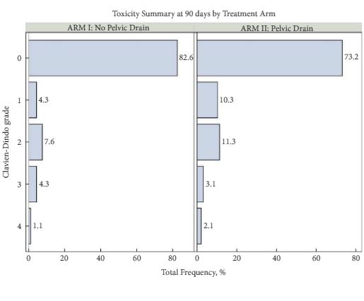
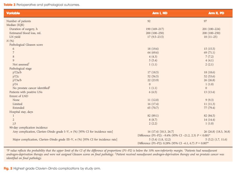
From 2012 to 2016, 189 patients were accrued to the study, with 92 patients allocated to the ND group and 97 to the PD group. Due to lower than expected accrual rates, accrual to the study was halted by regulatory entities, and we did not reach the intended accrual goal. The ND and PD groups were comparable for median PSA level (6.2 vs 5.8 ng/mL, P = 0.5), clinical stage (P = 0.8), D’Amico risk classification (P = 0.4), median lymph nodes dissected (17 vs 18, P = 0.2), and proportion of patients receiving an extended pelvic lymph node dissection (70.7% vs 79.4%, P = 0.3). Incidence of 90-day overall and major (Clavien–Dindo grade >III) complications in the ND group (17.4% and 5.4%, respectively) was not inferior to the PD group (26.8% and 5.2%, respectively; P < 0.001 and P = 0.007 for difference of proportions <10%, respectively). Symptomatic lymphocoele rates (2.2% in the ND group, 4.1% in the PD group) were comparable between the two arms (P = 0.7).

Conclusions

Incidence of adverse events in the ND group was not inferior to the group who received a PD. In properly selected patients, PD placement after RARP can be safely withheld without significant additional morbidity.

Read more articles of the week


Editorial: To drain or not to drain after RARP? That is the question

In the current issue of BJUI, the article by Chenam et al. [1] from the City of Hope Hospital clarifies an important surgical issue that most major uro-oncology centres face every day, but it also raises several other important surgical issues. The objective of the study was to determine if eliminating the routine placement of a pelvic drain after robot-assisted radical prostatectomy (RARP) affects the incidence of early (90-day) postoperative adverse events. RARP has become the new ‘gold standard’ approach for the surgical treatment of localized prostate cancer, and now comprises the majority of radical prostatectomies performed in the USA and the UK, with >80% of cases using this technique [2].

In this parallel-group, blinded, non-inferiority trial, patients who were planned to undergo RARP were randomized to one of two arms: no drain placement or pelvic drain placement. The primary endpoint was incidence of 90-day complications which, based on standard treatment using pelvic drain retrospectively, was estimated at 13%. The non-inferiority margin was set at 10%, and the planned sample size was 312. Despite stopping short of the intended number of patients to reach the accrual goal, it was impressive to read that the study was still well conducted, with robust statistical evidence that the no drain placement group was not inferior to the pelvic drain group. As a result, the authors conclude that pelvic drain can be safely omitted based on a clinical decision according to the surgeon’s discretion.

It has previously been demonstrated that the need for pelvic drain placement in RARP may be significantly less than in open prostatectomy techniques [3], and the concept of omitting the drain was initially presented more than 10 years ago [4]. But in any radical prostatectomy the rationale for placing a pelvic drain is potentially complex. Firstly, an anastomotic urine leak and subsequent urinoma or urinary peritonitis are the key historical concerns. The current reliable continuous running anastomosis now routinely possible with the robotic approach is far superior to the five to seven interrupted sutures inserted with the open or laparoscopic techniques, and hence the integrity is far more robust. Anastomotic leak rate is therefore generally very low at ~0.5–1% [2], and other non-randomized studies have previously shown that omission of pelvic drains is potentially safe [5].

Secondly, a drain may also assist with drainage of lymphatic leak after pelvic lymphadenectomy, preventing symptomatic lymphocele. The incidence of symptomatic lymphocele is ~2.5% in those undergoing RARP and extended pelvic lymph node dissection, as most lymphoceles are asymptomatic, but those that present late may be more at risk of infection in people with diabetes [6]. Another aspect that a small randomized controlled trial will not evaluate is the impact of the very occasional disaster, such as significant anastomotic disruption by a pelvic haematoma or a postoperative haemorrhage, and how that might be adverted by prior placement of a pelvic drain.

One of the few relevant problems with small randomized controlled trials in single centres is generalizability; results potentially only relate to very similar patient populations, i.e. those treated at high-volume and experienced centres, and cannot necessarily be extrapolated to other situations, such as RARPs performed by surgeons early on their learning curve or by those on fellowship/training programmes, or complex cases such as salvage RARP or when the anastomosis is technically challenging. This may include cases with significant bladder neck reconstruction, RARPs performed after TURP, or even cases with patients on steroids or other immunosuppressants. How necessary a pelvic drain is to the rapidly emerging Retzius-sparing RARPs remains to be seen, but judicious placement initially during the learning curve at least seems very sensible. Thus it would be wrong to conclude from the present study that pelvic drains are never indicated, as the authors also specify. Perhaps future larger studies may indicate more clearly those populations which do require pelvic drains in the form of an algorithm or decision-making tool.

It is clear that, as the emphasis shifts to enhanced recovery in the RARP population, pelvic drain placement may delay discharge and have a negative impact on the patient, such as increased anxiety and potential morbidity. In a public healthcare system such as the NHS, in which length of stay is an important cost variable, the present study empowers urologists to dispense with drains in the majority of cases. We believe the study was very well conducted and raises an important and a controversial topic. Overall, as we are all so much more familiar with standard RARP, it seems the time has come to omit the routine pelvic drain.

Salma GhanemBenjamin Namdarian and Ben Challacombe
Urology Centre, Guys Hospital, London, UK


Read the full article


References


2 BAUS UK Radical Prostatectomy Audit 2016. Available at: https:// www.baus.org.uk/_userfiles/pages/files/Publications/Audit/Radical% 20Prostatectomy%202016%20final%20analysis.pdf. Accessed November 2017


3 Danuser H, Di Pierro GB, Stucki P, Mattei A. Extended pelvic lymphadenectomy and various radical prostatectomy techniques: is pelvic drainage necessary? BJU Int 2013; 111: 9639


4 Sharma S, Kim HL, Mohler JL. Routine pelvic drainage not required after open or robotic radical prostatectomy. Urology 2007; 69: 3303


Article of the Week: Sentinel node biopsy for prostate cancer: report from a consensus panel meeting

Every Week the Editor-in-Chief selects an Article of the Week from the current issue of BJUI. The abstract is reproduced below and you can click on the button to read the full article, which is freely available to all readers for at least 30 days from the time of this post.

In addition to the article itself, there is an accompanying editorial written by a prominent member of the urological community. This blog is intended to provoke comment and discussion and we invite you to use the comment tools at the bottom of each post to join the conversation.

If you only have time to read one article this week, it should be this one.

Sentinel node biopsy for prostate cancer: report from a consensus panel meeting

Henk G. van der Poel* , Esther M. Wit*, Cenk Acar, Nynke S. van den Berg,Fijs W. B. van Leeuwen, Renato A. Valdes Olmos, Alexander Winter§,Friedhelm Wawroschek§, Fredrik Liedberg**, Steven Maclennan††and Thomas Lam†† On behalf of the Sentinel Node Prostate Cancer Consensus Panel Group members 


*Department of Urology, Netherlands Cancer Institute, Amsterdam, The Netherlands, Department of Urology, Eryaman Hospital, Ankara, Turkey, Department of Radiology, University of Leiden Medical Centre, Leiden, The Netherlands, §Klinikum Oldenburg, School of Medicine and Health Sciences, University Hospital for Urology, Oldenburg, Germany, Department of Urology, Skane University Hospital, Malmo, **Department of Translational Medicine Lund University, Lund, Sweden, and ††Academic Urology Unit, University of Aberdeen, Aberdeen, UK


Read the full article

Abstract

Objective

To explore the evidence and knowledge gaps in sentinel node biopsy (SNB) in prostate cancer through a consensus panel of experts.

Methods

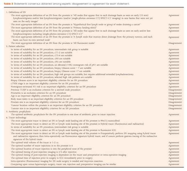
A two-round Delphi survey among experts was followed by a consensus panel meeting of 16 experts in February 2016. Agreement voting was performed using the research and development project/University of California, Los Angeles Appropriateness Methodology on 150 statements in nine domains. The disagreement index based on the interpercentile range, adjusted for symmetry score, was used to assess consensus and non-consensus among panel members.

aotw-aug-2017-4

Results

Consensus was obtained on 91 of 150 statements (61%). The main outcomes were: (1) the results from an extended lymph node dissection (eLND) are still considered the ‘gold standard’, and sentinel node (SN) detection should be combined with eLND, at least in patients with intermediate- and high-risk prostate cancer; (2) the role of SN detection in low-risk prostate cancer is unclear; and (3) future studies should contain oncological endpoints as number of positive nodes outside the eLND template, false-negative and false-positive SN procedures, and recurrence-free survival. A high rate of consensus was obtained regarding outcome measures of future clinical trials on SNB (89%). Consensus on tracer technology was only obtained in 47% of statements, reflecting a need for further research and standardization in this area. The low-level evidence in the available literature and the composition of mainly SNB users in the panel constitute the major limitations of the study.

Conclusions

Consensus on a majority of elementary statements on SN detection in prostate cancer was obtained.; therefore, the results from this consensus report will provide a basis for the design of further studies in the field. A group of experts identified evidence and knowledge gaps on SN detection in prostate cancer and its application in daily practice. Information from the consensus statements can be used to direct further studies.

Read more articles of the week

Editorial: Sentinel nodes in prostate cancer– are we chasing a ghost?

Van der Poel et al. [1] report the findings of a consensus panel meeting on sentinel node (SN) biopsy at the time of radical prostatectomy. The consensus process was based on a two-round Delphi survey on the current evidence and knowledge gaps on SN detection. Then, a number of experts among those who answered the survey were invited to further discuss these issues during a consensus meeting.

The authors are to be complimented on their effort to establish standard definitions, techniques, and reporting of outcomes of SN detection. Their results push the field forward and will make it possible to compare future data between institutions, individual surgeons, and techniques of SN detection. Nevertheless, although the authors admittedly represented a group of potentially biased experts, current limitations of SN detection were fairly acknowledged.

One thorny issue remains, the definition of SN and whether a true SN exists or not. The SN concept implies that one should reliably find tumour in the first echelon of drainage when tumour is present. The SN concept further assumes that if the SN is free of tumour, then so are the next drainage stations. Does that really happen in prostate cancer? We know from previous mapping studies that primary lymphatic landing sites of the prostate are heterogeneously localised, that drainage varies from site to site of tracer injection (and thus tumour location), and that drainage varies from patient to patient [2, 3]. Thus, prostate cancer does not fit the Halstedian paradigm of a pre-defined, stepwise, uniform pathway of metastatic spread. An additional setback of SN detection is that lymph nodes that contain a large tumour burden often fail to take up the tracer [2, 4]. One might critically argue that this limitation may be dependent on the tracer used. The consensus group could not conclusively agree on which tracer technology to use. Looking forward, imaging probes that target tumour-specific molecules may improve tumour detection during pelvic lymph node dissection (PLND). The prostate-specific membrane antigen (PSMA) represents a particularly promising marker in prostate cancer imaging. However, using 68Ga-PSMA-positron emission tomography/CT for the detection of lymph node metastases before radical prostatectomy, one group failed to observe the expected improvement and reported only 33% sensitivity [5]. Future studies will determine whether these advances in prostate cancer detection will translate into more precise targeted dissection of lymph node metastases.

Altogether, the consensus group concluded that extended PLND should remain the standard of care, at least in patients with intermediate- and high-risk prostate cancer. This conclusion is laudable, but to be fair, we have no level 1 evidence for this either. However, every oncology-oriented surgeon would agree that each potential positive lymph node should be found and removed during surgery with curative intent. In case of true low-risk prostate cancer, this is probably less of an issue, but it should be mentioned that this population is at very low risk of dying from prostate cancer, even if left untreated, and the indication for radical prostatectomy should be questioned rather than that for PLND. Furthermore, pathological Gleason score is underestimated in preoperative biopsies in ~30% of all cases [6], making the decision to perform PLND or not in low-risk disease difficult.

Again, the authors should be complimented for their thorough work. We still do not know whether the SN exists, but performing surgery with image-guidance provides quality control for completeness of resection and might help detect (positive?) lymph nodes outside of the extended PLND template. Indeed, up to 35% of prostate lymphatic drainage sites may remain outside the extended anatomical template [3]. SN detection might also teach us when and where to stop dissection to decrease potential morbidity of PLND without leaving tumour behind. Thus, chasing the ghost of the SN has made and makes us better and more meticulous cancer surgeons. At the same time, it is humbling that we have been chasing a ghost for so long without bringing up level 1 evidence.

George N. Thalmann and Daniel P. Nguyen


Department of Urology, University Hospital Bern, Bern, Switzerland


Read the full article


References

1 van der Poel HG, Wit EM, Acar C et al. Sentinel node biopsy for prostate cancer: report from a consensus panel meeting. BJU Int 2017; 120: 20411


4 Weckermann D, Dorn R, Holl G, Wagner T, Harzmann R. Limitations of radioguided surgery in high-risk prostate cancer. Eur Urol 2007; 51: 154958


© 2024 BJU International. All Rights Reserved.