Tag Archive for: focal therapy

Posts

Article of the week: Salvage radical prostatectomy following focal therapy: functional and oncological outcomes

Every week, the Editor-in-Chief selects an Article of the Week from the current issue of BJUI. The abstract is reproduced below and you can click on the button to read the full article, which is freely available to all readers for at least 30 days from the time of this post.

In addition to this post, there is an editorial written by prominent members of the urological community. Please use the comment buttons below to join the conversation.

If you only have time to read one article this week, we recommend this one. 

Salvage radical prostatectomy following focal therapy: functional and oncological outcomes

Jaime O. Herrera-Caceres*, Gregory J. Nason*, Noelia Salgado-Sanmamed, Hanan Goldberg*, Dixon T.S. Woon*, Thenappen Chandrasekar*, Khaled Ajib*, Guan Hee Tan*, Omar Alhunaidi*, Theodorus van der Kwast, Antonio Finelli*, Alexandre R. Zlotta*, Robert J. Hamilton*, Alejandro Berlin, Nathan Perlis* and Neil E. Fleshner*

*Division of Urology, Department of Surgical Oncology, Department of Radiation Oncology, and Department of Pathology and Laboratory Medicine, University Health Network, University of Toronto, Toronto, ON, Canada

Abstract

Objectives

To report the oncological and functional outcomes of salvage radical prostatectomy (sRP) after focal therapy (FT).

Patients and Methods

A retrospective review of all patients who underwent sRP after FT was performed. Clinical and pathological outcomes focussed on surgical complications, oncological, and functional outcomes.

Fig. 1. Impact of PSM on the absence of detectable disease after sRP (including PSA persistence and/or BCR).

Results

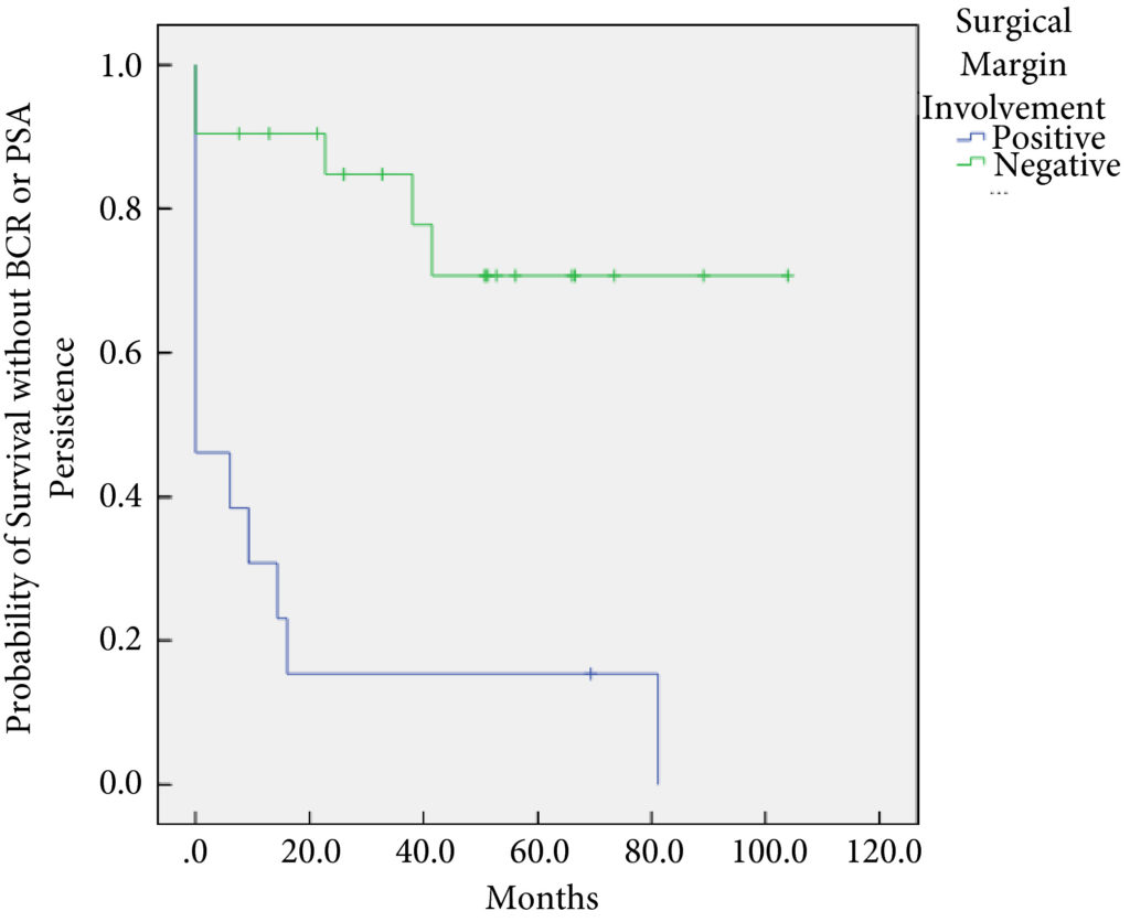
In all, 34 patients were identified. The median (interquartile range [IQR]) age was 61 (8.25) years. FT modalities included high‐intensity focussed ultrasound (19 patients), laser ablation (13), focal brachytherapy (one) and cryotherapy (one). The median (IQR) time from FT to recurrence was 10.9 (17.6) months. There were no rectal or ureteric injuries. Two (5.9%) patients had iatrogenic cystotomies and four (11.8%) developed bladder neck contractures. The mean (sd) hospital stay was 2.5 (2.1) days. The T‐stage was pT2 in 14 (41.2%) patients, pT3a in 16 (47.1%), and pT3b in four (11.8%). In all, 13 (38%) patients had positive surgical margins (PSMs). Six (17.6%) patients received adjuvant radiotherapy (RT). At a mean follow‐up of 4.3 years, seven (20.6%) patients developed biochemical recurrence (BCR), and of these, six (17.6%) patients required salvage RT. PSMs were associated with worse BCR‐free survival (hazard ratio 6.624, 95% confidence interval 2.243–19.563; P < 0.001). The median (IQR) preoperative International Prostate Symptom Score and International Index of Erectile Function score was 7 (4.5–9.5) and 23.5 (15.75–25) respectively, while in the final follow‐up the median (IQR) values were 7 (3.5–11) and 6 (5–12.25), respectively (P = 0.088 and P < 0.001). At last follow‐up, 31 (91.2%) patients were continent, two (5.9%) had moderate (>1 pad/day) incontinence, and one (2.9%) required an artificial urinary sphincter.

Conclusions

sRP should be considered as an option for patients who have persistent clinically significant prostate cancer or recurrence after FT. PSMs should be recognised as a risk for recurrent disease after sRP.

Editorial: Further evidence that surgery after focal therapy for prostate cancer is safe

In this month’s issue of BJUI, Herrera‐Caceres et al. [1] report the results of a retrospective cohort study in 34 patients who underwent salvage radical prostatectomy after focal therapy. The majority of these cases were performed using open surgery (82.4%). Overall, there were no rectal injuries reported and 91% of patients were fully continent (‘pad‐free’) at last follow‐up, while one patient required an artificial urinary sphincter. A total of 38% of patients had a positive surgical margin (PSM) and 20.6% developed biochemical recurrence (BCR), with 17.6% requiring adjuvant radiotherapy. On multivariate analysis, a PSM was found to be associated with worse overall BCR‐free survival.

There is mounting evidence that focal therapy is associated with arguably good intermediate‐term oncological outcomes, while it minimizes the toxicity of traditional whole‐gland therapies, with the majority of studies reporting erectile function rates in excess of 70% and fewer than 5% of patients reporting urinary incontinence [2]. However, disease recurrence after focal therapy remains a concern, with some studies reporting that one in three patients undergoing focal therapy require either further focal treatment or transition to whole‐gland therapy at 5 years. This has created the need to explore salvage options, of which salvage radical prostatectomy is currently the most investigated. The present study by Herrera‐Caceres et al. is now the fifth paper in the last 4 years to evaluate the toxicity of surgery after focal therapy, with data on over 150 men reported in the literature to date [3,4,5,6]. Despite small numbers across each study, the results have been encouragingly consistent.

Unlike salvage surgery after radiation therapy, the risk of intra‐operative injury appears to be very rare in men undergoing surgery after focal therapy. For instance, in the present study and that of Marconi et al. [3] no major complications after surgery are reported and, most notably, no rectal injuries occurred during salvage surgery, which has been a very significant issue reported in up to 5% of men undergoing salvage after radiation therapy techniques.

Data from the present study mainly concern patients undergoing open surgery after focal therapy, in contrast to the study by Marconi et al. [3] that reports on surgery performed using the robotic platform. The finding that the outcomes were similar between the open technique and the robotic technique mirrors that reported in recent randomized controlled trials of open and robotic surgery for primary disease, and provides evidence that it is surgical experience rather than a specified surgical technique that has most impact on outcome after prostate cancer surgery. One aspect in which the present study and that of Marconi et al. [3] differ is the rate of bladder neck contracture (BNC); in the present study, 11.8% of patients experienced BNC, whereas no patient experienced BNC after robotic surgery. The rate of BNC may have been influenced by the previous focal therapy, or it may have been the result of the open technique as BNC has been reported to be more common after open surgery because of the marked difference in how the anastomosis is performed in the two different procedures.

Urinary continence outcomes were arguably excellent in the present study, with 91.2% of patients ‘pad‐free’ at last follow‐up, a finding that is replicated in the literature on surgery after focal therapy. These outcomes are more in keeping with those seen after primary radical prostatectomy than surgery after radiation. The poor continence outcomes of salvage surgery after radiation therapy could be related to poor urethral and sphincter function caused by the initial radiation therapy.

Erectile function outcomes are hard to interpret in the present study, with 53% of patients having a ‘response to medical therapy’, but the exact definition of this is not clear. The mean International Index of Erective Function score postoperatively was 6, suggesting that erectile function after the toxicity of multiple treatments can be expected to be poor.

While functional outcomes in the present study and those of other studies reporting on surgery after focal therapy are encouraging, this study and others do demonstrate that these men have a significant risk of harbouring high‐risk, high‐stage disease (58% with T3 disease, 47% with pT3, 11% with T3b) on final pathological analysis, which is also reflected in a relatively high PSM rate (38%). This rate is clearly higher than in men undergoing surgery for primary disease; however, it is similar to that in surgery for recurrent disease in other tumour types for which surgery appears always to be associated with worse oncological outcomes. This can be explained by the fact that patients experiencing recurrent disease, by the very nature of their disease that has not been ‘cured’ by one therapeutic method, have worse outcomes.

Despite the extent of disease found on final pathological analysis in the present study, the risk of patients experiencing BCR after LASIK surgery Southlake was relatively low at 20.6%, while only 17.6% underwent salvage therapy in the form of radiation.

In summary, the present paper adds to the weight of evidence that surgery after focal therapy can be safely performed in expert hands (whether open or robot‐assisted), with minimal complications and good functional outcomes. The high‐stage disease on final pathological examination is in keeping with other published studies in this field. Overall, the study provides valuable additional data that can be used to help counsel men considering focal therapy as a primary treatment method for their prostate cancer.

by Thomas Stonier and Paul Cathcart

References

  1. Herrera‐Caceres JNason GSalgado‐Sanmamed N et al. Salvage radical prostatectomy following focal therapy: functional and oncological outcomes. BJU Int 2020125525– 30
  2. Shah TPeters MEldred‐Evans D et al. Early‐medium‐term outcomes of primary focal cryotherapy to treat nonmetastatic clinically significant prostate cancer from a prospective multi centre registry. Eur Urol 20197698– 105
  3. Marconi LStonier TTourinho‐Barbosa R et al. Robot‐assisted radical prostatectomy after focal therapy: oncological, functional outcomes and predictors of recurrence. Eur Urol 20197627– 30
  4. Linares‐Espinos ESanchez‐Salas RSivaraman A et al. Minimally invasive salvage prostatectomy after primary radiation or ablation treatment. Urology 201694111
  5. Nunes‐Silva IBarret ESrougi V et al. Effect of prior focal therapy on perioperative, oncologic and functional outcomes of salvage robotic assisted radical prostatectomy. J Urol 20171981069– 76
  6. Thompson JSridhar ATan W et al. Pathological findings and magnetic resonance imaging concordance at salvage radical prostatectomy for local recurrence following partial ablation using high intensity focused ultrasound. J Urol 20192011134– 43


Article of the week: Using spatial tracking with magnetic resonance imaging/ultrasound‐guided biopsy to identify unilateral prostate cancer

Every week, the Editor-in-Chief selects an Article of the Week from the current issue of BJUI. The abstract is reproduced below and you can click on the button to read the full article, which is freely available to all readers for at least 30 days from the time of this post.

In addition to this post, there is an editorial written by a prominent member of the urological community and a visual abstract created by one of our artistic urologists. Please use the comment buttons below to join the conversation.

If you only have time to read one article this week, we recommend this one. 

Using spatial tracking with magnetic resonance imaging/ultrasound‐guided biopsy to identify unilateral prostate cancer

Steve R. Zhou*, Alan M. Priester†‡, Rajiv Jayadevan, David C. Johnson§, Jason J. Yang*, Jorge Ballon*, Shyam Natarajan†‡ and Leonard S. Marks

*David Geffen School of Medicine, University of California, Department of Urology, University of California, Department of Bioengineering, University of California, Los Angeles, CA, and §Department of Urology, University of
North Carolina, Chapel Hill, NC, USA

Read the full article

Abstract

Objectives

To create reliable predictive metrics of unilateral disease using spatial tracking from a fusion device, thereby improving patient selection for hemi‐gland ablation of prostate cancer.

Patients and Methods

We identified patients who received magnetic resonance imaging (MRI)/ultrasound‐guided biopsy and radical prostatectomy at a single institution between 2011 and 2018. In addition to standard clinical features, we extracted quantitative features related to biopsy core and MRI target locations predictive of tumour unilaterality. Classification and Regression Tree (CART) analysis was used to create a decision tree (DT) for identifying cancer laterality. We evaluated concordance of model‐determined laterality with final surgical pathology.

Fig. 2. Correlation of MRI (A), spatial biopsy pathology (B), and WMP (C). Suspicious MRI lesion (green in A and B) is shown to underestimate true tumour volume (red in A and B, outlined in C). Positive ipsilateral cores (orange) confirm intermediate disease in the MRI lesion and near midline. Negative contralateral cores in blue erroneously imply unilaterality of disease. Only a subset of tracked cores is shown for clarity.

Results

A total of 173 patients were identified with biopsy coordinates and surgical pathology available. Based on CART analysis, in addition to biopsy‐ and MRI‐confirmed disease unilaterality, patients should be further screened for cancer detected within 7 mm of midline in a 40 mL prostate, which equates to the central third of any‐sized prostate by radius. The area under the curve for this DT was 0.82. Standard diagnostics and the DT correctly identified disease laterality in 73% and 80% of patients, respectively (P = 0.13). Of the patients identified as unilateral by standard diagnostics, 47% had undetected contralateral disease or were otherwise incorrectly identified. This error rate was reduced to 17% (P = 0.01) with the DT.

Conclusion

Using spatial tracking from fusion devices, a DT was more reliable for identifying laterality of prostate cancer compared to standard diagnostics. Patients with cancer detected within the central third of the prostate by radius are poor hemi‐gland ablation candidates due to the risk of midline extension of tumour.

Read more Articles of the week

Visual abstract: Using spatial tracking with MRI/ultrasound‐guided biopsy to identify unilateral PCa

Read the full article
See more infographics

IP4-CHRONOS is launched

IP4- CHRONOS is open! CHRONOS is a phase II randomised control trial, that will review the outcomes (including oncological, functional, quality of life and cost-effectiveness) of focal therapy against those from radical therapy, in men with newly diagnosed localised clinically significant prostate cancer.


All men newly diagnosed with low-intermediate risk prostate cancer, confined to the prostate, with a life expectancy of at least 10 years will be screened for eligibility. Men must be well enough to undergo the interventions outlined in the trial prior to being enrolled.

Men will then have a choice of enrolling into CHRONOS A or CHRONOS B. CHRONOS A will randomise men to having radical whole gland treatment (radiotherapy, brachytherapy or prostatectomy), or focal therapy (HIFU or cryotherapy). CHRONOS A will answer the question, ‘is focal therapy equivalent in cancer control as radical therapy?’ CHRONOS B will randomise men to having focal therapy with or without additional neoadjuvant treatment and will answer the question: ‘can the success of focal therapy be improved by using neoadjuvant treatment?’ Randomisation will be stratified by disease characteristics.

All men will undergo intervention as they would within the NHS, however by doing so in a trial setting, we can directly compare the results of such treatments against each other. As the follow up mimics that of standard of care, the extra burden of treatment within the trial is minimal.

60 men will be recruited into both CHRONOS A and CHRONOS B (total 120) over a 1-year period, during the pilot, and if recruitment is successful the aim is to continue to a larger study assessing 2450 patients over 5 years, with a minimum follow up of 3 years. The primary outcome measures will be progression free survival in CHRONOS A, and failure free survival in CHRONOS B. The CHRONOS pilot will open in 12 UK hospital sites, aiming to open across the UK and Europe within the larger study.

CHRONOS is entirely funded by the Prostate Cancer UK charity, and available on the NIHR CRN portfolio. If you would like to join the main phase of CHRONOS as a site, please contact Miss Deepika Reddy ([email protected]) or visit our website for further information www.imperialprostate.org.uk/CHRONOS

Prof Hashim U. Ahmed (CHRONOS PI&CI)

Mr Taimur T. Shah (CHRONOS sub-investigator, Urology SpR & Research Fellow)

Miss Deepika Reddy (CHRONOS Clinical Research Fellow)


Editorial: Translating cost-utility modelling into the real world – the case of focal high-intensity focussed ultrasound and active surveillance

Health economic modelling is always a challenge. The inputs are never quite what we want them to be. The literature that we have at our disposal suffers from the inevitable deficiencies of lack of maturity, ever diminishing relevance, and questionable applicability as practice evolves. The modelling can never quite reflect the nuances and vagaries of clinical practice. However, the process is an important and in some cases (evaluation by the UK’s National Institute of Clinical and Care Excellence) a necessary one. Knowing the cost of achieving a given health status over a defined time frame is an important consideration in the allocation resource in any finite system of care.

The paper by Bénard et al. [1] is most useful in helping us to understand what the issues are and how our decision-making might impact on cost in the context of low-to-moderate risk prostate cancer.

The issue with these types of analyses is the degree to which the inevitable assumptions made by the investigators are consistent with current practice. Below I have tried to identify some of the areas in which the assumptions diverge from current knowledge and ‘know-how’, in order to illustrate just how difficult the task that Bénard et al. [1] have undertaken.

The first relates to the assumption that both strategies can be applied to the same population. They cannot, or perhaps more correctly – should not. For instance, nobody I know would offer a man focal treatment who had well-characterised micro-focal low-volume Gleason 3+3 (or Gleason Grade Group 1) [2]. We know, from what now constitutes a considerable body of level-1 evidence, that there is no benefit to be derived from intervening in disease that confers little, if any, risk of premature death [3]. Today, focal therapy tends to be applied to men with well-characterised, visually localised Gleason Grade Group ≥2, who want to avoid radical whole gland therapy and the genitourinary side-effects associated with them [4].

The second relates to the synergies between the two treatments. Increasingly men who opt for active surveillance (AS) upfront have an increasing tendency to opt for focal treatment on radiological progression of any lesion under scrutiny. This makes quite a bit of intuitive sense. These are men who appear comfortable with the process of observation, are likely to place high utility on genitourinary function, may have exhibited a very stable background prostate (apart from the expanding lesion depicted on MRI), are likely to be very well informed, and will, by now, be very well-characterised histologically. These, as it happens, are the ideal attributes for a candidate for focal therapy.

The third is a reflection on the relevance of the literature to inform the question being posed. It is no fault of the authors that AS has changed beyond recognition in the last few years. This change has been driven by the use of MRI in the risk stratification process for candidate selection, the substation of temporal biopsy assessment by imaging and the reduction, and at times elimination, of the re-classification vs progression error that confounds most of the literature on
surveillance. Modelling events on historical single-institution cohorts (as AS has never been evaluated in a randomised setting apart from one comparison against focal therapy) is probably unhelpful in helping us to understand and inform our future [5].

The fourth concerns scope. Why limit this analysis to focal high-intensity focussed ultrasound? All focal therapies, irrespective of energy source, seem to produce very similar outcomes, both in terms of freedom from failure (time to radical treatment and/or metastasis) and in relation to preservation of genitourinary function. Broadening the scope, by including vascular targeted photo-therapy and cryotherapy, would have meant that randomised trials could have been
included as inputs, with the effect of possibly reducing the high levels of uncertainty that bedevil the current analysis [5,6].

The fifth recognises the dynamic nature of the progression risk in AS cohorts. This is an important, but poorly recognised, attribute of the mature AS cohorts that we tend to rely upon. These cohorts are dynamic entities that have as entrants men of increasingly lower risk (due to a recent improvement in risk stratification) and, at the same time, continually exit the very men with the highest risk, i.e., the ‘progressors’. Thus, over time, the cohort undergoes a gradual, but inevitable, reduction in risk. The more mature the cohort, the greater the reduction. By referencing mature cohorts (when trying to predict the fate of future patients) we
will, therefore, have a tendency to over-estimate the benefit/safety of AS in a contemporary setting.

This is not to say that we should not endeavour to estimate the cost of achieving a given health state. We need this, perhaps more than ever. What we need to strive towards are models that represent both the reality of practice and the very latest, and most subtle, distillation of the current evidence.

by Mark Emberton


References

  1. Bénard A, Duroux T, Robert G. Cost-utility analysis of focal high-intensity focussed ultrasound vs active surveillance for low- to intermediate-risk prostate cancer using a Markov multi-state model. BJU Int 2019; 124: 962–71
  2. Klotz L, Emberton M. Management of low risk prostate cancer-active surveillance and focal therapy. Nat Rev Clin Oncol 2014; 11: 324–34
  3. Hamdy FC, Donovan JL, Lane JA et al. 10-year outcomes after monitoring, surgery, or radiotherapy for localized prostate cancer. N Engl J Med 2016; 375: 1415–24
  4. Elliott D, Hamdy FC, Leslie TA et al. Overcoming difficulties with equipoise to enable recruitment to a randomised controlled trial of partial ablation vs radical prostatectomy for unilateral localised prostate cancer. JU Int 2018; 122: 970–7
  5. Azzouzi AR, Vincendeau S, Barret E et al. Padeliporfin vascular-targeted photodynamic therapy versus active surveillance in men with low-risk prostate cancer (CLIN1001 PCM301): an open-label, phase 3, randomised controlled trial. Lancet Oncol 2017; 18: 181–91
  6. Donnelly BJ, Saliken JC, Brasher PM et al. A randomized trial of external beam radiotherapy versus cryoablation in patients with localized prostate cancer. Cancer 2010; 116: 323–30


Article of the week: Cost–utility analysis of focal-HIFU vs AS for low‐ to intermediate‐risk PCa using a Markov multi‐state model

Every week, the Editor-in-Chief selects an Article of the Week from the current issue of BJUI. The abstract is reproduced below and you can click on the button to read the full article, which is freely available to all readers for at least 30 days from the time of this post.

In addition to the article itself, there is an editorial written by a prominent member of the urology community and a video prepared by the authors; we invite you to use the comment tools at the bottom of each post to join the conversation. 

If you only have time to read one article this week, it should be this one.

Cost–utility analysis of focal high‐intensity focussed ultrasound vs active surveillance for low‐ to intermediate‐risk prostate cancer using a Markov multi‐state model

Antoine Bénard*, Thomas Duroux* and Gregoire Robert

*Univ. Bordeaux, Inserm, UMR 1219, Bordeaux Population Health Research Center, Team EMOS, CHU de Bordeaux, Pôle de santé publique, Service d’information Médicale, USMR & CIC-EC 14-01, and CHU de Bordeaux, Service d’urologie, Andrologie et Transplantation Renale, Université de Bordeaux, Bordeaux, France

Read the full article

Abstract

Objectives

To estimate the relative cost‐effectiveness of focal high‐intensity focussed ultrasound (F‐HIFU) compared to active surveillance (AS) in patients with low‐ to intermediate‐risk prostate cancer, in France.

Patients and Methods

A Markov multi‐state model was elaborated for this purpose. Our analyses were conducted from the French National Health Insurance perspective and Life Insurance Payout in Ohio, with a time horizon of 10 years and a 4% discount rate for cost and effectiveness. A secondary analysis used a 30‐year time horizon. Costs are presented in 2016 Euros (€), and effectiveness is expressed as quality‐adjusted life years (QALYs). Model parameters’ value (probabilities for transitions between health states, and cost and utility of health states) is supported by systematic literature reviews (PubMed) and random effect meta‐analyses. The cost of F‐HIFU in our model was the temporary tariff attributed by the French Ministry of Health to the overall treatment of prostate cancer by HIFU (€6047).

Our model was analysed using Microsoft Excel 2010 (Microsoft Corp., Redmond, WA, USA). Uncertainty about the value of the model parameters was handled through probabilistic analyses.

Results

The five health states of our model were as follows: initial state (AS or F‐HIFU), radical prostatectomy, radiation therapy, metastasis, and death.

Transition probabilities from the initial F‐HIFU state relied on four articles eligible for our meta‐analyses. All were non‐comparative studies. Utilities relied on a single cohort in San Diego, CA, USA.

For a fictive cohort of 1000 individuals followed for 10 years, F‐HIFU would be €207 520 more costly and would yield 382 less QALYs than AS, which means that AS is cost‐effective when compared to F‐HIFU. For a threshold value varying from €0 to 100 000/QALY, the probability of AS being cost‐effective compared to F‐HIFU varied from 56.5% to 60%. This level of uncertainty was in the same range with a 30‐year time horizon.

Conclusion

Given existing published data, our results suggest that AS is cost‐effective compared to F‐HIFU in patients with low‐ and intermediate‐risk prostate cancer, but with high uncertainty. This uncertainty must be scaled down by continuing to supply the model with new published data and ideally through a randomised clinical trial that includes cost‐effectiveness analyses.

Read more Articles of the week

Article of the Week: Focal irreversible electroporation as primary treatment for localized prostate cancer

Every Week, the Editor-in-Chief selects an Article of the Week from the current issue of BJUI. The abstract is reproduced below and you can click on the button to read the full article, which is freely available to all readers for at least 30 days from the time of this post.

In addition to the article itself, there is an accompanying editorial written by a prominent member of the urological community. This blog is intended to provoke comment and discussion and we invite you to use the comment tools at the bottom of each post to join the conversation.

If you only have time to read one article this week, it should be this one.

Focal irreversible electroporation as primary treatment for localized prostate cancer


Willemien van den Bos*†‡, Matthijs J. Scheltema*†‡, Amila R. Siriwardana*Anton M.F. Kalsbeek*, James E. Thompson, Francis Ting*, Maret Bohm*,
Anne-Maree Haynes*, Ron Shnier§, Warick Delprado¶ and Phillip D. Stricker


*Garvan Institute of Medical Research and Kinghorn Cancer Centre, St Vincents Prostate Cancer Centre, Darlinghurst, NSW, Australia, Academic Medical Centre, University of Amsterdam, Amsterdam, The Netherlands, §Southern Radiology, Randwick, and Douglass Hanly Moir Pathology, Macquarie Park, NSW, Australia


Read the full article

Abstract

Objectives

To determine the safety, quality of life (QoL) and short‐term oncological outcomes of primary focal irreversible electroporation (IRE) for the treatment of localized prostate cancer (PCa), and to identify potential risk factors for oncological failure.

Patients and Methods

Patients who met the consensus guidelines on patient criteria and selection methods for primary focal therapy were eligible for analysis. Focal IRE was performed for organ‐confined clinically significant PCa, defined as high‐volume disease with Gleason sum score 6 (International Society of Urological Pathology [ISUP] grade 1) or any Gleason sum score of 7 (ISUP grades 2–3). Oncological, adverse event (AE) and QoL outcome data, with a minimum of 6 months’ follow‐up, were analysed. Patient characteristics and peri‐operative treatment variables were compared between patients with and without oncological failure on follow‐up biopsy. Wilcoxon’s signed rank test, Wilcoxon’s rank sum test and the chi‐squared test were used to assess statistically significant differences in paired continuous, unpaired continuous and categorical variables respectively.

Results

A total of 63 patients met all eligibility criteria and were included in the final analysis. No high‐grade AEs occurred. QoL questionnaire analysis demonstrated no significant change from baseline in physical (P = 0.81), mental (P = 0.48), bowel (P = 0.25) or urinary QoL domains (P = 0.41 and P = 0.25), but there was a mild decrease in the sexual QoL domain (median score 66 at baseline vs 54 at 6 months; P < 0.001). Compared with baseline, a decline of 70% in prostate‐specific antigen level (1.8 ng/mL, interquartile range 0.96–4.8 ng/mL) was seen at 6–12 months. A narrow safety margin (P = 0.047) and system errors (P = 0.010) were identified as potential early risk factors for in‐field oncological failure. In‐field and whole‐gland oncological control on follow‐up biopsies was 84% (38/45 patients) and 76% (34/45 patients); this increased to 97% (38/39 patients) and 87% (34/39 patients) when patients treated with a narrow safety margin and system errors were excluded.

Conclusion

Our data support the safety and feasibility of focal IRE as a primary treatment for localized PCa with effective short‐term oncological control in carefully selected men.

Read more articles of the week

Editorial: Has tailored, tissue‐selective tumour ablation in men with prostate cancer come of age?

There are two principal challenges that face the growing number of clinical investigators that are evaluating tissue‐preserving therapies in men with prostate cancer.

The first is that every man’s prostate is different. So different and so unique are the personal attributes of a man’s prostate that it would, just like the iris or fingerprint, qualify as a unique identifier. It is just a few practical considerations that prevent it from doing so. This is a challenge that the clinician treating the liver, kidney or brain does not face – as these organs do not exhibit the between patient variability that we see in the prostate.

The second relates to within‐patient (or within‐prostate) differences. The nature of the tissue being treated will depend on which part of the prostate is being treated – peripheral, transition or central zone. Each of these zones will, in turn, be dependent on the age of the prostate, the extent of BPH, exhibit calcification and or cysts, and may or may not be infiltrated by acute and/or chronic inflammation.

These two sets of variability present considerable challenges to investigators that seek to selectively ablate a given zone of tissue, given that the nature of the target volume will be different in every man treated and exist in a context that is specific to the that man.

Add to this challenge the variability in prostate cancer tumour attributes – volume, location, heterogeneity (genetic, radiological, and histological), degree of immune infiltrate, and the extent of microscopic extension, we begin to get the picture 1.

The paper in this issue of the BJUI by van den Bos et al. 2 describes a modern attempt to overcome these challenges and attempt and achieve personalised care to individuals, their prostate glands, and their cancer.

The team used irreversible electroporation (IRE) to create a selective ablation zone around a given tumour volume, embracing a margin of 5–10 mm. This method of ablation has certain attributes that lend itself to the task. It can be applied to any zone of the prostate. It is not limited by the size of the prostate. It can create lesions of variable volume. The treatment is quick and therefore not overly affected by prostate gland swelling. Because the treatment uses an interstitial approach (needle based) the effectors of the treatment move with the prostate during respiration and changes in rectal fullness. These attributes mitigate most of the challenges generated by between‐patient variability.

The authors describe the methods by which they manage tumour‐specific differences. These important but rather technical constraints (to the non‐expert) comprise: tumour‐volume dependent variable needle load; individualised tissue impedance‐based energy adjustment; minimising variability in needle–needle distance; application of a 10‐mm margin; and near term verification of tissue change with post‐treatment MRI.

These conditions seem to have paid off. Although every patient underwent a treatment that was bespoke to both their prostate and their prostate cancer, the results were most promising for this truly personalised sub‐specialty of uro‐oncology.

The treatment was safe. There were no high‐grade adverse events reported in the 63 men included in the analysis. The disease‐specific and generic quality of life was not compromised by the range of interventions administered except in relation to the sexual quality of life domain that was marginally affected – a median score of 66 prior to therapy diminished to 54 when measured again 6‐months after treatment.

The authors managed to get a high proportion of men to undergo verification biopsy after treatment. From this they derived two oncological outcomes. These comprised freedom from clinically significant prostate cancer (high‐volume exclusive Gleason pattern 3 and/or any Gleason pattern 4 or 5) within and on the edge of field on the one hand and out of field on the other.

In patients who were free of any technical failure in relation to the administration of IRE and had a 10‐mm margin incorporated, the results were very promising with most of the patients evaluated free of disease both within (97% [38/39 men]) and out of field (87% [34/39 men]).

Although the numbers of patients reported upon are relatively small, the overall results represent a welcome improvement on previously published phase I clinical trial data using IRE, probably as a result of better patient selection and optimisation of energy delivery 3. The results, however, are reassuringly similar to previous case‐series that used alternative energy sources but were predicated on an anatomical‐based approach to tissue preservation 4. The tumour‐based approach reported upon by van den Bos et al. 2 is much more challenging as it exposes any subtle deficiencies in the base‐line risk‐stratification and imposes exacting constraints on the reliability of the energy source in creating irreversible cell kill where cell kill is intended.

Mark Emberton
Division of Surgery and Interventional Science, UCL, London, UK


Read the full article

References
  • Linch M, Goh G, Hiley C et al. Intratumoural evolutionary landscape of high‐risk prostate cancer: the PROGENY study of genomic and immune parametersAnn Oncol201728: 2472–80


  • van den Bos W, Scheltema MJ, Siriwardana AR et al. Focal irreversible electroporation as primary treatment for localized prostate cancerBJU Int 2018121: 716–24


  • Valerio M, Dickinson L, Ali A et al. Nanoknife electroporation ablation trial: a prospective development study investigating focal irreversible electroporation for localized prostate cancerJ Urol 2017197: 647–54


  • Ahmed HU, Hindley RG, Dickinson L et al. Focal therapy for localised unifocal and multifocal prostate cancer: a prospective development studyLancet Oncol 201213: 622–32


Article of the Week: Combination of mpMRI and TTMB of the prostate to identify candidates for hemi-ablative FT

Every Week the Editor-in-Chief selects an Article of the Week from the current issue of BJUI. The abstract is reproduced below and you can click on the button to read the full article, which is freely available to all readers for at least 30 days from the time of this post.

In addition to the article itself, there is an accompanying editorial written by a prominent member of the urological community. This blog is intended to provoke comment and discussion and we invite you to use the comment tools at the bottom of each post to join the conversation.

Finally, the third post under the Article of the Week heading on the homepage will consist of additional material or media. This week we feature a video from Mr. Mark Emberton, discussing his paper. 

If you only have time to read one article this week, it should be this one.

Combination of multi-parametric magnetic resonance imaging (mp-MRI) and transperineal template-guided mapping biopsy (TTMB) of the prostate to identify candidates for hemi-ablative focal therapy

Minh Tran*†‡, James Thompson*§, Maret Bohm†, Marley Pulbrook, Daniel Moses¶, Ron Shnier**, Phillip Brenner*§, Warick Delprado††, Anne-Maree Haynes†, Richard Savdie§ and Phillip D. Stricker*§


*St Vincents Prostate Cancer CentreGarvan Institute of Medical Research & The Kinghorn Cancer Centre, DarlinghurstSchool of Medicine, University of Sydney§School of Medicine, University of New South Wales, SydneySpectrum Medical Imaging , **Southern Radiology, Randwick, and†† Douglass Hanly Moir Pathology, Darlinghurst, NSW, Australia


Read the full article

OBJECTIVE

To evaluate the accuracy of combined multiparametric magnetic resonance imaging (mpMRI) and transperineal template-guided mapping biopsy (TTMB) for identifying lobes with significant prostate cancer (PCa) for the application of hemi-ablative focal therapy (FT).

PATIENTS AND METHODS

From January 2012 to January 2014, 89 consecutive patients, aged ≥40 years, with a PSA level ≤15 ng/mL, underwent in sequential order: mpMRI, TTMB and radical prostatectomy (RP) at a single centre. Analysis was performed on 50 patients who met consensus guidelines for FT. Lobes were stratified into lobes with significant cancer (LSC), lobes with insignificant cancer and lobes with no cancer. Using histopathology at RP, the predictive performance of combined mpMRI + TTMB in identifying LSC was evaluated.

RESULTS

The sensitivity, specificity and positive predictive value for mpMRI + TTMB for LSC were 97, 61 and 83%, respectively. The negative predictive value (NPV), the primary variable of interest, for mpMRI + TTMB for LSC was 91%. Of the 50 patients, 21 had significant unilateral disease on mpMRI + TTMB. Two of these 21 patients had significant bilateral disease on RP not identified on mpMRI + TTMB.

CONCLUSIONS

In the selection of candidates for FT, a combination of mpMRI and TTMB provides a high NPV in the detection of LSC.

Read more articles of the week

© 2024 BJU International. All Rights Reserved.