Tag Archive for: male

Posts

Article of the Week: Does tadalafil improve ejaculatory dysfunction?

Every week the Editor-in-Chief selects the Article of the Week from the current issue of BJUI. The abstract is reproduced below and you can click on the button to read the full article, which is freely available to all readers for at least 30 days from the time of this post.

In addition to the article itself, there is an accompanying editorial written by a prominent member of the urological community. This blog is intended to provoke comment and discussion and we invite you to use the comment tools at the bottom of each post to join the conversation.

Finally, the third post under the Article of the Week heading on the homepage will consist of additional material or media. This week we feature a video of  Darius Paduch discussing his paper.

If you only have time to read one article this week, it should be this one.

Effects of 12 weeks of tadalafil treatment on ejaculatory and orgasmic dysfunction and sexual satisfaction in patients with mild to severe erectile dysfunction: integrated analysis of 17 placebo-controlled studies

Darius A. Paduch*†, Alexander Bolyakov*†, Paula K. Polzer‡ and Steven D. Watts‡

*Department of Urology and Reproductive Medicine,Weill Cornell Medical College, New York, NY, †Consulting Research Services, Inc., Red Bank, NJ, and ‡Lilly Research Laboratories, Eli Lilly, Indianapolis, IN, USA


Weill Cornell Medical College Press Release

OBJECTIVES

• To compare effects of tadalafil on ejaculatory and orgasmic function in patients presenting with erectile dysfunction (ED).

• To determine the effects of post-treatment ejaculatory dysfunction (EjD) and orgasmic dysfunction (OD) on measures of sexual satisfaction.

PATIENTS AND METHODS

• Data from 17 placebo-controlled 12-week trials of tadalafil (5, 10, 20 mg) as needed in patients with ED were integrated.

• EjD and OD severities were defined by patient responses to the International Index of Erectile Function, question 9 (IIEF-Q9; ejaculation) and IIEF-Q10 (orgasm), respectively.

• Satisfaction was evaluated using the intercourse and overall satisfaction domains of the IIEF and Sexual Encounter Profile question 5.

• Analyses of covariance were performed to compare mean ejaculatory function and orgasmic function, and chi-squared tests evaluated differences in endpoint responses to IIEF-Q9 and IIEF-Q10.

RESULTS

• A total of 3581 randomized subjects were studied.

• Treatment with tadalafil 10 or 20 mg was associated with significant increases in ejaculatory and orgasmic function (vs placebo) across all baseline ED, EjD, and OD severity strata.

• In the tadalafil group, 66% of subjects with severe EjD reported improved ejaculatory function compared with 36% in the placebo group (P < 0.001).

• Similarly, 66% of the tadalafil-treated subjects (vs 35% for placebo; P < 0.001) with severe OD reported improvement.

• Residual severe EjD and OD after treatment had negative impacts on sexual satisfaction.

• Limitations of the analysis include its retrospective nature and the use of an instrument (IIEF) with as yet unknown performance in measuring treatment responses for EjD and OD.

CONCLUSIONS

• Tadalafil treatment was associated with significant improvements in ejaculatory function, orgasmic function and sexual satisfaction.

• Proportions of subjects reporting improved ejaculatory or orgasmic function were ª twofold higher with tadalafil than with placebo.

• These findings warrant corroboration in prospective trials of patients with EjD or OD (without ED).


Read Previous Articles of the Week

Dr Paduch’s commentary on tadalafil and ejaculatory dysfunction


Effects of 12 weeks of tadalafil treatment on ejaculatory and orgasmic dysfunction and sexual satisfaction in patients with mild to severe erectile dysfunction: integrated analysis of 17 placebo-controlled studies

Darius A. Paduch*†, Alexander Bolyakov*†, Paula K. Polzer‡ and Steven D. Watts‡

*Department of Urology and Reproductive Medicine,Weill Cornell Medical College, New York, NY, †Consulting Research Services, Inc., Red Bank, NJ, and ‡Lilly Research Laboratories, Eli Lilly, Indianapolis, IN, USA

Read the full article
OBJECTIVES

• To compare effects of tadalafil on ejaculatory and orgasmic function in patients presenting with erectile dysfunction (ED).

• To determine the effects of post-treatment ejaculatory dysfunction (EjD) and orgasmic dysfunction (OD) on measures of sexual satisfaction.

PATIENTS AND METHODS

• Data from 17 placebo-controlled 12-week trials of tadalafil (5, 10, 20 mg) as needed in patients with ED were integrated.

• EjD and OD severities were defined by patient responses to the International Index of Erectile Function, question 9 (IIEF-Q9; ejaculation) and IIEF-Q10 (orgasm), respectively.

• Satisfaction was evaluated using the intercourse and overall satisfaction domains of the IIEF and Sexual Encounter Profile question 5.

• Analyses of covariance were performed to compare mean ejaculatory function and orgasmic function, and chi-squared tests evaluated differences in endpoint responses to IIEF-Q9 and IIEF-Q10.

RESULTS

• A total of 3581 randomized subjects were studied.

• Treatment with tadalafil 10 or 20 mg was associated with significant increases in ejaculatory and orgasmic function (vs placebo) across all baseline ED, EjD, and OD severity strata.

• In the tadalafil group, 66% of subjects with severe EjD reported improved ejaculatory function compared with 36% in the placebo group (P < 0.001).

• Similarly, 66% of the tadalafil-treated subjects (vs 35% for placebo; P < 0.001) with severe OD reported improvement.

• Residual severe EjD and OD after treatment had negative impacts on sexual satisfaction.

• Limitations of the analysis include its retrospective nature and the use of an instrument (IIEF) with as yet unknown performance in measuring treatment responses for EjD and OD.

CONCLUSIONS

• Tadalafil treatment was associated with significant improvements in ejaculatory function, orgasmic function and sexual satisfaction.

• Proportions of subjects reporting improved ejaculatory or orgasmic function were ª twofold higher with tadalafil than with placebo.

• These findings warrant corroboration in prospective trials of patients with EjD or OD (without ED).

Patient with De Novo Adenosquamous Carcinoma of the Prostate

We report a case of ASC arising spontaneously in a 54 year old male with no previous risk factors.

Authors: Love, Matthew; Storey, Barckley; Alatassi, Houda; Tonkin, Jeremy
Corresponding Author: Love, Matthew


Abstract
Primary adenosquamous cell carcinoma of the prostate (ASC) is an exceedingly rare and aggressive form of prostate cancer, making up <1% of all diagnoses. Since its initial description by Thompson in 1942, there have been fewer than 30 cases reported in the literature. Recent reports of age-adjusted incidence rates of ASC have been shown to be around 0.03 cases per million people per year, making it less prevalent than pure squamous cell carcinoma, an exceedingly rare subtype in itself. While the majority of these tend to arise subsequent to endocrine or radiation treatment with squamous differentiation, approximately one-third of cases have arisen in a de novo setting. We report a case of ASC arising spontaneously in a 54 year old male with no previous risk factors.

Introduction
Primary adenosquamous cell carcinoma of the prostate (ASC) is an exceedingly rare and aggressive form of prostate cancer. Since its initial description by Thompson in 1942, there have been fewer than 30 cases reported in the literature [1]. While the majority of these tend to arise subsequent to endocrine or radiation treatment with squamous differentiation, approximately one-third of cases have arisen in a de novo setting [2]. We report a case of ASC arising spontaneously in a 54 year old African American male with no previous risk factors.

Case Report
A 54 year old African American male was referred to the University of Louisville Department of Urology following detection of an elevated PSA of 20 ng/mL on annual screening. He denied hematuria, dysuria, ejaculatory issues, or lower urinary tract symptoms at the time of presentation. Past medical history was noncontributory and there was no history of prostate cancer or any other malignancies in his family. On digital rectal examination the patient was noted to have a 40 gram prostate that was firm, non-tender, and with no discernible nodules. Repeat PSA was obtained and was found to be 46.7 ng/mL and he was subsequently scheduled for an ultrasound guided prostate biopsy.

Standard 12 core template prostate biopsy revealed adenosquamous carcinoma of the prostate, Gleason 4+4=8 in 7/12 cores and involving more than 50% of each core (See Figure 1 and Figure 2). As a result, an extensive metastatic workup was performed. Bone scan revealed multiple metastatic sites including the patient’s right rib, pubic symphysis, and iliac crest. CT abdomen/pelvis was obtained showing extensive retroperitoneal lymphadenopathy. Chest X-ray was negative for disease.

Due to the extensive metastatic nature of the disease, the patient opted for hormone deprivation therapy and an elective orchiectomy was performed. He was then referred to medical oncology for chemotherapeutic intervention.

Discussion/Conclusion
ASC of the prostate is among one of the rarest and most aggressive subtypes of prostate cancer making up <1% of all diagnoses [3]. Recent reports of age-adjusted incidence rates of ASC have been shown to be around 0.03 cases per million people per year, making it less prevalent than pure squamous cell carcinoma, an exceedingly rare subtype in itself [4].

The underlying mechanism for the progression and development of ASC is unknown and debated; however, most theories contend that differentiation is triggered by the various treatment modalities rather than de novo development [5]. While over two thirds of the cases of ASC originate in patients with previously diagnosed adenocarcinoma of the prostate who have been treated with additional endocrine/radiation therapy, it is extremely rare to find a primary case of this particular subtype[5]. While some believe that hormonal treatment and radiotherapy induce squamous metaplasia in the glandular cells of adenomatous prostate cancer, others contend that ASC develops de novo from divergent differentiation from epithelial stem cell lines within the prostatic cells [6] [7]. Due to the infrequent occurrence of this disease there are very few available studies to examine the mechanism behind this metaplastic process and research is currently undergoing.

ASC is defined by the presence of both glandular and squamous components on histological examination. The squamous elements of ASC constitute on average of 40% of the tumor volume but can range anywhere from 5-95% [8]. This wide range of cell types is reflected in ASC variability through immunohistochemical staining. Stains such as PSA, PSAP, and low molecular weight keratin (CAM5.3) are commonly found only in the glandular components of ASC and therefore patients with a large squamous fraction can have normal serum levels of PSA, possibly further delaying diagnosis [3]. Similarly, the squamous portion can stain positive for high molecular weight keratin (AE3), but this can vary depending on the amount of tissue that has squamous components involved (See Table 1) [9]. Additionally, glandular components have a tendency to be more high grade with an average Gleason score of >6, however, this is also a controversial point as some have suggested that Gleason scoring should not be applied to this subtype [8].

ASC tends to follow the traditional metastatic pathways similar to adenocarcinoma of the prostate, starting with local invasion and then spreading to bone and other distant soft tissue sites. However, one notable difference is that unlike standard prostatic adenocarcinoma, bone metastatic sites are characteristically osteolytic rather than osteoblastic in nature [3].

ASC is an extremely aggressive subtype of prostate cancer and in most cases is found to have widely metastasised at time of diagnosis, suggesting that this disease has a tendency to disseminate early. Most cases reported in the literature presented in individuals who were found to be in urinary retention and the pathological diagnosis was made on TURP specimens [8]. The disease is highly resistant to radiation and chemotherapy and in those individuals fortunate enough to be diagnosed early with localized/regional disease, prostatectomy shows some survival advantage [2]. It is not clear whether hormone ablation is an effective treatment modality, as some authors suggest an early response while others have noted that patients are refractory to hormone deprivation. Long term survival is extremely poor. Wang et al, evaluating SEER data and isolating for patients with an ASC diagnosis, found that the median cancer specific survival was 16 months [2]. For patients who presented with distant disease, the 6 month survival rate was only 20% with all dying within one year of diagnosis.

While literature on the subject of ASC is limited, it appears that the best initial treatment for this particular type of cancer is aggressive surgical intervention in patients with regionally restricted disease. However, due to the highly aggressive nature of this disease in most cases, such as this one, patients present with widely disseminated disease and are relegated to a regimen of hormone ablation and chemotherapy. Currently there are no recommended chemotherapeutic regimens targeted at ASC.

087Figure1


Fig. 1

087Figure2


Fig. 2

References
1. Thompson GJ. Transurethral resection of malignant lesions of the prostate gland. JAMA, 1942;120: 1105-9.
2. Wang J, Wang FW, LaGrange CA, et al. Clinical features and outcomes of 25 patients with primary adenosquamous cell carcinoma of the prostate. Rare Tumors, 2010; 2(3): e47.
3. Humphrey PA. Histological variants of prostatic carcinoma and their significance. Histopathology, 2012; 60(1): 59-74.
4. Marcus DM, Goodman M, Jani AB, et al. A comprehensive review of incidence and survival in patients with rare histological variants of prostate cancer in the United States from 1973 to 2008. Prostate Cancer and Prostatic Disease, 2012. doi: 10.1038/pcan.2012.4.
5. Mazzucchelli R, Lopez-Beltran A, Cheng L, et al. Rare and unusual histological variants of prostatic carcinoma: clinical significance. BJU International, 2008; 102: 1369–1374.
6. Baydar DE, Kosemehmetoglu K, Akdogan B, et al. Prostatic Adenosquamous Carcinoma Metastasizing to Testis. The Scientific World Journal, 2006; 6: 2491–2494.
7. Egilmez T, Bal N, Guvel S, et al. Adenosquamous carcinoma of the prostate. Int J Urol, 2005; 12(3): 319-21.
8. Parwani AV, Kronz JD, Genega EM, et al. Prostate carcinoma with squamous differentiation: an analysis of 33 cases. Am J Surg Pathol, 2004; 28(5): 651-7.
9. Gattuso P, Carson HJ, Candel A, et al. Adenosquamous carcinoma of the prostate. Hum Pathol, 1995; 26(1): 123-6.


Table 1

Capture


Immunohistochemicalstaining patterns of adenocarcinomas and squamous cell carcinomas of the prostate.


Date added to bjui.org: 14/12/2012

DOI: 10.1002/BJUIw-2012-087-web


Segmental testicular infarction mimicking a testis tumor in a 64-year-old man

We report a case in which a 64-year-old man presented with a painful left hemiscrotum and focal mass in the left testis that mimicked a testicular tumor.

Authors: R.A.L. Jacobs1, E. Degreef2, K. De Laet1

1. Department of Urology, Maxima Medical Center, Veldhoven, The Netherlands
2. Department of Pathology and Medical Microbiology (PAMM), Eindhoven, The Netherlands

Corresponding Author: R.A.L. Jacobs, Department of Urology, Maxima Medical Center, Veldhoven, The Netherlands.Email: [email protected]

Introduction
Segmental testicular infarction (STI) is an uncommon testicular condition. We report a case in which a 64-year-old man presented with a painful left hemiscrotum and focal mass in the left testis that mimicked a testicular tumor.


Case report
 
A 64-year-old man with no relevant medical history presented to his general practitioner with acute left hemiscrotal pain and slight swelling of the left epididymis. He was treated with antibiotics for acute epididymitis. One week later, the patient was referred to the urology department because of persistent left sided scrotal pain.
There was no history of trauma and the patient experienced no fever or chills. He had not noted dysuria, hematuria or lower urinary tract symptoms. In the previous few months he had lost 11 kilograms in weight, which according to the patient, was due to his son’s death from melanoma.
Clinically, both testicles were normally positioned. The scrotal skin was devoid of erythema or thickening. The right testicle and epididymis were normal and non tender. The left epididymis was tender to palpation, but not enlarged. A firm non tender mass was palpated deep in the left testis. No pathological supraclavicular lymph nodes, abdominal masses or gynaecomastia were detected.
Ultrasonography revealed a heterogenous hypoechogenic oval shaped lesion in the upper third of the left testis with a size of 9.6mm x 1.8mm, suspicious of a tumor. Color Doppler ultrasound showed an avascular central zone in the lesion (Figure 1).


Figure 1. Color Doppler ultrasound showed an avascular central zone in the lesion
a) 


echo testis 1b) 
echo testis 2
Several hypoechogenic spots were observed, adjacent to the lesion. There were no signs of invasion of the tunica vaginalis. The right testis and both epididymes were normal on ultrasound.
Tumor markers for testicular cancer were within their normal ranges: αFP 3.3 μg/l (normal <7 μg/L), β-HCG <0.10 IU/l (normal: <3 IU/l) and LDH 240 U/l (normal: <248 U/l). The white blood cell count was 7.8.109/l (normal range: 4.0-10.0.109/l), creatinine 67 μmol/l  (normal 64-104 μmol/l), hemoglobin 10.1 mmol/l (normal 8.5-11.0 mmol/l) and hematocrit was 0.46 l/l (normal 0.40-0.50 l/l). Dipstick analysis of the urine was normal. A CT-scan of the chest and abdomen did not reveal any abnormalities.
Based on the clinical picture and ultrasound findings, a testicular tumor was suspected and a left inguinal exploration was performed. Correction of a small left indirect inguinal hernia was performed simultaneously. At exploration, the left testis appeared normal. No decolorization was noticed. A firm mass deep in the parenchyma was palpable. An inguinal orchiectomy was then performed. No complications occured and the patient made an uneventful recovery.
Pathological examination revealed an area of extensive infarction of the seminiferous tubules. In the surrounding testicular tissue no signs of intraepithelial germ cell neoplasia were present (Figure 2).


Figure 2. No signs of intraepithelial germ cell neoplasia presentT11-39855


Discussion
 
Unlike global testicular infarction, segmental testicular infarction (STI) is an uncommon testicular condition resulting from partial ischemia of the testis. STI commonly affects males between the second and fourth decades of life.1 However, several cases of STI in children have been reported.[1-3]
STI usually presents with acute scrotal pain and may resemble orchido-epididymitis or torsion of the spermatic cord. The condition can, however, present without pain. Sometimes swelling of the testis is present. In early stages palpation of the testis is normal, however, in more advanced cases an indurated testicular mass can be detected.[4]
Partial ischaemia of the testis is thought to be secondary to venous thrombosis, for which various local and systemic disorders have been reported as causal factors. Such factors include sickle cell anemia, polycythemia, vasculitis, iatrogenic vascular injury, trauma, arterial intimal fibroplasia and previous epididymo orchitis.[5-8] Another possible mechanism reported only recently by Adachi et al. is cholesterol embolism-associated STI.[8] However, the majority of cases have been reported as idiopathic.[5]
In this case the patient was initially treated for acute epididymitis with antibiotics by his general physician. Although it may be possible that this patient developed an STI secondary to epididymitis, our patient had no clinical, laboratory, or echographical signs for this condition. In addition, there was no history of systemic disease or genital trauma. In this  case therefore, the STI could be considered idiopathic.
The majority of cases of STI reported in the literature underwent a (partial) orchiectomy because of the suspicion of malignancy or the need for histology for definitive diagnosis.[5] However, a conservative approach to STI is possible and safe if the diagnosis is reached with a high degree of certainty.[9,10] In this patient we had not reached a high degree of certainty. The age of the patient, the acute onset of pain and negative tumor markers were suggestive of a non malignant condition. However, acute scrotal symptoms may occur in patients with testicular tumors.[11] On the other hand, weight loss of 11 kilograms and the firm mass palpable within the parenchyma were more suggestive of a possible malignancy.
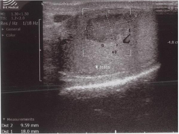
Although ultrasound is the first choice diagnostic tool in evaluating scrotal pain, distinguishing the hypoechogenic lesion from a testicular tumor on ultrasound was difficult in this case. This difficulty in distinguishing between testicular infarction and testicular tumor, especially in small sized lesions, is also reported by other authors.[4,12] STI in adults usually is wedge shaped on ultrasound as is demonstrated by the study of Fernández-Pérez et al.[13]  In our patient, the lesion was oval shaped. Several studies have also reported a round morphology. [5,6,13]
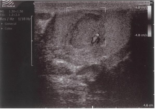
Color Doppler ultrasound showed an avascular central zone with several peri-lesional spots with low colour signals. However, on Color Doppler ultrasound it can be difficult to detect increased blood flow in tumors less than 2 cm in size.[14] In any case, the presence of hypervascularity is not specific for the diagnosis of malignancy, because of the frequent hypovascular pattern of testicular tumors.[15] Therefore, an intratesticular tumor could not be excluded in our patient despite the absence of blood flow in the center of the hypoechogenic lesion.
When a discrepancy between clinical and ultrasonographic findings exists, MRI with contrast may provide additional information over ultrasound, occasionally showing a well-circumscribed hypo-intense area with an enhanced rim, indicating an STI.[13] Nevertheless, definite exclusion of a testicular tumor is not always possible because MRI may demonstrate different findings for different stages of infarction, showing lesions that are lacking the typical features previously mentioned.[16] The use of contrast enhanced ultrasound in the diagnosis of STI seems promising but requires further investigation.[6]
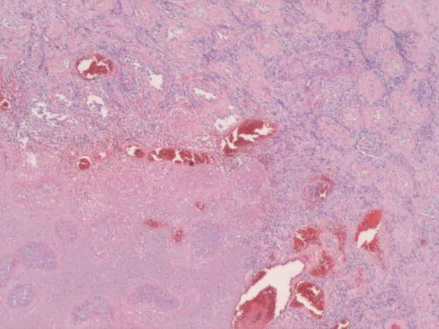
When STI cannot be diagnosed based on the clinical picture and radiologic findings, histological examination is necessary. In most studies STI is histologically confirmed following radical or partial orchiectomy.[4,8,16,19,20] However, in certain cases sparing of the testis is desired and a less invasive way for obtaining a histological confirmation is needed. Perioperative frozen section examination has proven to be accurate for distinguishing benign from malignant lesions, allowing the testis to be spared. In addition, there is no evidence that oncological safety is impaired with this technique.[17] Several studies have showed the feasibility of this method of confirming histology.[1,12,21] In the case of nonpalpable or small hypoechogenic lesions, intraoperative ultrasound guided needle localization with microsurgical exploration has proven to be a safe and effective approach for microsurgical removal of the lesion and obtaining histology. This allows maximal preservation of the testicular parenchyma and its vasculature.[22,23] In this case frozen section examination was not performed, because the patient did not   want to take any risk of the tumor being missed on the frozen section. In addition, because of his age our patient was not troubled by the fact of having one testis for the rest of his life.
As previously mentioned, conservative management has been proven to be possible and safe in highly selected patients where a diagnosis of STI has been made with a high degree of certainty. According to the literature, patients clinically supected to have for STI, with negative tumor markers and Doppler ultrasound and contrast enhanced MRI indicating a STI, are suitable for conservative management with scrotal ultrasound follow-up. Several studies report the development of the ultrasound appearance of STI during follow up ranging from one day to five years.[6,7,9,10] In these studies, follow-up ultrasound demonstrated no progression, or a gradual reduction in size of the lesion, with reflectivity and vascularity essentially unchanged or diminished. This could differentiate STI from a testicular tumor.
In conclusion, differential diagnosis between STI and testicular maligancy remains difficult. STI is therefore mostly detected after (partial) orchiectomy. Some features may suggest this benign condition; a hypovascular wedge-shaped lesion, in patients with normal tumor markers. In highly selected patients,  diagnosis with a high degree of certainty allows conservative treatment with strict follow-up ultrasound. However, if the clinical picture and radiologic imaging are inconclusive or unclear, histologic confirmation cannot be avoided. In that case explorative surgery together with (microsurgical) frozen section examination should be performed, especially in patients in whom sparing of the testis is desired. In the future, further studies are needed to better diagnose this benign condition.


References
 
1) Kim HK, Goske MJ, Bove KE, Minovich E. Segmental testicular infarction in a young man simulating a testicular tumor. Pediatr Radiol. 2009;39:400–402
2) Fukuda S, Takahashi T, Kumori K, Takahashi Y, Yasuda K, Kasai T, Yamaguchi S. Idiopathic testicular infarction in a boy initially suspected to have acute epididymo-orchitis associated with mycoplasma infection and Henoch-Schönlein purpura. J Pediatr Urol. 2009;5:68-71
3) Barber TD, Al-Omar O, Poulik J, McLorie GA. Testicular infarction in a 12-year-old boy with Wegener’s granulomatosis. Urology 2006;67:846.e9-10
4) Ruibal M, Quintana JL, Fernández G, Zungri E. Segmental testicular infarction.
J Urol.2003;170:187-188.
5) Gianfrilli D, Isidori AM, Lenzi A. Segmental testicular ischaemia: presentation, management and follow-up. Int J Androl. 2009;32:524-31
6) Bertolotto M, Derchi LE, Sidhu PS, Serafini G, Valentino M, Grenier N, Cova MA. Acute segmental testicular infarction at contrast-enhanced ultrasound: early features and changes during follow-up. AJR Am J Roentgenol. 2011;196:834-41
7) Bilagi P, Sriprasad S, Clarke JL, Sellars ME, Muir GH, Sidhu PS. Clinical and ultrasound features of segmental testicular infarction: six-year experience from a single centre. Eur Urol. 2007;17:2810-2818
8) Adachi S, Tsutahara K, Kinoshita T, Hatano K, Kinouchi T, Kobayashi M, Inoue H, Takada T, Hara T, Yamaguchi S. Segmental testicular infarction due to cholesterol embolism: not the first case, but the first report. Pathol Int. 2008;58:745-8
9) Madaan S, Joniau S, Klockaerts K, DeWever L, Lerut E, Oyen R, Van Poppel H. Segmental testicular infarction: conservative management is feasible and safe. Eur Urol. 2008;53:441-445
10) Madaan S, Joniau S, Klockaerts K, DeWever L, Lerut E, Oyen R, Van Poppel H. Segmental testicular infarction. Conservative management is feasible and safe: part 2. Eur Urol. 2008;53:656-658
11) Guthrie JA, Fowler RC. Ultrasound diagnosis of testicular tumours presenting as epididymal disease. Clin Radiol. 1992;46:397-400
12) Calcagno C, Gastaldi F. Segmental testicular infarction following herniorrhaphy and varicocelectomy. Urol Int. 2007;79:273-275
13) Fernández-Pérez GC, Tardáguila FM, Velasco M, Rivas C, Dos Santos J, Cambronero J, Trinidad C, San Miguel P. Radiologic findings of segmental testicular infarction. AJR Am J Roentgenol. 2005;184:1587-1593
14) Venugopal S, Schoeman D, Damola A, Hamid B, Powell C. Acute scrotal pain with a twist. Ann R Coll Surg Engl. 2010;92:24-26
15) Horstman WG, Melson GL, Middleton WD, Andriole GL. Testicular tumors: findings with color Doppler US. Radiology 1992;185:733-737
16) Kodama K, Yotsuyanagi S, Fuse H, Hirano S, Kitagawa K, Masuda S. Magnetic resonance imaging to diagnose segmental testicular infarction. J Urol. 2000;163:910–911
17) Kirkham AP, Kumar P, Minhas S, Freeman AA, Ralph DJ, Muneer A, Allen C. Targeted testicular excision biopsy: when and how should we try to avoid radical orchiectomy? Clinic Radiol. 2009;64:1158-1165
18) Sharma SB, Gupta V. Segmental testicular infarction. Indian J Pediatr. 2005;72:81-82
19) Secil M, Kocyigit A, Aslan G, Kefi A, Ozdemir I, Tuna B, Yorukoglu K. Segmental testicular infarction as a complication of varicocelectomy: sonographic findings. J Clin Ultrasound. 2006;34:143-145
20) Flanagan JJ, Fowler RC. Testicular infarction mimicking tumour on scrotal ultrasound – a potential pitfall. Clin Radiol. 1995;50:49-50
21) Hidalgo J, Rodríguez A, Canalias J, Muntané MJ, Huerta MV, Carrasco N, Vesa J. Segmental testicular infarction vs testicular tumour: the usefulness of the excisional frozen biopsy. Arch Esp Urol. 2008;61:92-93
22) Hopps CV, Goldstein M. Ultrasound guided needle localization and microsurgical exploration for incidental nonpalpable testicular tumors. J Urol. 2002;168:1084-1087
23) Kravets FG, Cohen HL, Sheynkin Y, Sukkarieh T. Intraoperative sonographically guided needle localization of nonpalpable testicular tumors. AJM Am J Roentgenol. 2006;186:141-143


Date added to bjui.org: 18/06/2012

DOI: 10.1002/BJUIw-2012-003-web


Report of the management of bilateral, plurifocal sertoli cell adenomas

A twenty two year old male was referred a urology outpatient clinic for the evaluation of persistent scrotal discomfort following a culture proven E. Coli urinary tract infection(UTI). 

Authors: Moran, Diarmaid; Thomas, Arun; Grainger, Ronald
 
Corresponding Author: Diarmaid Moran, St James Hospital, Urology, Dublin, Ireland.   Email: [email protected]

Case Report
 
A twenty two year old male was referred to our urology outpatient clinic for the evaluation of persistent scrotal discomfort following a culture proven E. Coli urinary tract infection(UTI). His initial UTI had been successfully treated by his General Practitioner with oral ciprofloxacin.  The patient had no significant medical or psycho-social history, was sexually inactive and had no family history of note. Other than vague scrotal discomfort that was persistent for six weeks he had no genito-urinary symptoms.
General and abdominal examination were normal. Specifically his height and weight were within the normal range, he had no gynaecomastia, had normally distributed body and pubic hair and had no excessive skin freckling around his head area.  Examination of his scrotum revealed small testes bilaterally. The right testicle had a small, palpable and tender nodule in the interpolar region. Examination of the left testicle was normal.
A  testicular ultrasound demonstrated multiple hypoechoic lesions within both testes (Fig. 1).
 

Figure 1. A testicular ultrasound demonstrated multiple hypoechoic lesions within both testes


The largest lesion (palpable) measured 1 x 0.7cm. There was no associated increase in vascularity and both epididymides were normal in appearance with no ultrasonic features of epididymo-orchitis. The radiological findings were reported a consistent with lymphoma or less likely multifocal seminoma.
A follow-up CT scan of his neck, thorax, abdomen and pelvis demonstrated no evidence of para-aortic lymphadenopathy or metastatic disease. Testicular tumour markers were all within the normal range. The case was discussed both pre and post operatively at our institution’s multi-disciplinary team meeting.
The patient underwent right testicular exploration via an inguinal approach. Intra-operative examination of the right testis revealed a small testis with a 10mm interpolar nodule corresponding to clinical and radiological findings. The nodule was locally excised and sent for frozen section evaluation.  Grossly, this 10 x 10 x 8mm nodule had a glistening outer surface and when sectioned had a smooth, cream surface.  Histological examination showed a benign sertoli cell hamartoma (adenoma) with no evidence of intratubular germ cell neoplasia. Two further ultrasound guided biopsies of the same testis were performed. Repeated frozen section analysis of these separate lesions demonstated atrophic seminiferous tubules with focally prominent Leydig cells, pathologically typical of a cryptorchid testis.


Fig.2. Sertoli cell hamartoma stained with hematoxylin and eosin stain at 20x (A), 100x (B), 200x (C) and 400x (D)and magnification


Both testes were preserved and the patient made an uncomplicated post operative recovery. At a six month review,  all genitourinary symtoms had resolved, with no evidence of testicular atrophy or endocrine dysfunction. Serum testosterone was normal. Follow-up clinical and ultrasonic examination findings of both testes  at 6 weeks and 6 months remained unchanged. The patient performs regular self examination and attends for annual review.


Discussion
 
Testicular sertoli tumours, first described by Teilum in 1944, account for 1% of all testicular neoplasms and 17% of non germ cell tumours. The majority of sertoli tumours are benign but 10% to 22% have malignant potential with less than 50 cases worldwide [1,2].  Five histological variants are recognised; classic sertoli tumour, large cell calcifying,  sclerosing, heterongenous and not otherwise specified (NOS). While sertoli cell tumours were initially reported to present most commonly in the first decade of life,  many of these early descriptions occurred before the recognition of testicular juvenile granulosa tumours in 1979. The dramatic reduction in the reporting of sertoli cell tumours in children since then suggests that many of these early reports may have been incorrectly classified [3]. Sertoli cell tumours may still occur in children, but presentation in the 3rd to 5th decade is more typical.  Sertoli cell tumours occur in both testes equally, usually presenting as a gradually enlarging testicular mass or as an incidental finding on ultrasound. While hormonal abnormalities are infrequent, gynaecomastia is sometimes seen (5% in benign / 12% in malignant tumours) [4]. Only 11 cases of bilateral sertoli cell tumours are reported in the literature[5-15]. One case of bilateral, plurifocal sertoli cell tumours has been documented previously, occuring in a single cryptorchid testes of a 14 year old girl with testicular feminisation syndrome [7]. Other anecdotal reported conditions associated with sertoli cell tumours include cryptorchidism, Peutz-Jaeger syndrome, the Carney complex and the aquired immunodeficiency syndrome [16]. Systemic clinical features of these associated conditions or systemic effects from hormonally active sex cord-stromal tumours may be the presenting feature which prompts the patient (or their parents) to seek medical attention. In this case, apart from the testicular abnormalities described, our patient had a normal clinical examination at presentation with no other systemic features to suggest the presence of an associated syndrome.
As with most testicular tumours, the standard treatment of unilateral sex chord scrotal tumours is radical inguinal orchidectomy. It permits a definitive pathologic diagnosis and provides local tumour treatment. However the European Association of Urology clinical guidelines suggest that testis-sparing surgery may be considered as an alternative treatment modality in certain circumstances provided surgical rules and principles are respected [17]. This form of treatment should be considered in; a tumour in a solitary testes with normal pre-operative testosterone levels, synchronous bilateral testicular tumours, metachronous contralateral tumours, tumour volume of less than 2 cm or less than 30% of the testicular volume, and infertile / monorchid patients. Secondary orchidectomy can always be performed if the final pathology reveals a non-stromal (e.g. germ cell) tumour.
Our case showed no pre or intra-operative evidence of malignancy. Therefore we elected to perform testis sparing surgery. The rationale for pursuing this clinical management course included consideration of the patient’s age (22 years) and the detrimental impact (both physical and psychological) of performing bilateral radical orchidectomies on a patient in this age group. By preserving both testicles, we have maximised his future fertility potential and have preserved endocrine, exocrine and sexual functions of the testis.
Due to the rarity of the condition and the lack of follow-up in most reported cases, the EAU guidelines on testicular cancer [17] does not make specific recommendations regarding the appropriate follow-up for patients with sertoli-cell tumours. However these guidelines do suggest that an individualised follow-up plan, tailored to the clinical needs of the patient, be implemented. Our patient has had a six month surveillance scrotal ultrasound which has demonstrated no change in comparison to his initial examination and will continue to be reviewed at our urology outpatient department on an annual basis.


Conclusion
 
Management of benign but potentially malignant bilateral testicular tumours in a young adult male is challenging. A balance between preserving testicular function and optimizing oncological control has to be considered. Without evidence of malignancy, testicular preserving surgery should be considered. The patient needs to be fully informed regarding the nature of the condition and the management options available. An individualised post intervention surveillance strategy needs to implemented to provide optimal patient care.


References
1. Krege S et al. European consensus conference on diagnosis and treatment of germ cell cancer: a report of the second meeting of the European Germ Cell Cancer Consensus Group (EGCCCG): part II. Eur Urol 2008 Mar; 53 (3):497-513.
2. Souchon R, Schmoll HJ, Krege S, for the German Testicular Cancer Study Group (eds). Qualitätsicherung in der Onkologie. 1st edn. München-Bern-Wien-New York- Zuckschwerdt, 2002. German Cancer Society: Evidence-based guideline for the assessment and treatment of testicular tumours.
3. Young RH, Rosenberg AE, Clement PB. Sertoli cell tumors of the testis, not otherwise specified: a clinicopathologic analysis of 60 cases. Mod Pathol. 1997; 10 (12):1228-32.
4. McGlynn KA, Devesa SS, Sigurdson AJ, Brown LM, Tsao L, Tarone RE. Trends in the incidence of testicular germ cell tumours in the United States. Cancer 2003; 97 (1): 63-70.
5. Brown B, Ram A, Clayton P, Humphrey G. J Pediatr Surg. 2007; 42 (9): 13-5.Conservative management of bilateral Sertoli cell tumors of the testicle in association with the Carney complex: a case report.
6. Baksu A, Kabukcuoglu F, Baksu B, Goker N. Sertoli cell adenoma and serous cyst in a patient with androgen insensitivity syndrome. Eur J Obstet Gynecol Reprod Biol. 2004 10; 114 (1): 104-7.
7. Chatelain D, Ricard J, Ghighi C, Colombat M, Leclercq F, Cordonnier C, Pouzac M, Sevestre H, Gontier MF. Plurifocal testicular hamartomas and testicular feminization syndrome. Ann Pathol. 2000; 20 (6): 605-8.
8. Drevelengas A, Kalaitzoglou I, Destouni E, Skordalaki A, Dimitriadis A. Bilateral Sertoli cell tumor of the testis: MRI and sonographic appearance. Eur Radiol. 1999; 9 (9): 1934.
9. White MD, Loughlin MW, Kallakury BV, Ross JS, Mandell J. Bilateral large cell calcifying Sertoli cell tumor of the testis in a 7-year-old boy. J Urol. 1997 158 (4): 1547-8.
10. Noszian IM, Balon R, Eitelberger FG, Schmid N. Bilateral testicular large-cell calcifying sertoli cell tumor and recurrent cardiac myxoma in a patient with Carney’s complex. Pediatr Radiol. 1995; 25 Suppl. 1: S236-7.
11. Dreyer L, Jacyk WK, du Plessis DJ. Bilateral large-cell calcifying Sertoli cell tumor of the testes with Peutz-Jeghers syndrome: a case report. Pediatr Dermatol. 1994; 11 (4): 335-7.
12. Niewenhuis JC, Wolf MC, Kass EJ. Bilateral asynchronous Sertoli cell tumor in a boy with the Peutz-Jeghers syndrome. J Urol. 1994; 152 (4):1246-8.
13. Schumacher A, Rohde E. A rare case of bilateral Sertoli-Leydig cell tumor. Zentralbl Gynakol. 1991; 113 (23):1323-6.
14. Gutirrez Bos JL, Garijo Ayemsa F, Ladrn Gil C, Sandoval F, Picatoste Pati J. Bilateral Sertoli-Leydig cell tumor with heterologous elements: report of an unusual case and review of the literature. Arch Esp Urol.1987; 40 (7): 523-5.
15. Fligiel Z, Kaneko M, Leiter E.  Bilateral Sertoli cell tumor of testes with feminizing and masculinizing activity occurring in a child. Cancer. 1976; 38 (4): 1853-8.
16. Robert O. Petersen, Isabell A. Sesterhenn, Charles J. Davis. Urologic Pathology. Vol 3, 2009, page 366
17. P. Albers , W. Albrecht, F. Algaba, C. Bokemeyer, G. Cohn-Cedermark, K. Fizazi, A. Horwich, M.P. Laguna. Guidelines on testicular cancer. European Association of Urology (EAU) clinical guidelines 2010.

 
Date added to bjui.org: 16/08/2011 


DOI: 10.1002/BJUIw-2011-047-web


Polyorchidism in children

We report a case of a very rare right-sided polyorchidism in a child, review the reported pediatric cases and discuss the management of this anomaly. 


Authors: Dushi, Gezim; Ramseyer, Pascal; Meyrat, Blaise; Frey, Peter. CHUV, Pediatric Urology, Lausanne, Switzerland.
Corresponding Author: Peter Frey, CHUV, Pediatric Urology, Lausanne, Switzerland.  Email: [email protected]

Introduction
Polyorchidism, a rare anomaly, signifies presence of more than two testes. Since the first case, found on a routine autopsy by Blasius in 1670 [1], further 191 cases in a mixed adult and pediatric population were reported until recently [2]. Most of the cases presented were left sided triorchidism.
After literature review, we could not find any specific analysis of polyorchidism in the pediatric population. We therefore report a case of a very rare right-sided polyorchidism in a child, review the reported pediatric cases and discuss the management of this anomaly.


Case report
A 13 year-old-boy presented at the out-patient clinic for investigation of a painless soft tissue mass in the right scrotum. The ultrasound (fig. 1) revealed two normal homogenous structures, being testes, in the right scrotum. The cranial one was bigger than the caudal and measured 1.8x2x2.7cm compared to the caudal, measuring 1.3×1.4×1.6cm. An epididymis attached to each testis was seen. There was only one testis on the left side, which was normal on ultrasound. MRI (fig.2) showed that the epididymi of each right-sided testis were attached to each other and only one vas deferens was seen. This presentation of polyorchidism is classified according to Leung [3] as type 3, to Singer [4] as type 1, to Thum [5] as type II and to Bergholz [6] as type A3.
The tumor markers alpha-fetoprotein, prostate-specific antigen and beta-human chorionic gonadotropin were negative. There was no surgery performed. Patients were followed-up, every 6 months, clinical examination was uneventful and annual ultrasound was normal 3 years after the diagnosis of polyorchidism. Clinical and ultrasound examination will be further performed in the future.


Figure 1. Ultrasound: Presence of two homogenous testes. The caudal testis has its own epidydimis (circle or arrow)


Figure 2. MRI: Sagittal view T2-weighted. Double testes on the right, sharing the same vas deferens. (arrow)


Literature review
 
Methods
Using the combinations of key words “polyorchidism”,” triorchidism”, “supernumerary testes” “boy”, “child” we searched the electronic databases MEDLINE® and EMBASE®. Histologically proven cases of polyorchidism or cases diagnosed by imaging techniques were included in our study, the latest entry being in February 2011.


Results
47 cases of polyorchidism were reported in children aged from 1 month to 16 years ( mean 6.8 years). Only one case of bilateral testicular duplication was reported.
There is a predominant 83% left-side presentation. 21 (42.5%) were discovered during surgical intervention for testicular maldescent, hernia in three (6.5%), testicular torsion in two (4%), pain in two (4%) cases, hydrocele in one (2%) and scrotal lymphangioma in one (2%) case. In 18 (38%) children the polyorchidism was found during the evaluation of a palpable scrotal mass from which 10 (55%) were operated. Presence of a seminoma and a teratoma was reported in two respective cases (4%).


Discussion
Polyorchidism is a rare congenital malformation. The left-sided predominance encountered in adults  (91.65% of cases) [2] was confirmed in the pediatric population where it was found in 83% of cases. Earlier reports of abnormalities associated with polyorchidism showed an association of 40% with maldescended testis, 30% with hernia, 15% with torsion, 9% with hydrocele and 6% with tumors in a mixed adult and pediatric population [7]. In a more recent meta-analysis by Bergholz [2] the reported percentages were almost equal.
In the pediatric group, however, in the literature this was shown to be true only in the case of maldescended testis (42.5%) and tumors (4%). In all other associated anomalies the percentages were much lower. In children, scrotal pain leading to the discovery of polyorchidism was low (4%) and comparable to the one reported in the mixed population [2].
The reported presence of painless scrotal mass in children was high (38%), which is in accordance with earlier reported results [3] but in contrary to the recent meta-analysis [2] where only 16% of the patients presented with this feature.
With the advances in imaging techniques (US and MRI) the former practice of removing the supernumerary testes has changed. The conservative surveillance of polyorchidism in cases with a normal radiological appearance and negative tumor-markers has started to be accepted, especially in the pediatric population [8-13].
Despite this fact, we found that 55% of cases of polyorchidism in children, diagnosed by ultrasound for a painless scrotal mass, were operated on.
There are mainly two possible classifications of polyorchidism in the literature: the etiological [3,5] and, since the etiology is not clearly understood, the anatomical [4,6] classification was developed.
We propose a rather simple pragmatic approach in children with the aim to preserve their reproductive capacity and to avoid potential operation-related complications.
This management strategy is similar to the one published by Khedis et al [14], however,  it has been simplified and in particular suggests an ultrasound to be performed on a regular and compulsory basis. Also it differs from the strategy suggested by Repetto et al [8], as we do not suggest performing repeated MRI.
In cases of  polyorchidism recognised by the presence of a scrotal mass, and clearly identified by imaging techniques and with negative tumor markers, we propose conservative treatment, performing clinical and ultrasound examination at 6 monthly and yearly intervals respectively.
In cases of  polyorchidism discovered during surgical intervention, we advocate orchidectomy if the supernumerary testis is atrophic, separated from the normal testis, or without connection to the vas deferens. These were formerly classified according to Leung as type 1, to Singer as type 2, to Thum as type I and to Bergholz as type B1 and 2.
In all the other cases, formerly classified according to Leung type 2,3 and 4, to Singer as type 1; to Thum as type II and III, and to Bergholz as type A1,2 and 3 we propose conservative management and clinical follow-up as described above.
Conclusions: The optimal management of polyorchidism is still a matter of debate. After literature review in children we propose whenever possible conservative management, however, these patients should be very closely followed up.


References
 
1.  Ahlfeld F. Die Missbildungen des Menschen. Grunow, Leipzig 1880: 126.
2. Bergholz R, Wenke K. J Urol.  2009  182 : 2422-2427.
3. Leung AK. Am Fam Physician. 1988  38 : 153-156.
4. Singer BR, Donaldson JG, Jackson DS. Urology.  1992  39 : 384-388.
5. Thum G. Polyorchidism: case report and review of literature. J  Urol.  1991 145 : 370-372. 6. Bergholz R, Koch B, Spieker T, Lohse K. Polyorchidism: a case report and classification.J Pediatr Surg.  2007 42 : 1933-1935.
7. Yeniyol CÖ, Nergiz N, Tuna A. Abdominal polyorchidism:A case report and review of the literature. Int Urol Nephrol. 2004  36: 407-408.
8. Repetto P, Ceccarelli P, Bianchini A, Durante V, Biondini D, Cacciari A. Three small testes in left hemiscrotum: a rarer case of polyorchidism. J Pediatr Surg.  2010 45: E21-E23.
9. Savas M, Yeni E Ciftci H, Cece H, Topal U, Utangac MM.  Polyorchidism:  a three-case report and review of literature. Andrologia.  2009  42 : 57-61.
10. Danrad R, Ashker L, Smith W. Polyorchidism: Imaging may denote reproductive potential of accessory testicle. Pediatr Radiol.  2004  34 : 492-494.
11.Hunald FA, Rakototiana AF, Razafimanjato N, Tsiaviry P, Ahmad A, Rantomalala HYH. Un cas de polyorchidie: revue de la littérature.  Arch Pediatr.  2008  15: 1430-1432
12. Bhogal HR, Palit A, Prasad KK. Conservative management of polyorchidism in a young man: a case report and review of literature. Pediatr Surg Int.   2007  23 : 689-691.
13. Park HY, Moon HS. Polyorchidism.  Kor J Urol.  2005 46 : 536-538.
14. Khedis M, Nohra J, Dierickx L et al. Urol Int. 2008 80: 98-101.

 
Date added to bjui.org: 02/08/2011 


DOI: 10.1002/BJUIw-2011-043-web


© 2024 BJU International. All Rights Reserved.