Tag Archive for: multi-parametric MRI

Posts

RE: National implementation of multi-parametric MRI for prostate cancer detection – recommendations from a UK consensus meeting

Letter to the Editor

National implementation of multi-parametric magnetic resonance imaging for prostate cancer detection – recommendations from a UK consensus meeting [1]

Dear Sir,

Appaya et al report on an expert consensus meeting regarding the implementation of multi-parametric magnetic resonance imaging (mpMRI) for prostate cancer detection [1]. A key item related to ‘who can request an mpMRI’ for patients with suspicion of prostate cancer. The panel unanimously agreed that GPs should not be able to. This is perhaps unsurprising, given the panel was composed entirely of specialists. The authors did consider inviting a GP: however, their assumption was that other than for this question a GP would have little to add. We believe this was a critical omission, as improving access to diagnostic testing in primary care could improve prostate cancer diagnosis,  urology outpatient workloads, patient experiences, and outcomes.

The vast majority of cancer diagnoses, including prostate, occur in symptomatic patients presenting to primary care [2]. GPs already have direct access to diagnostic testing for several other cancer types in the NHS – all endorsed by NICE guidance – including gastroscopy, colonoscopy, flexible sigmoidoscopy, MRI head, ultrasound, and CT abdomen. Each can be requested in primary care, with GPs retaining clinical responsibility for the investigation findings [3].

An oft-raised concern with GP direct access to diagnostic tests is that it will lead to inappropriate referrals. However, a recent systematic review of direct access cancer testing in primary care found no significant difference in pooled cancer conversion rate between GP and specialist requests (except for gastroscopy) and no significant difference in the appropriateness of referrals. Time from referral to testing was shorter with GP direct access testing and patient satisfaction was high [4].

If implementation of pre-biopsy mpMRI truly does reduce the need for biopsy in 27% of men, as suggested from the PROMIS [5] and PRECISION [6] trials, then direct access testing in primary care could significantly reduce referrals for suspected prostate cancer. At a time of limited NHS resources and more ambitious targets for cancer diagnosis and treatment times, this could ease the pressure on Urology departments across the UK.

There are several unanswered questions with regard to the optimal use of pre-biopsy mpMRI: for example, which men to test, how to safely follow-up mpMRI-negative patients, and MRI capacity. Including GPs in these discussions would not just be courteous: it may find better answers.

Dr Samuel Merriel1, Dr Fiona Walter2, Prof Willie Hamilton3

Clinical Research Fellow, University of Exeter

2 Principle Researcher in Primary Care Cancer Research, University of Cambridge

3 Professor of Primary Care Diagnostics, University of Exeter

References

  1. Appayya MB, Adshead J, Ahmed HU, Allen C, Bainbridge A, Barrett T, et al. National implementation of multi-parametric magnetic resonance imaging for prostate cancer detection – recommendations from a UK consensus meeting. BJU Int. 2018;122(1):13–25.
  2. Emery JD, Shaw K, Williams B. The role of primary care in early detection and follow-up of cancer. Nat Rev Clin Oncol. 2014;11:38–48.
  3. National Collaborating Centre for Cancer. Suspected cancer [Internet]. NICE. London; 2015.
  4. Smith CF, Tompson AC, Jones N, Brewin J, Spencer EA, Bankhead CR, et al. Direct access cancer testing in primary care: a systematic review of use and clinical outcomes. Br J Gen Pract. 2018;(August):1–10.
  5. Ahmed HU, El-Shater Bosaily A, Brown LC, Gabe R, Kaplan R, Parmar MK, et al. Diagnostic accuracy of multi-parametric MRI and TRUS biopsy in prostate cancer (PROMIS): a paired validating confirmatory study. Lancet [Internet]. 2017;389(10071):815–22.
  6. Kasivisvanathan V, Rannikko AS, Borghi M, Panebianco V, Mynderse LA, Vaarala MH, et al. MRI-Targeted or Standard Biopsy for Prostate-Cancer Diagnosis. N Engl J Med [Internet]. 2018.

Reply by the authors

I thank Dr Samuel Merriel and Dr Fiona Walter for writing in about the role of the GP in requesting prostate MRI studies. Indeed, we reported that our consensus panel unanimously agreed that GPs should not be requesting prostate MRI studies [1]. Their letter in follow-up of this offers an opportunity to explain further this recommendation.

We adopted a UCL-RAND based methodology to conduct the consensus meeting. Whilst we would have liked to have broader representation on the panel, one of the key metrics necessitates that a minimum proportion of participants be able to answer a particular question for that question to be valid. Further broadening of the panel risked delivery of this.

Nonetheless, we agree that GP representation in designing and implementing healthcare is invaluable and should not be ignored and could have all of the benefits for prostate cancer diagnostics that are pointed out by Dr Merriel and Dr Walter.

To clarify the consensus panels discussion on this topic; the panel felt that within the current climate there remained many areas that needed standardisation and improvement if we are to realise the benefits highlighted in PROMIS [2] and PRECISION [3] e.g. diagnostic quality of scans, training of radiologists to report scans all the way through to a consensus from urologists of how to manage patients with a specific scan result.  The panel did not believe that GPs were incapable of managing direct referral services for prostate MRI, only that this needed to be introduced in a controlled fashion if we were to be successful in implementing multi-parametric MRI whilst maintaining its performance and value. The panel felt that direct GP referral should therefore be re-discussed once mechanisms to maintain scan quality and standards of reporting were realised across the UK.

Indeed, GPs should be included in the discussion of which men to test, how to follow-up mp-MRI negative patients and those related to MRI capacity – all of these assume that one is looking at a test that is correctly set-up and reported to a specific standard. Perhaps a consensus on management of patients amongst urologists and primary care physicians is a warranted next step.

Shonit Punwani1

1Centre for Medical Imaging, University College London, UK.

References

  1. Appayya MB, Adshead J, Ahmed HU et al. National implementation of multi-parametric magnetic resonance imaging for prostate cancer detection – recommendations from a UK consensus meeting. BJU Int 2018; 122:13-25.
  2. Ahmed HU, El-Shater Bosaily A, Brown LC, Gabe R, Kaplan R, Parmar MK, et al. Diagnostic accuracy of multi-parametric MRI and TRUS biopsy in prostate cancer (PROMIS): a paired validating confirmatory study. Lancet [Internet]. 2017;389(10071):815–22.
  3. Kasivisvanathan V, Rannikko AS, Borghi M, Panebianco V, Mynderse LA, Vaarala MH, et al. MRI-Targeted or Standard Biopsy for Prostate-Cancer Diagnosis. N Engl J Med [Internet]. 2018.


RE: National implementation of multi-parametric MRI for prostate cancer detection – recommendations from a UK consensus meeting

Letter to the Editor

National implementation of multi-parametric magnetic resonance imaging for prostate cancer detection – recommendations from a UK consensus meeting [1]

Dear Sir,

We congratulate the authors for their efforts in standardising prostate mpMRI. However, we are concerned that the consensus as reported may place substantial pressure on the diagnostic pathway in a rapidly evolving field. Most departments in the West of Scotland and the UK are striving to offer a routine pre-biopsy MRI service, witnessing doubling of the demand on scanning and radiologist time [2], aggravating the nationwide shortage of radiologists [3].

A standard reporting system is vital to the consistent implementation of diagnostic pathways. PI-RADS version 2 focuses on the standardisation of reading, setting out where and when to use each sequence, and has been widely implemented [4]. This, however, has taken time and its learning curve is ongoing within local hospitals [5]. Its main advantage over a clinico-radiological Likert impression is the reduced flexibility of interpretation of radiological parameters, giving those gaining experience in the field a clear set of definitions to work to. We are concerned that enforcing a second system so quickly will discourage radiologists during their learning curves. The panel is entirely correct in stating that even with the use of Likert system, the reporting will be influenced by PI-RADS criteria. While the urology and uro-radiology communities have widely embraced PI-RAD reporting, official switch to Likert reporting may introduce unnecessary reporting subjectivity before individual radiologists are fully experienced with the use of PI-RADS. Furthermore, with the expectation of PI-RADS v3 in the near future, it may be better to adopt PI-RADS v2 now, and compare PI-RADS v3 and Likert system in due course.

The panel describes dynamic contrast enhancement (DCE) as essential component and we agree that mpMRI incorporating the use of contrast is well established, with particular impact on the distinction between PIRADS 3 and 4 lesions. However, a short 9 minute biparameteric MR protocol was as good as a longer and more elaborate protocol using DCE in detecting clinically significant prostate cancer [6]. In NHS Greater Glasgow and Clyde (NHSGGC), we adopted a pragmatic approach, selecting patients for whom contrast may be particularly informative, i.e. those with hip replacement surgery and those who had previous negative biopsies. A contrast MRI prostate scan takes at least 10 minutes longer, and requires nominated radiologist supervision, reducing the overall capacity by 20%. In a 12 month period (2017-2018), 2,333 diagnostic MR prostate scans (20% with contrast) were performed within NHSGGC. For full adoption of contrast prostate MR, it will be necessary to increase the imaging capacity by ~100 supervised imaging lists per annum. Pharmacokinetic modelled DCE parameters (and not visually inspected start of enhancement) have the greatest potential to detect aggressive disease [7,8]. However this approach is currently not deemed feasible for widespread UK use, due to the lack of suitable standardized software.

In summary, we welcome the consensus recommendations for a unified prostate cancer imaging diagnostic strategy, but propose a pragmatic evolving approach that is sympathetic to the constraints of local and regional resources, both in terms of imaging capacity and uro-radiology reporting expertise.

Elizabeth Day1, Amit Patel2, John Morrison2, Thomas Hambrock3, Hing Y Leung1,4

1 Department of Urology, NHS Greater Glasgow and Clyde, Glasgow, G12 0XH

2 Department of Radiology, NHS Greater Glasgow and Clyde, Glasgow, G12 0XH

3 Department of Radiology, The Christie NHS Foundation Trust, Manchester M20 4BX

4 CRUK Beatson Institute for Cancer Research, Glasgow G61 1BD


References

  1. Appayya MB, Adshead J, Ahmed HU et al. National implementation of multi-parametric magnetic resonance imaging for prostate cancer detection – recommendations from a UK consensus meeting. BJU Int 2018; 122:13-25.
  2. Day E, Nalagatla S, Shin JS et al. Triaging patients to primary biopsy or prostate MRI based on digital rectal examination improves the detection rate of TRUS biopsy and avoids unnecessary biopsies. JCU 2018.
  3. The Royal College of Radiologists. Scottish patients at risk from radiologist shortages. 2018.
  4. Padhani AR, Weinreb J, Rosenkrantz AB et al. Prostate Imaging-Reporting and Data System Steering Committee: PI-RADS v2 Status Update and Future Directions. Eur Urol 2018.
  5. Hansen NL, Koo BC, Gallagher FA et al. Comparison of initial and tertiary centre second opinion reads of multiparametric magnetic resonance imaging of the prostate prior to repeat biopsy. Eur Radiol 2017; 27:2259-2266.
  6. Kuhl CK, Bruhn R, Krämer N et al. Abbreviated Biparametric Prostate MR Imaging in Men with Elevated Prostate-specific Antigen. Radiology 2017; 285:493-505.
  7. Vos EK, Litjens GJ, Kobus T et al. Assessment of prostate cancer aggressiveness using dynamic contrast-enhanced magnetic resonance imaging at 3 T. Eur Urol 2013; 64:448-55.
  8. Hambrock T, Vos P, Hulsbergen-van de Kaa C et al. Prostate cancer: computer-aided diagnosis with multiparametric 3-T MR imaging–effect on observer performance. Radiology 2013; 266:521-30

Reply by the authors

I read with great interest the letter composed by Day et al regarding our article: National implementation of multi-parametric magnetic resonance imaging for prostate cancer detection – recommendations from a UK consensus meeting (Appayya et al [1]).

The authors of the letter highlight several important points that were indeed discussed in detail at the consensus meeting.

The first being the pressure on diagnostic services that is expected to result from the implementation of a strategy adopting multi-parametric MRI prior to biopsy. We concur that there is a real risk that without additional funding and the specific training of specialist radiologists in prostate MRI, our aspirations to implement pre-biopsy MR will meet limited success. The consensus panel did agree that this remains an area where national support and prioritisation will be a key driver of success or failure.

The second point raised by Day et al was in regard to recommendation for the use of Likert verses PI-RADS version 2 reporting systems. This again was a well debated item at the consensus meeting. To clarify the consensus discussion, the panel felt that the PI-RADS v2 system was a good system to use when training to report prostate MRI, specifically as it has a very rigid set of definitions. Indeed, the intention of the panel in recommending Likert reporting was not to disregard the PI-RADS system, but to highlight that when sufficiently experienced we also use other factors in scoring that were not as yet incorporated into PI-RADS v2. Indeed, many experienced radiologists knowingly/unknowingly do not adhere to strict PI-RADS v2 scoring. For example, PI-RADS v2 recommends that clinical details (including PSA) should not influence interpretation yet it is almost ubiquitous that we report in light of the PSA and are now recommending PSA density measures to help us guide practice. Furthermore, many centres in the UK do not perform DCE MRI, yet report score as a PI-RADS v2– although DCE MRI as Day et al point out is a key component of the PI-RADS v2 scoring system. Any reports produced for patients that have had previous treatment cannot be scored by PI-RADS v2 criteria, yet there are ongoing examples of radiologists stating a PI-RADS v2 score within such reports.

In reality, Day et al are correct in pointing out that PI-RADS v3 may indeed resolve many of these issues.  PI-RADS v3 was not as far developed when the consensus meeting took place.

With regard to a bi-parametric verses multi-parametric approach; I would agree that most significant tumours will be detected with a bi-parametric MRI and that it will be a small minority that would benefit from DCE MRI. The difficulty to date has been that DCE MRI applications are very varied across studies and have generally concentrated on the use of pharmacokinetic parameters – which has limited studies looking at the value of DCE-MRI. Indeed, such protocols are difficult to themselves apply. The panel felt that a high resolution short DCE protocol that lasted 3 minutes could be implemented with visual inspection of the early arterial phase image for the detection of early enhancement indicative tumour. They also acknowledged that further research is required to establish the benefits of DCE MRI.

I would like to thank Day et al for their letter, as it is important to highlight these areas from the consensus paper in order (i) to recognise where national support is required, (ii) to help clarify any areas which more detailed description could not be provided within the paper, and (iii) to continue to recognise areas where research and further technique refinement is required.

Shonit Punwani1

1Centre for Medical Imaging, University College London, UK.

References

  1. Appayya MB, Adshead J, Ahmed HU et al. National implementation of multi-parametric magnetic resonance imaging for prostate cancer detection – recommendations from a UK consensus meeting. BJU Int 2018; 122:13-25.


Multiparametric MRI – Is the result convincing for AS patients?

Sir,

We read with interest the recent ‘Article of the Month’ by Park et al. in which they concluded that multi-parametric 3T-MRI can be used to predict adverse pathological features and to assess eligibility of patients for active surveillance (AS), in those initially meeting the PRIAS criteria [1]. Nevertheless, we would urge a degree of caution before widespread adoption of this strategy in patient selection for AS.

Firstly, it is accepted that there are false positives with multi-parametric MRI, with the addition of contrast only leading to a minor increase in accuracy, due to increased sensitivity being offset by reduced specificity [2]. Thus, it is imperative to at least make some effort to correlate tumour site on MRI with site on histopathology, which the authors acknowledge was not performed in their study.

Whilst realising that substantial technical difficulties arise in the correlation of imaging with radical prostatectomy specimens, we believe that, as a minimum, tumour side on MRI should be compared to tumour side on histopathology. This is relatively straightforward and could have been performed by Park et al., since 41% of the patients in the study were pathological stage T2a/b.

For example, an audit of 76 patients suitable for AS at our unit (Wirral University Teaching Hospital, UK), but electing for mapping transperineal template guided saturation biopsy, revealed that 53 patients had undergone MRI with diffusion weighted and 23 patients full multi-parametric dynamic contrast enhanced imaging, using a 1.5 T scanner. When analysed without correlation to tumour side the sensitivity was 83%, specificity was 68% and positive predictive value was 79%. However, when analysed with respect to tumour side on MRI with tumour side on histopathology the result becomes 73%, 61%, 79% respectively.

Park et al. concludes that MRI can be used to assess the eligibility of patients with PCa for AS, which is not backed up by their data. With only 11.7% exhibiting no tumour visible on imaging, the investigation will only exclude a relatively small proportion of patients from AS, whereas in the 88.3% with visible cancer on imaging, 50.2% did not have their cancer upgraded and 47.9% had favourable disease on final histology of the whole specimen. Thus, the authors have demonstrated a statistical significant correlation between identification of a lesion on MRI and the risks of upgrading and unfavourable disease, but not demonstrated that multi-parametric 3T MRI is a clinically useful investigation in this setting. In essence, introduction of MP-MRI would be likely to exclude more patients suitable for AS than those with adverse pathology. It seems more likely that it can only be built into a nomogram, rather than a stand-alone assessment tool.

Read the article

Debashis Sarkar*, Nijel J Parr**
*Research Fellow Urology, **Consultant Urologist, Wirral University Teaching Hospital, Upton, UK

Correspondence: Debashis Sarkar, Research Fellow Urology, Wirral University Teaching Hospital,
Upton, UK. e-mail: [email protected]

References

  1. Park BH, Jeon HG, Choo SH et al. Role of multiparametric 3.0-Tesla magnetic resonance imaging in patients with prostate cancer eligible for active surveillance. BJU Int 2014; 113: 864–870
  2. Tanimoto A, Nakashima J, Kohno H, Shinmoto H and Kuribayashi S. Prostate cancer screening: The clinical value of diffusion-weighted imaging and dynamic MR imaging in combination with T2-weighted imaging. J Magn Reson Imaging 2007; 25: 146–152


A novel deformable MR-US registration system

Image-directed, tissue-preserving focal therapy of prostate cancer: a feasibility study of a novel deformable magnetic resonance-ultrasound (MR-US) registration system

Louise Dickinson*, Yipeng Hu, Hashim U. Ahmed*, Clare Allen§, Alex P. Kirkham§, Mark Emberton* and Dean Barratt

Departments of *Urology and §Radiology, University College London Hospitals NHS Foundation Trust, Division of Surgery and Interventional Sciences and Centre for Medical Image Computing and Department of Medical Physics and Bioengineering, Univeristy College London, London, UK

Read the full article
OBJECTIVE

• To evaluate the feasibility of using computer-assisted, deformable image registration software to enable three-dimensional (3D), multi-parametric (mp) magnetic resonance imaging (MRI)-derived information on tumour location and extent, to inform the planning and conduct of focal high-intensity focused ultrasound (HIFU) therapy.

PATIENTS AND METHODS

• A nested pilot study of 26 consecutive men with a visible discrete focus on mpMRI, correlating with positive histology on transperineal template mapping biopsy, who underwent focal HIFU (Sonablate 500®) within a prospective, Ethics Committee-approved multicentre trial (‘INDEX’).

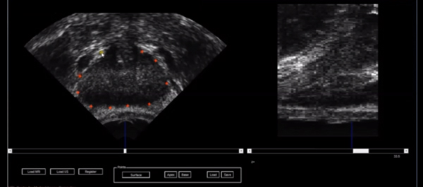
• Non-rigid image registration software developed in our institution was used to transfer data on the location and limits of the index lesion as defined by mpMRI.

• Manual contouring of the prostate capsule and histologically confirmed MR-visible lesion was performed preoperatively by a urologist and uro-radiologist.

• A deformable patient-specific computer model, which captures the location of the target lesion, was automatically generated for each patient and registered to a 3D transrectal ultrasonography (US) volume using a small number (10–20) of manually defined capsule points.

• During the focal HIFU, the urologist could add additional sonications after image-registration if it was felt that the original treatment plan did not cover the lesion sufficiently with a margin.

RESULTS

• Prostate capsule and lesion contouring was achieved in <5 min preoperatively. The mean (range) time taken to register images was 6 (3–16) min.

• Additional treatment sonications were added in 13 of 26 cases leading to a mean (range) additional treatment time of 45 (9–90) s.

CONCLUSION

• Non-rigid MR-US registration is feasible, efficient and can locate lesions on US.

• The process has potential for improved accuracy of focal treatments, and improved diagnostic sampling strategies for prostate cancer.

• Further work on whether deformable MR-US registration impacts on efficacy is required.

© 2024 BJU International. All Rights Reserved.