Tag Archive for: positron-emission tomography

Posts

Article of the week: 68Ga‐PSMA PET/CT predicts complete biochemical response from RP and lymph node dissection in intermediate‐ and high‐risk PCa

Every week, the Editor-in-Chief selects an Article of the Week from the current issue of BJUI. The abstract is reproduced below and you can click on the button to read the full article, which is freely available to all readers for at least 30 days from the time of this post.

In addition to the article itself, there is an editorial written by a prominent member of the urological community. These are intended to provoke comment and discussion and we invite you to use the comment tools at the bottom of each post to join the conversation. 

If you only have time to read one article this month, it should be this one.

Gallium‐68‐prostate‐specific membrane antigen (68Ga‐PSMA) positron emission tomography (PET)/computed tomography (CT) predicts complete biochemical response from radical prostatectomy and lymph node dissection in intermediate‐ and high‐risk prostate cancer


Pim J. van Leeuwen*, Maarten Donswijk, Rohan Nandurkar, Phillip Stricker§¶Bao Ho**, Stijn Heijmink††, Esther M.K. Wit*, Corinne Tillier*, Erik van Muilenkom*, Quoc Nguyen§, Henk G. van der Poel* and Louise Emmett§**


*Department of Urology, Department of Nuclear Medicine, The Netherlands Cancer Institute, Amsterdam, The Netherlands, Faculty of Medicine, University of New South Wales Sydney, §The Australian Prostate Cancer Research Centre-NSW, The Garvan Institute of Medical Research, St Vincents Clinic, **Department of Theranostics and Nuclear Medicine, St Vincents Hospital Sydney, Sydney, New South Wales, Australia and ††Department of Radiology, The Netherlands Cancer Institute, Amsterdam, The Netherlands

Abstract

Objective

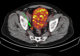
To determine the value of gallium‐68‐prostate‐specific membrane antigen (68Ga‐PSMA)‐11 positron emission tomography (PET) /computed tomography (CT) in men with newly diagnosed prostate cancer.

Patients and methods

We analysed results of 140 men with intermediate‐ and high‐risk prostate cancer. All men underwent 68Ga‐PSMA‐11 PET/CT and multiparametric magnetic resonance imaging (mpMRI) before radical prostatectomy (RP) with extended pelvic lymph node (LN) dissection. For each patient, the clinical and pathological features were recorded. Prostate‐specific antigen (PSA) was documented at staging scan, and after RP, at a median (interquartile range) of 110 (49–132) days. A PSA level of ≥0.03 ng/mL was classified as biochemical persistence (BCP). Logistic regression was performed for association of clinical variables and BCP.

Results

In these 140 patients with intermediate‐ and high‐risk prostate cancer, 27.1% had PSMA PET/CT‐positive findings in the pelvic LNs. Sensitivity and specificity for detection of LN metastases were 53% and 88% (PSMA PET/CT) and 14% and 99% (mpMRI), respectively. The overall BCP rate was 25.7%. The BCP rate was 16.7% in men who were PSMA PET/CT LN‐negative compared to 50% in men who were PSMA PET/CT LN‐positive (P < 0.05). The presence of PSMA‐positive pelvic LNs was more predictive of BCP after RP than cT‐stage, PSA level, and the Gleason score, adjusted for surgical margins status.

Conclusions

68Ga‐PSMA‐11 PET/CT is highly predictive of BCP after RP, and should play an important role informing men with intermediate‐ or high‐risk prostate cancer.


Editorial: Preoperative PSMA‐targeted PET imaging: more than just a tool for prostate cancer staging?

The presence of lymph node metastases at the time of prostate cancer diagnosis has significant implications for treatment. According to current guidelines from the National Comprehensive Cancer Network, men with positive lymph nodes on initial staging imaging should be offered treatment with androgen deprivation (± abiraterone) along with consideration for external beam radiation therapy [1]. In contrast, men with clinically localised high‐ or very‐high‐risk prostate cancer have the option of undergoing radical prostatectomy. Unfortunately, currently available diagnostic imaging modalities (i.e. contrast‐enhanced CT and MRI) fall short in their ability to accurately identify lymph node metastases, which are often small and difficult to discern from other structures within the pelvis. Thus, there exists a conundrum: if we cannot accurately detect lymph node involvement, how can we appropriately manage it?

In this edition of the BJUI, Leeuwen et al. [2] report on the utility of molecular imaging with 68Ga‐PSMA‐11 positron emission tomography (PET)/CT in the preoperative staging of men with prostate cancer. To date, the greatest clinical utility of PSMA‐targeted PET has been in the management of men with biochemically recurrent prostate cancer [3]. In the present study by Leeuwen et al. [2], 140 patients with newly diagnosed intermediate‐ or high‐ risk prostate cancer underwent 68Ga‐PSMA‐11 PET/CT before radical prostatectomy with extended pelvic lymph node dissection. Surgical pathology served as the reference standard to which findings on 68Ga‐PSMA‐11 PET/CT were compared. In total, 27.1% of men were found to have radiotracer uptake in their pelvic lymph nodes, resulting in a sensitivity of 53% and a specificity of 88%. In contrast, multiparametric MRI had a sensitivity of only 14%, albeit with a higher specificity of 99%. These findings are in line with prior studies evaluating the diagnostic performance of PSMA‐targeted PET imaging for preoperative prostate cancer staging [4]. Of greater interest, however, is the authors’ observation that positivity on 68Ga‐PSMA‐11 PET/CT was strongly associated with postoperative PSA persistence (i.e. failure to cure). More specifically, after controlling for Gleason score, surgical margin status, and preoperative PSA level, positivity on PET/CT had an odds ratio of 5.87 (95% CI 1.30–26.59) for biochemical persistence. Furthermore, men with pN1 disease and a positive preoperative PET/CT (i.e. true positives) were over three times more likely to experience biochemical persistence than patients with pN1 disease and negative imaging (71.4% vs 21.4%). Thus, PSMA‐targeted PET not only stands to inform clinical staging, but also has the potential to offer independent prognostic information.

A future line of investigation is to explore the biological basis of the authors’ observation regarding PSMA as a prognostic marker. One explanation is that PET/CT identified men with higher volume lymph node metastases (a known prognostic factor), whilst patients with smaller more curable nodes were negative on imaging. After all, the authors state that the imaging test did not detect any pathologically positive lymph nodes <2 mm. Furthermore, only 27% of positive lymph nodes between 2 and 4 mm showed radiotracer uptake. Unfortunately, the authors did not account for differences in the volume of nodal metastases in their analysis. A second possible explanation for the authors’ observation is that PSMA is upregulated through the same signaling pathways that drive an aggressive prostate cancer phenotype, allowing for PSMA expression to provide prognostic information independent of tumour volume. Indeed, others have previously shown that PSMA expression, as measured by immunohistochemistry, corresponds with increasing tumour grade, stage and risk of biochemical failure [5]. Of course, these concepts are not mutually exclusive and further investigation is needed in order for PSMA‐targeted imaging to be rationally applied as a prognostic test.

References

  1. NCCN Clinical Practice Guidelines in Oncology: Prostate Cancer (Version 4.2018)2018. Accessed November 2018. Available at: https://www.nccn.org/professionals/physician_gls/pdf/prostate.pdf.
  2. Leeuwen, PJDonswijk, MNandurkar, R et al. Gallium‐68‐prostate‐specific membrane antigen (68Ga‐PSMA) positron emission tomography (PET)/computed tomography (CT) predicts complete biochemical response from radical prostatectomy and lymph node dissection in intermediate‐ and high‐risk prostate cancer. BJU Int 201912462– 8
  3. Han, SWoo, SKim, YJSuh, CHImpact of 68Ga‐PSMA PET on the management of patients with prostate cancer: a systematic review and meta‐analysis. Eur Urol 201874179– 90
  4. Gorin, MARowe, SPPatel, HD et al. Prostate specific membrane antigen targeted 18F‐DCFPyL positron emission tomography/computerized tomography for the preoperative staging of high risk prostate cancer: results of a prospective, phase II, single center study. J Urol 2018199126– 32
  5. Minner, SWittmer, CGraefen, M et al. High level PSMA expression is associated with early PSA recurrence in surgically treated prostate cancer. Prostate 201171281– 8


sLND for Prostate Cancer Nodal Recurrence: #urojc September 2014 summary

The September 2014 edition of the International Urology Journal Club (#urojc) returned to familiar territory – prostate cancer. In particular, the discussion focused on salvage lymph node dissection following radical prostatectomy. For the second time (first in July 2014), two journal articles were selected. Both were kindly made available to open access by The Journal of Urology (@JUrology).

The first paper from the Mayo Clinic by Karnes et al., titled ‘Salvage Lymph Node Dissection (sLND) for Prostate Cancer Nodal Recurrence Detected by 11C-Choline Positron Emission Tomography/Computed Tomography (PET/CT)’, reported on a retrospective single-surgeon series of 52 men who underwent salvage lymph node dissection for nodal recurrence post radical prostatectomy. Median follow-up was 20 months. Three-year Biochemical recurrence (BCR)-free survival rate was 45.5% (PSA <0.2). Metastatic/systemic progression-free and cancer-specific survival rates were 46.9% and 92.5% respectively. They concluded that sLND may delay further progression of disease but highlighted the need for randomised controlled trials.

The second paper from German group Tilki et al., titled ‘Salvage Lymph Node Dissection for nodal recurrence of prostate cancer after Radical Prostatectomy’, also reported on a retrospective series of 58 patients who underwent sLND for nodal recurrence on PET/CT post radical prostatectomy. Median follow-up was 39 months. All but 1 patient had BCR. Five-year clinical recurrence-free and cancer-specific survival rates were 35.9% and 71% respectively.  Tilki et al. concluded that while most patients had BCR, sLND may delay ADT and clinical recurrence in selected cases.

A common sentiment shared during the discussion related to the lack of randomised evidence for sLND:

There were some serious concerns about the methodology and results from the two articles:

Discussions quickly shifted away from the two articles to the actual clinical question of sLND in oligometastatic disease and delay to ADT. Matthew Katz provided useful links to the use of stereotactic radiation therapy.

Issues surrounding sLND training and the paradigm shift in recent years were also highlighted:

Opinions were divided on the question of surgical morbidity versus the potential increase in time to ADT:

Pop culture references were in vogue this month. An article by the Mayo Clinic on the 11C-Choline PET scan sparked the linked exchange:

Some take home messages pertained to the uncertainty regarding patient selection and the role of sLND in the broader multidisciplinary arena of prostate cancer treatment:

The winner of the Best Tweet Prize is Brian Chapin (@ChapinMD) for his tweet above.  We thank the Journal of Clinical Urology for supporting this month’s prize by way of a one year electronic subscription to their journal.  We also thank the Journal of Urology for supporting this month’s discussion by way of allowing time limited open access of both articles.

Staying true to form, this month’s edition of #urojc provided a forum for lively international discussion. We look forward to next month’s installment and especially encourage trainees to make use of this excellent educational opportunity.


Isaac Thangasamy is a second year Urology Trainee currently working at the Royal Brisbane and Women’s Hospital, Brisbane, Australia. He is passionate about education and social media. Follow him on Twitter @iThangasamy


Article of the week: How useful is FDG-PET/CT in managing carcinoma invading bladder muscle?

Every week the Editor-in-Chief selects the Article of the Week from the current issue of BJUI. The abstract is reproduced below and you can click on the button to read the full article, which is freely available to all readers for at least 30 days from the time of this post.

In addition to the article itself, there is an accompanying editorial written by a prominent member of the urological community. This blog is intended to provoke comment and discussion and we invite you to use the comment tools at the bottom of each post to join the conversation.

Finally, the third post under the Article of the Week heading on the homepage will consist of additional material or media. This week we feature a video of Ms Mertens and Prof Horenblas discussing their findings.

If you only have time to read one article this week, it should be this one.

Impact of 18F-fluorodeoxyglucose (FDG)-positron-emission tomography/computed tomography (PET/CT) on management of patients with carcinoma invading bladder muscle

Laura S. Mertens, Annemarie Fioole-Bruining*, Erik Vegt, Wouter V. Vogel, Bas W. van Rhijn and Simon Horenblas

Departments of Urology, *Radiology and Nuclear Medicine, The Netherlands Cancer Institute, Antoni van Leeuwenhoek Hospital, Amsterdam, The Netherlands

Read the full article
OBJECTIVE

• To evaluate the clinical impact of 18F-fluorodeoxyglucose (FDG)-positron-emission tomography/computed tomography (PET/CT) scanning, compared with conventional staging with contrast-enhanced CT imaging (CECT).

PATIENTS AND METHODS

• The FDG-PET/CT results of 96 consecutive patients with bladder cancer were analysed. Patients included in this study underwent standard CECT imaging of the chest and abdomen/pelvis <4 weeks before FDG-PET/CT.

• Based on the original imaging reports and recorded tumour stage before and after FDG-PET/CT imaging, the preferred treatment strategies before FDG-PET/CT and after FDG-PET/CT were determined for each patient using an institutional multidisciplinary guideline. One of the following treatment strategies was chosen: (i) local curative treatment; (ii) neoadjuvant/induction chemotherapy; or (iii) palliation.

• The changes in management decisions before and after FDG-PET/CT were assessed.

RESULTS

• The median (range) interval between CECT and FDG-PET/CT was 0 (029) days.

• In 21.9% of the patients, stage on FDG-PET/CT and CECT were different. Upstaging by FDG-PET/CT was more frequent than downstaging (19.8 vs 2.1%).

• Clinical management changed for 13.5% of patients as a result of FDG-PET/CT upstaging. In eight patients, FDG-PET/CT detected second primary tumours. This led to changes of bladder cancer treatment in another four of 96 patients (4.2%).

• All the management changes were validated by tissue confirmation of the additional lesions.

CONCLUSIONS

• FDG-PET/CT provides important additional staging information, which influences the treatment of carcinoma invading bladder muscle in almost 20% of cases.

• Patient selection for neoadjuvant/induction chemotherapy was improved and futile attempts at curative treatment in patients found to have metastases were avoided.


Read Previous Articles of the Week


Editorial: Is FDG-PET/CT ready for prime time?

Fluorodeoxyglucose positron-emission tomography (FDG PET)/computed tomography (CT) in bladder cancer

In this month’s issue Mertens et al. [1] present a retrospective analysis of the clinical impact of fluorodeoxyglucose positron-emission tomography (FDG PET)/CT in 96 patients with muscle-invasive bladder cancer. Muscle invasion is present in ≈30% of patients presenting with bladder cancer and is associated with a higher incidence of nodal and metastatic disease than non-muscle-invasive tumours [2]. Accurate staging in this patient group will influence management decisions to proceed to local therapies, to instigate neoadjuvant treatment before local therapy, or to offer palliative chemotherapy where there is imaging evidence and subsequent confirmation of metastatic disease [2].

While there have been a few previous studies investigating FDG PET or FDG PET/CT for staging bladder cancer [3-7], with reported sensitivities and specificities ranging from 60 to 81% and 67 to 94% respectively, to date there are few data describing the impact on clinical management. A recent FDG PET/CT study of 57 patients with bladder cancer [3] reported that management was changed in 68% of cases after PET suggesting that FDG PET/CT has a substantial impact on the management of these patients. However, most patients in that study underwent FDG PET/CT for a suspected recurrence (72%) and the remainder for initial staging (21%) or post-treatment monitoring (chemotherapy or radiotherapy; 7%); 44% of patients had metastatic disease.

In the study reported by Mertens et al. [1], clinical data obtained in 96 patients during the patients’ clinical pathway were reviewed retrospectively. FDG PET/CT staging with standard contrast-enhanced CT was discordant in 22% of cases (21 patients), where PET/CT predominantly upstaged patients, consistent with the previous reports [3, 4]. After PET/CT, the treatment recommendations changed in 13.5% (13 patients) due to disease upstaging. In seven of the 13 patients treatment recommendations altered from local to palliative, due to the presence of metastatic disease, and in the remaining six of the 13 patients, neoadjuvant treatment was recommended in addition to planned local therapy. In another four patients management changed as a consequence of detecting other incidental primary tumours with FDG PET/CT.

However, the final clinical impact of FDG PET/CT may be less. When actual treatment changes were recorded, in only eight of these 13 patients were the recommendations implemented, due to patient co-morbidity or patient wishes in the remainder, e.g. FDG PET/CT changed actual treatment in only 8% in this study (eight of 96 patients). Including the four patients in whom incidental other primary tumours were discovered, the management impact of FDG PET/CT was 12.5%.

There is no doubt that from current published data and supported by this study by Mertens et al. [1] that FDG PET/CT improves staging in bladder cancer due to its higher sensitivity for metastatic disease. However, the actual change in management is relatively low and more prospective data will be required to confirm its clinical and cost effectiveness in terms of outcome, both in a single and multicentre setting.

Vicky Goh* and Gary Cook*
*Division of Imaging Sciences and Biomedical Engineering, King’s College London, Department of Radiology, and Clinical PET Imaging Centre, Guy’s and St Thomas’ Hospitals NHS Foundation Trust, London, UK

Read the full article

References

  1. Mertens L, Fioole-Bruining A, Vegt E, Vogel W, van Rhijn B, Horenblas S. Impact of 18F-fluorodeoxyglucose (FDG)-positron-emission tomography/computed tomography (PET/CT) on management of patients with carcinoma invading bladder muscle. BJU Int 2013; 112: 729–734
  2. Kaufman DS, Shipley WU, Feldman AS. Bladder cancer. Lancet 2009; 374: 239–249
  3. Apolo AB, Riches J, Schoder H et al. Clinical value of fluorine-18 2-fluoro-2-deoxy-d-glucose positron emission tomography/computed tomography in bladder cancer. J Clin Oncol 2010; 28: 3973–3978
  4. Kibel AS, Dehdashti F, Katz MD et al. Prospective study of [18F] Fluorodeoxyglucose positron emission tomography/computed tomography for staging of muscle-invasive bladder carcinoma. J Clin Oncol 2009; 27: 4314–4320
  5. Anjos DA, Etchebehere EC, Ramos CD, Santos AO, Albertotti C, Camargo EE. 18F-FDG PET/CT delayed images after diuretic for restaging invasive bladder cancer. J Nucl Med 2007; 48: 764–770
  6. Drieskens O, Oyen R, Van Poppel H, Vankan Y, Flamen P, Mortelmans L. FDG-PET for preoperative staging of bladder cancer. Eur J Nucl Med Mol Imaging 2005; 32: 1412–1417
  7. Kosuda S, Kison PV, Greenough R, Grossman HB, Wahl RL. Preliminary assessment of fluorine-18 fluorodeoxyglucose positron emission tomography in patients with bladder cancer. Eur J Nucl Med 1997; 24: 615–620

Video: Upstage, downstage: the spotlight on FDG-PET/CT for managing bladder cancer

Impact of 18F-fluorodeoxyglucose (FDG)-positron-emission tomography/computed tomography (PET/CT) on management of patients with carcinoma invading bladder muscle

Laura S. Mertens, Annemarie Fioole-Bruining*, Erik Vegt, Wouter V. Vogel, Bas W. van Rhijn and Simon Horenblas

Departments of Urology, *Radiology and Nuclear Medicine, The Netherlands Cancer Institute, Antoni van Leeuwenhoek Hospital, Amsterdam, The Netherlands

Read the full article
OBJECTIVE

• To evaluate the clinical impact of 18F-fluorodeoxyglucose (FDG)-positron-emission tomography/computed tomography (PET/CT) scanning, compared with conventional staging with contrast-enhanced CT imaging (CECT).

PATIENTS AND METHODS

• The FDG-PET/CT results of 96 consecutive patients with bladder cancer were analysed. Patients included in this study underwent standard CECT imaging of the chest and abdomen/pelvis <4 weeks before FDG-PET/CT.

• Based on the original imaging reports and recorded tumour stage before and after FDG-PET/CT imaging, the preferred treatment strategies before FDG-PET/CT and after FDG-PET/CT were determined for each patient using an institutional multidisciplinary guideline. One of the following treatment strategies was chosen: (i) local curative treatment; (ii) neoadjuvant/induction chemotherapy; or (iii) palliation.

• The changes in management decisions before and after FDG-PET/CT were assessed.

RESULTS

• The median (range) interval between CECT and FDG-PET/CT was 0 (029) days.

• In 21.9% of the patients, stage on FDG-PET/CT and CECT were different. Upstaging by FDG-PET/CT was more frequent than downstaging (19.8 vs 2.1%).

• Clinical management changed for 13.5% of patients as a result of FDG-PET/CT upstaging. In eight patients, FDG-PET/CT detected second primary tumours. This led to changes of bladder cancer treatment in another four of 96 patients (4.2%).

• All the management changes were validated by tissue confirmation of the additional lesions.

CONCLUSIONS

• FDG-PET/CT provides important additional staging information, which influences the treatment of carcinoma invading bladder muscle in almost 20% of cases.

• Patient selection for neoadjuvant/induction chemotherapy was improved and futile attempts at curative treatment in patients found to have metastases were avoided.

© 2024 BJU International. All Rights Reserved.