Tag Archive for: renal stones

Posts

Residents’ podcast: NICE guidelines – renal and ureteric stones

Nikita Bhatt is a Specialist Trainee in Urology in the East of England Deanery and a BURST Committee member @BURSTUrology

NICE Guideline – Renal and ureteric stones: assessment and management

Context

Renal and ureteric stones usually present as an acute episode with severe pain, although some stones are picked up incidentally during imaging or may present as a history of infection. The initial diagnosis is made by taking a clinical history and examination and carrying out imaging; initial management is with painkillers and treatment of any infection.

Ongoing treatment of renal and ureteric stones depends on the site of the stone and size of the stone (less than 10 mm, 10 to 20 mm, greater than 20 mm; staghorn stones). Options for treatment range from observation with pain relief to surgical intervention. Open surgery is performed very infrequently; most surgical stone management is minimally invasive and the interventions include shockwave lithotripsy (SWL), ureteroscopy (URS) and percutaneous stone removal (surgery). As well as the site and size of the stone, treatment also depends on local facilities and expertise. Most centres have access to SWL, but many use a mobile machine on a sessional basis rather than a fixed‐site machine, which has easier access during the working week. The use of a mobile machine may affect options for emergency treatment, but may also add to waiting times for non‐emergency treatment.

Although URS for renal and ureteric stones is increasing (there has been a 49% increase from 12,062 treatments in 2009/10, to 18,066 in 2014/15 [Hospital Episode Statistics data]), there is a trend towards day‐case/ambulatory care, with this increasing by 10% to 31,000 cases a year between 2010 and 2015. The total number of bed‐days used for renal stone disease has fallen by 15% since 2009/10. However, waiting times for treatment are increasing and this means that patient satisfaction is likely to be lower.

Because the incidence of renal and ureteric stones and the rate of intervention are increasing, there is a need to reduce recurrences through patient education and lifestyle changes. Assessing dietary factors and changing lifestyle have been shown to reduce the number of episodes in people with renal stone disease.

Adults, children and young people using services, their families and carers, and the public will be able to use the guideline to find out more about what NICE recommends, and help them make decisions. These recommendations apply to all settings in which NHS‐commissioned care is provided.


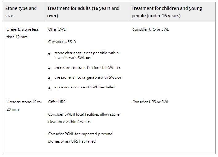
Table 2.Surgical treatment (including SWL) of ureteric stones in adults, children and young people Abbreviations: PCNL, percutaneous nephrolithotomy; SWL, shockwave lithotripsy; URS, ureteroscopy.


BJUI Podcasts now available on iTunes, subscribe here https://itunes.apple.com/gb/podcast/bju-international/id1309570262


NICE Stone Guidelines 2019

The NICE (National Institute For Health And Care Excellence) “Renal and ureteric stones: assessment and management” guideline NG118 was published on-line on Tuesday 8th January 2019 and appeared on the BJUI website on Friday 18th January.

NICE guidelines are based on the best available evidence for the treatment of the specific clinical condition evaluated (i.e. from randomised controlled trials) and aim to provide recommendations that will improve the quality of healthcare within the NHS. As such, the need for a particular guideline is determined by NHS England, and NICE commissions the NGC produce it. The renal and ureteric stone guidelines are comprised a series of evidence reports, each based on the PICO system for a systematic review, covering the breadth of stone management in patients with symptomatic and asymptomatic renal or ureteric stones from initial diagnosis and pain management, through the much debated subject of medical expulsive therapy, to a comprehensive assessment of the surgical treatment of stone disease, including pre- and post- treatment stenting. Follow up imaging, dietary intervention and metabolic investigations have also been reviewed and analysed in detail. These reports are summarised in what is referred to as “The NICE Guideline”, and which is published in the BJUI itself in the February issue (Volume 123, Issue 2, February 2019).  The guideline uses the term “offer” to indicate a strong recommendation with the alternative “consider” to indicate a less robust evidence base, with both terms chosen to highlight the need for patient-centred discussion and shared decision making. Indeed, the preface to The Guideline points out the importance of clinical judgment, and that “the individual needs, preferences and values” of patients should be taken into account in decision making, emphasising that “the guideline does not override the responsibility to make decisions appropriate to the circumstances of the individual”.

We have written these blogs to highlight the individual reports, which can be downloaded from NICE at www.nice.org.uk, and to stimulate some thoughts and comments about their implications for the management of stone patients in the UK and internationally. 

Daron Smith and Jonathan Glass
Institute of Urology, UCH and Guys and St Thomas’ Hospitals
London, January 15th 2019


Daron Smith Commentary

Considering the patient journey to begin with acute ureteric colic, the first recommendation is that a low-dose non-contrast CT should be performed within 24 hours of presentation (unless a child or pregnant) [Evidence Review B, a 73 page document analysing 5224 screened articles, of which 13 were of sufficient quality to be included in the review]. Their pain management should be with NSAIDs as first line pain relief, i.v. paracetamol as second line and opioids as third line, but antispasmodics should not be used [Evidence Review E, a 227 page document for which 1685 articles were screened, of which 38 were of sufficient quality to be included in the review]. Somewhat contentiously for UK practice, given the SUSPEND findings, is that alpha blockers should be considered for patients with distal ureteric stones less than 10 mm [Evidence Review D, a 424 page document for which 1351 articles were screened, of which 71 were of sufficient quality to be included in the review].

As far as stone interventions are concerned, observation was deemed to be reasonable for asymptomatic stones, especially if less than 5mm, that ESWL should be offered for renal stones less than 10mm and PCNL offered for those greater than 20mm with those in between having all options to be considered. Ureteric stones less than 10mm should be offered ESWL (unless unlikely to be cleared within 4 weeks, or contraindicated, or previously failed) whereas ureteric stones larger than 10mm should be offered URS. These conclusions were drawn from 2459 articles of which 66 were of sufficient quality to be included and summarised [Evidence Review F, a 369 page document]. Perhaps the most important aspect for change in practice relate to the use of stents (both before and after treatment) and the timing of definitive intervention (i.e. without a prior temporising JJ stent). Specifically, the guidance recommends patients with uncontrolled pain, or where the stone is deemed unlikely to pass spontaneously, should have definitive treatment within 48 hours [Evidence Review G, a 39 page document based on 3234 screened articles of which 3 were of sufficient quality to be included in the review]. Stents should not be inserted before ESWL for either renal or ureteric stones [Evidence Review H, a78 page document for which 1630 articles were screened, 7 being sufficiently high quality to be included in the review]. Patients who undergo URS for stones less than 20mm should not have a post-operative stent placed as a matter of routine [Evidence Review I, a 107 page document  derived from 1630 screened articles of which 17 were of sufficient quality to be included in the review]. Clearly individual circumstances (ureteric trauma, need for second phase procedure, infection, risk of renal insufficiency) apply to this decision. Given that currently a URS is reimbursed at £2,172, and stent removal as £1,018, perhaps it is time that the treatment episode is remunerated as a combined £3,190, thereby encouraging stent-less procedures instead of stented ones…

Once the treatment is complete, the optimum frequency of follow-up imaging was assessed, comparing monitoring visits less than 6 monthly against 6 monthly and with rapid access/review on request, a strategy that includes no follow up at all for asymptomatic patients [presented in the 29 page Evidence Review J, in which 2385 articles were screened, but none of which were of sufficient quality to be included in the review]. No specific recommendations could therefore be made, other than the need to specifically evaluate the effectiveness of 6 monthly reviews for three years in future research. Of course, if preventative management were more effective, then imaging review would become less important… The guidelines have also reviewed the non-surgical options to avoid stone recurrence [summarised in Evidence Review K – “prevention of recurrence” – a 141 page document in which 3187 articles were screened, of which 19 were of sufficient quality to be included in the review and Evidence Review C, an 81 page document in which 1785 articles were screened, of which 10 were of sufficient quality to be included in the review]. These advised a fluid intake of 2.5 to 3 litres of water per day (with added lemon juice) and that dietary sodium intake should be restricted but calcium intake should not. As far as medical therapy is concerned, potassium citrate and thiazide diuretics should be considered in patients with calcium oxalate stones and hypercalciuria respectively.

In the final aspect of the pathway for stone patients, the clinical and cost effectiveness of metabolic investigations including stone analysis, blood and urine tests (serum calcium and uric acid levels, and urine volume, pH, calcium, oxalate, citrate, sodium, uric acid and cystine) were compared to the outcomes achieved with no metabolic testing following treatment as appropriate for any recurrent stones. Outcomes sought included stone recurrence and need for any intervention, the nature of any metabolic abnormality detected, Quality of life and Adverse events related to the tests or treatment [reported in the 36 page Evidence Review A, in which 933 articles were screened, but which none were of sufficient quality to be reviewed]. A formal research study to evaluate the clinical and cost effectiveness of a full metabolic assessment compared with standard advice alone in people with recurrent calcium oxalate stones was recommended. Following comments in the review process, the guidelines have recommendation that serum calcium should be checked, and biochemical stone analysis considered.

In addition to these individual topic reports, a 49 page evidence review summaries the research methodology and provides an extensive glossary of terms, and a 73 page “Costing analysis of surgical treatments” provides the information regarding the cost effectiveness of the treatments, such as the estimates that 1000 URS procedures and follow up would cost £3,328,895 compared with £961,376 for 1000 ESWL treatments and follow up.

In conclusion, the NICE Guideline Renal and ureteric stones: assessment and management (NG118) is a 33 page summary of over 1700 pages of evidence and analysis. It is therefore an example of where the parts are very much greater than the sum: there is an enormous wealth of high quality data presented in the eleven Evidence Reviews, which are like individual handbooks of contemporary stone management, almost exclusively based on Level 1 Randomised Controlled Trial Evidence. At a time when Brexit dominates national and international news, this is a British Export that we can be proud of.

The real test, of course, will be in the delivery of these ideals, and it is likely that the goal of treating symptomatic patients with ureteric stones within 48 hours will be difficult to achieve. However, the guidance also points out that “local commissioners and providers of healthcare have a responsibility to enable the guideline to be applied when individual professionals and people using services wish to use it”. Along with the GIRFT report, the NICE guidelines are key drivers for change not just in the way that stone patients are managed by their urologist, but in the way that they are treated by the system. Who does not want to be able to treat a patient in pain, with a definitive intervention (be it ESWL or URS) within 48 hours, and without the need for a stent for either the patient or Urologist to worry about. That is the goal that these guidelines have set us; achieving that would be something that Endourologists can be very proud of, and our patients will be extremely grateful for. Are we up for the challenge?

DS
London, January 2019

Jonathan Glass Commentary

The NICE Stone Guidelines – clarification or confusion?

This guideline covers assessing and managing renal and ureteric stones.
It aims
to improve the detection, clearance and prevention of stones, so reducing
pain and
anxiety, and improving quality of  life’.

This is the opening paragraph of the recently produced NICE guidelines on the management of urinary tract stones.  The guidelines have been produced in the context of existing guidelines produced by the European Association of Urology and the American Urological Association pre-existing, and one hoped that these guidelines would add something for the treatment of stone disease in the UK to justify the expenditure spent producing them.  I write these comments in full recognition of the terms of reference to which NICE adheres in producing a set of guidelines.

I, with other members of the committee of the Section of Endourology of BAUS wrote a response to the draft guidelines and we are delighted that the committee has changed some aspects of the published guidelines as a result of our (and other contributions) to the consultation process.  I must record however that what follows is a personal opinion, and not that of the committee.

These guidelines do refer to patients with a single stone.  That of course immediately means that they have limited application to many of our patients who have multiple stones at first presentation.

The draft guidelines, which are in the public domain, stated ‘Do not use opioids’ in the treatment of ureteric colic.  Although this has been changed to ‘Do not offer opioids to adults, children and young people with suspected renal colic unless both NSAIDs and intravenous paracetamol are contraindicated or have not been effective’ this still potentially leaves patient in severe pain for too long.  Our first duty as doctors is to relieve pain.  In my view, as a doctor caring for stone patients but also as an individual who has suffered ureteric colic, if opioids are needed, they should be given in a timely manner.

The recommendations on medical expulsive therapy are unusual at best and arguably a little bizarre and confusing to the British urologist.  There is good evidence from a large UK study – the SUSPEND trial – that alpha blockers have little role to play in improving stone passage.  This is the best level 1 evidence in the use of alpha blockers in stone disease.  The study was sponsored by the NIHR and as such was truly independent, was statistically robust, and randomised.  A representation was made to the guidelines committee by the Aberdeen group that published the study following distribution of the draft guidelines pointing out the robust nature of their study and the less than robust nature of the studies that made up the meta-analysis from which the guideline was derived. I would suggest that this guideline puts British urologists in a situation of huge uncertainty about how we advise our patients in this regard.  Do we tell our patients the best evidence shows one course of action – not to use alpha blockers, but the NICE guidelines suggest another path?  (I am pleased however that the administration of nifedipine, the use of which appeared in the draft guidelines, was removed from the final document).

The recommendation about pre-stenting children with staghorn stones prior to lithotripsy is arguably an historical perspective.  Children with staghorn stones should be considered for primary percutaneous surgery. The recommendation in the guideline possibly reflects review of papers in a field where treatments and approaches to care have changed considerably in the last 10 years.  I recognise that robust level 1A evidence is lacking for these interventions.  It could indeed be argued that a guideline stating ‘consider ESWL, ureteroscopy or PCNL’ for stones 10-20mm and for stones greater than 20mm or staghorn stones is of limited use. Complex patients require bespoke care individualised to the patient in front of the clinician, taking in to account the stone and all other factors with respect to the patient other than the stone.

Suggesting treatment within 48 hours of presentation of patients with ureteric stones including lithotripsy will put urologists under huge pressure.  Patients could hold up these guidelines and demand care.  Treatment within 48 hours is often unnecessary, has huge cost implications, may well be unachievable and could lead to excessive intervention.  To introduce it successfully, given that most stones present to district general hospitals, would suggest that NICE is calling for a lithotripter in every DGH, and in so doing, suggests the death of the mobile lithotripsy service; alternatively it will require the rapid and streamlined transfer of patients to stone centres for intervention.  Either way the cost implications of this are considerable.  I am certainly an advocate for the clinically appropriate timely treatment of stone patients but producing guidelines that are possibly unrealistic and impossible to implement might be considered a missed opportunity.

The recommendation to not offer routine stenting to patients undergoing ureteroscopy is controversial.  As clinicians we understand the symptoms caused by stents.  We also know the risk of sepsis following any stone intervention, the pain from stones obstructing the ureter and the oedema generated by ureteroscopy in the unstented ureter.  Sepsis from urological disease is life threatening. These guidelines allow the legal justification of leaving a ureter unstented post ureteroscopy.  I don’t know and can’t always predict which patients are going to go septic post intervention.  Stents in this scenario save lives but proving that with level 1A evidence is nigh on impossible.  I have concerns that this recommendation is potentially harmful and may be dangerous.  We accept that many patients have interventions and procedures that may appear unnecessary to protect the few where it is life saving.  This is true of nasogastric tubes following major surgery, of patients having a radical prostatectomy, of the placement of the nephrostomy tube following percutaneous surgery.  It is also true of stents after ureteroscopy.

The metabolic considerations are a little odd.  Sending the stone for analysis is only something that should be considered in these guidelines, and yet recommendations are made – based on the stone analysis.  Similarly, there are no recommendations for metabolic testing beyond taking a serum calcium, and yet treatments are recommended for patients with hypocitraturia or hypercalciuria with no suggestion when and in whom these conditions should be sought and diagnosed.

Is this an opportunity lost?  Do these recommendations justify the considerable cost in time and money that NICE has put in?  Are these guidelines potentially harmful – and will they result in the justification of stones not being sent for analysis, the inappropriate use of alpha blockers, obstructed infected kidneys after ureteroscopy and a serum creatinine never being sent.

I have a healthy scepticism for medicine by committee. The MDT discusses treatments for prostate cancer and makes recommendations without the patient being present.  I am not sure this process has relieved me of my scepticism.  ‘This guideline… aims to improve the detection, clearance and prevention of stones, so reducing pain and anxiety, and improving quality of life’.  Read them, and decide for yourselves whether these aims have been met and the expense producing them justified.

JG
London, January 2019


Re: “Super-mini percutaneous nephrolithotomy (SMP) versus retrograde intrarenal surgery for the treatment of 1-2 cm lower-pole renal calculi: an international multicentre randomized controlled trial”

Letter to the Editor

Super-mini percutaneous nephrolithotomy (SMP) versus retrograde intrarenal surgery for the treatment of 1-2 cm lower-pole renal calculi: an international multicenter randomized controlled trial

Dear Sir,

We are interested to read the study by Zeng et al. [1] and appreciate the authors for their study showing that super-mini-percutaneous nephrolithotomy (SMP) was more effective than retrograde intrarenal surgery (RIRS) to treat 1-2 cm lower-pole renal calculi in terms of a better stone-free rate and lesser auxiliary rate. However, some issues still require clarification in this study.

The study validated that SMP was more effective than RIRS to treat 1-2 cm lower-pole renal calculi. However, the results should be explained with caution. As we known, pyelocaliceal anatomy seriously influences the performances of flexible ureteroscopy, while no obvious affection for SMP [2]. This parameter should be evaluated before patients were randomized into RIRS groups. Although there were no significant differences in characteristics of lower-pole spatial anatomical between the two groups, it would be more reasonable and reliable to compare the efficacy between SMP and RIRS, after excluding the patients with unfavorable pyelocaliceal anatomy such as an acute infundibulopelvic angle, or a long lower-pole calyx infundibulum.

The authors set a definition of stone-free status as no residual fragments of ≥0.3 cm on KUB and ultrasound at 1-day and on CT scan at 3-months after operation. We are curious to know how the authors managed when the results were different between KUB and ultrasound. As the primary endpoint was the stone-free rate at 3-months after surgery, we would like to know the size of the residual fragments in the 6.2% (5/80) of patients in SMP and 17.5% (14/80) in RIRS. Do they need another auxiliary procedure? All those information is important for drawing the final conclusion.

The study was an international multicentre, prospective, randomized, non-blinded controlled study, conducted at 10 academic medical centers in China, India, and Turkey. The authors said the two groups had similar baseline characteristics, but we would like to know if it was so for each center. If not, did it affect the final results when exclude those centers?

In conclusion, Zeng et al. made a laudable effort and have provided a level-1 evidence comparing the two modalities for treating 1-2 cm lower-pole renal calculi, but the authors’ conclusion should be taken with caution until more reasonable prospective studies with large sample size have been conducted. We look forward to the authors’ reply to clarify our queries.


Conflict of interest

The authors declare that they have no conflicts of interests.


Huiming Jiang1*, Nanhui Chen1

1Department of Urology, Meizhou People’s Hospital (Huangtang Hospital), Meizhou Hospital Affiliated to Sun Yat-sen University, No.63, Huang Tang Road, Meizhou 514031, Guangdong, People’s Republic of China.


References

  1. Zeng G, Zhang T, Agrawal M et al. (2018) Super-mini percutaneous nephrolithotomy (SMP) vs retrograde intrarenal surgery for the treatment of 1-2 cm lower-pole renal calculi: an international multicentre randomised controlled trial. BJU international. 2018;122(6):1034-1040. doi: 10.1111/bju.14427
  2. Geavlete P, Multescu R, Geavlete B (2008) Influence of pyelocaliceal anatomy on the success of flexible ureteroscopic approach. Journal of endourology 22 (10):2235-2239. doi:10.1089/end.2008.9719


Read the article


Article of the Month: Surgical outcomes of PCNL and results of stone analysis

Every Month the Editor-in-Chief selects an Article of the Month from the current issue of BJUI. The abstract is reproduced below and you can click on the button to read the full article, which is freely available to all readers for at least 30 days from the time of this post.

In addition to the article itself, there is an accompanying editorial written by a prominent member of the urological community. This blog is intended to provoke comment and discussion and we invite you to use the comment tools at the bottom of each post to join the conversation.

If you only have time to read one article this week, it should be this one.

Surgical outcomes of percutaneous nephrolithotomy in 3402 patients and results of stone analysis in 1559 patients

Syed Adibul Hasan Rizvi*, Manzoor Hussain*, Syed Hassan Askari*, Altaf Hashmi*,Murli Lal* and Mirza Naqi Zafar


*Departments of Urology, and Pathology, Sindh Institute of Urology and Transplantation, Civil Hospital, Karachi, Pakistan
Read the full article

Abstract

Objective

To report our experience of a series of percutaneous nephrolithotomy (PCNL) procedures in a single centre over 18 years in terms of patient and stone characteristics, indications, stone clearance and complications, along with the results of chemical analysis of stones in a subgroup.

Patients and Methods

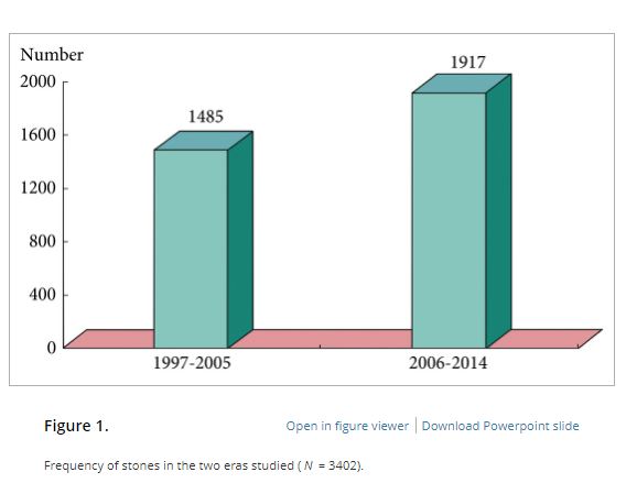
We retrospectively analysed the outcomes of PCNL in 3402 patients, who underwent the procedure between 1997 and 2014, obtained from a prospectively maintained database. Data analysis included patients’ age and sex, laboratory investigations, imaging, punctured calyx, duration of operation, volume of irrigation fluid, radiation exposure time, blood transfusion, complications and stone-free status at 1-month follow-up. For the present analysis, outcomes in relation to complications and success were divided in two eras, 1997–2005 and 2006–2014, to study the differences.

Results

Of the 3402 patients, 2501 (73.5%) were male and 901 (26.5%) were female, giving a male:female ratio of 2.8:1. Staghorn (partial or complete) calculi were found in 27.5% of patients, while 72.5% had non-staghorn calculi. Intracorporeal energy sources used for stone fragmentation included ultrasonography in 917 patients (26.9%), pneumatic lithoclast in 1820 (53.5%), holmium laser in 141 (4.1%) and Lithoclast® master in 524 (15.4%). In the majority of patients (97.4%) a 18–22-F nephrostomy tube was placed after the procedure, while 69 patients (2.03%) underwent tubeless PCNL. The volume of the irrigation fluid used ranged from 7 to 37 L, with a mean of 28.4 L. The stone-free rate after PCNL in the first era studied was 78%, vs 83.2% in the second era, as assessed by combination of ultrasonography and plain abdominal film of the kidney, ureter and bladder. The complication rate in the first era was 21.3% as compared with 10.3% in the second era, and this difference was statistically significant. Stone analysis showed pure stones in 41% and mixed stones in 58% of patients. The majority of stones consisted of calcium oxalate.

Conclusions

This is the largest series of PCNL reported from any single centre in Pakistan, where there is a high prevalence of stone disease associated with infective and obstructive complications, including renal failure. PCNL as a treatment method offers an economic and effective option in the management of renal stone disease with acceptable stone clearance rates in a resource-constrained healthcare system.

Read more articles of the week

Editorial: Management of urolithiasis in South Asia

The article by Rizvi et al. [1] makes a great read. The authors deserve credit for their work and the data presented. A few points merit mention to summarise and put the article in perspective.

First, the authors present a mammoth database from a public sector hospital in Pakistan. In the initial era, as noted by the authors, they adopted extracorporeal shockwave lithotripsy (ESWL) as their mainstay for treating stones. ESWL as the least invasive, safe and readily available method remained the preferred option initially. However, stones seen in South Asia differ from those in the West. In this geographical area, the stone bulk is large and often not amenable to ESWL. In the subsequent period, the authors changed to percutaneous surgery. The reason for this shift, apart from large stone burden, may also have been influenced by local facto required to be travelled by patients to reach a healthcare facility and the lack of resources and infrastructure in remote locations. In such situations, the treatment option that offers rapid, safe, and efficacious results would be preferred. These criteria are fulfilled with the percutaneous approach to renal stones and this is what the authors did!

Second, it is worthwhile noting that that the need for embolisation and/or nephrectomy is a miniscule number in this series [1]. This emphasises the importance of the basic tenet in percutaneous renal surgery that a perfect initial access is the secret to successful percutaneous removal of stones. It should be noted that in this large series the complications across all Clavien–Dindo complication grades reduced as the authors ascended the learning curve.

Third, we feel the major limitation of this study [1] was the means of assessing the stone-free rate. The authors used a combination of ultrasonography and plain abdominal radiograph of the kidneys, ureters and bladder. As acknowledged by the authors this could have possibly overestimated the stone-free rates and skewed the data and interpretation. The authors can substantiate these findings in further prospective studies.

Fourth, the paper exemplifies that stone composition, choice of approach, and patient preferences vary from region to region globally. The findings in the study [1] are similar to the results of Desai et al. [2] from India.

Last but not the least, the AUA guidelines [3] state that the optimal strategy for stone management must take into consideration patient health and economic outcomes. Stone-free requirement is global but economic implications are regional. In this context, the treatment options for similar sized stones may vary for a particular patient located in Europe or Asia. Hence, we feel this paper could be considered as a benchmark for future multicentre trials investigating treatment options and strategies for urolithiasis in South Asia.

Mahesh R. Desai and Arvind P. Ganpule
Department of Urology, Muljibhai Patel Urological Hospital, Nadiad, Gujarat, India


Read the full article


References


1 Rizvi SAHussain MAskari SHHashmi ALal MZafar MN. Surgical outcomes of percutaneous nephrolithotomy in 3402 patients and results of stone analysis in 1559 patients from a single centre in Pakistan. BJUInt 2017; 120: 7029


2 Desai MJain PGanpule ASabnis RPatel SShrivastav PDevelopments in technique and technology: the effect on the results of percutaneous nephrolithotomy for staghorn calculi. BJU Int 2009; 104:5428


3 Assimos DKrambeck AMiller NL et al. Surgical management of stones: American Urological Association/Endourological Society Guideline. Available at: https://www.auanet.org/guidelines/surgical-management-of-stones-(aua/endourological-society-guideline-2016). Accessed August 2017


Article of the Week: Preoperative JJ stent placement to treat ureteric and renal stones

Every Week the Editor-in-Chief selects an Article of the Week from the current issue of BJUI. The abstract is reproduced below and you can click on the button to read the full article, which is freely available to all readers for at least 30 days from the time of this post.

In addition to the article itself, there is an accompanying editorial written by a prominent member of the urological community. This blog is intended to provoke comment and discussion and we invite you to use the comment tools at the bottom of each post to join the conversation.

Finally, the third post under the Article of the Week heading on the homepage will consist of additional material or media. This week we feature a video from Dr. Marianne Schmid and Dr. Atiqullah Aziz, discussing their Editorial.

If you only have time to read one article this week, it should be this one.

Preoperative JJ stent placement in ureteric and renal stone treatment: results from the Clinical Research Office of Endourological Society (CROES) ureteroscopy (URS) Global Study

Dean Assimos, Alfonso Crisci*, Daniel Culkin, Wei Xue, Anita Roelofs§, Mordechai Duvdevani, Mahesh Desai** and Jean de la Rosette†† on behalf of the CROES URS Global Study Group


Department of Urology, University of Alabama at Birmingham School of Medicine, Birmingham, AL, USA, *Department of Urology, Careggi Hospital, Florence, Italy, Department of Urology, The University of Oklahoma Health Sciences Center, Oklahoma City, OK, USA, Department of Urology, Renji Hospital, Shanghai, China, §Department of Urology, Rijnstate Hospital, Arnhem, The Netherlands, ††Department of Urology, AMC University Medical Centre, Amsterdam, The Netherlands, Department of Urology, Hadassah Ein-Kerem University Hospital, Jerusalem, Israel, and **Department of Urology, Muljibhai Patel Urological Hospital, Nadiad, India


Read the full article

Objective

To compare outcomes of ureteric and renal stone treatment with ureteroscopy (URS) in patients with or without the placement of a preoperative JJ stent.

Patients and Methods

The Clinical Research Office of the Endourological Society (CROES) URS Global Study collected prospective data for 1 year on consecutive patients with ureteric or renal stones treated with URS at 114 centres around the world. Patients that had had preoperative JJ stent placement were compared with those that did not. Inverse-probability-weighted regression adjustment (IPWRA) was used to examine the effect of preoperative JJ stent placement on the stone-free rate (SFR), length of hospital stay (LOHS), operative duration, and complications (rate and severity).

Results

Of 8 189 patients with ureteric stones, there were 978 (11.9%) and 7 133 patients with and without a preoperative JJ stent, respectively. Of the 1 622 patients with renal stones, 590 (36.4%) had preoperative stenting and 1 002 did not. For renal stone treatment, preoperative stent placement increased the SFR and operative time, and there was a borderline significant decrease in intraoperative complications. For ureteric stone treatment, preoperative stent placement was associated with longer operative duration and decreased LOHS, but there was no difference in the SFR and complications. One major limitation of the study was that the reason for JJ stent placement was not identified preoperatively.

AprAOTW5

Conclusions

The placement of a preoperative JJ stent increases SFRs and decreases complications in patients with renal stones but not in those with ureteric stones.

Read more articles of the week

Editorial: Some like it safe

Since the implementation of ureteroscopy (URS) about 100 years ago, technological as well as peri-operative management improvements have made URS the treatment of choice for ureteric and renal stones. Depending on stone location and size, stone-free rates of up to 100% have been reported in combination with low peri-operative complications and short hospital stay. Endoscopic therapy of stone disease, e.g. (primary) URS, reflects the zeitgeist: minimally invasive, fast, efficient and economic. There is, however, still a lack of consensus on the question of preoperative stenting in stone management strategies. The underlying aim of preoperative stenting is to cause passive dilation of the ureter, allowing easier access to the upper urinary tract during a secondary URS.

In the current issue of the BJUI, Assimos et al. [1] report higher stone-free rates, as well as fewer complications, with the use of a JJ stent before URS in patients with renal stones, and their article, therefore, supports the previously reported findings of retrospective series with observational, multicentre evidence [2]. Their findings do not, however, corroborate improved outcomes for prestented patients with ureteric stones [1]. Notably, more stents overall were placed in the presence of renal stones compared with ureteric stones (36.4 vs 11.9%). Primary URS may not always be feasible because of anatomical abnormalities, a narrow ureteric lumen, complex ureteric path or previous instrumentation [3]. In such cases, further manipulation risking trauma and potentially long-term stricture formation should be avoided and ureteric stent placement is necessary before further therapy. Indeed, it is possible that renal stone treatment may be associated with increased ureteric manipulation; therefore, passive ureteric dilation might be helpful and facilitate endocopic access to the renal pelvis. A direct elective prestenting in these patients followed by a secondary URS, therefore, warrants discussion. Although prestenting is an additional procedure, it has the potential to lower healthcare costs by decreasing complications rates, operating time and re-operation rates, especially for large proximal stones [4].

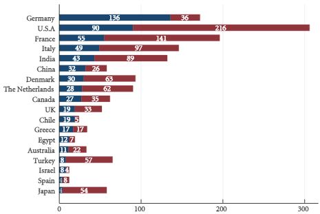
Routine preoperative stenting is not necessarily recommended by current guidelines [5]; however, the management of pre-URS stent placement is left to institutional and international practice patterns. Indeed, as shown in Figs 1 and 2 of the present paper [1], the incidence of preoperative JJ stenting varied tremendously by country. Whereas the large majority of patients with ureteric (88.1%) as well as renal (63.6%) stones were treated without a stent, in Germany, for example, >50% of patients were stented before URS. Also in China, Chile, Egypt and Israel, a higher percentage of patients with ureteric stones primarily received a JJ stent.

The use of stents is known to cause discomfort, pain and urinary symptoms, and therefore can represent significant health problems for the patient. These negatively affect daily activities, work capacity and quality of life [6]. Other disadvantages of a stenting may be higher costs associated with the need for interval procedure(s) and additional hospital stay(s). In addition, as recently reported, preoperative ureteric stent placement was not an independent predictor of stone-free status after flexible URS for renal stone removal [7].

Although there is no consensus or definite recommendation for pre-URS stenting, it should be considered and discussed with the patient when obtaining preoperative consent, especially for purely elective, non-urgent cases and in the presence of renal stones.

Long-term outcomes will show whether or not pre-URS stenting makes a difference with regard to the formation of ureteric strictures. Finally, surgical strategies need to weigh carefully the benefits to the patients and improved outcomes against cost-effectiveness.

Read the full article
Atiqullah Aziz, and Marianne Schmid
Department of Urology, University Medical Centre Hamburg- Eppendorf, Hamburg, Germany


References


3 Cetti RJ, Biers S, Keoghane SR. The difcult ureter: what is the incidence of pre-stenting? Ann R Coll Surg Engl 2011; 93: 313


4 Chu L, Farris CA, Corcoran AT, Averch TD. Preoperative stent placement decreases cost of ureteroscopy. Urology 2011; 78: 30913 


5 Turk C, Petřík A, Sarica K et al. EAU guidelines on interventional treatment for urolithiasis. Eur Urol 2015; doi: 10.1016/j.eururo.2015. 07.041. [Epub ahead of print]


6 Joshi HB, Newns N, Stainthorpe A, MacDonagh RP, Keeley FX JrTimoney AG. Ureteral stent symptom questionnaire: development and validation of a multidimensional quality of life measure. J Urol 2003; 169: 10604


Video: Some like it safe

Preoperative JJ stent placement in ureteric and renal stone treatment: results from the Clinical Research Office of Endourological Society (CROES) ureteroscopy (URS) Global Study

Dean Assimos, Alfonso Crisci*, Daniel Culkin, Wei Xue, Anita Roelofs§, Mordechai Duvdevani, Mahesh Desai** and Jean de la Rosette†† on behalf of the CROES URS Global Study Group


Department of Urology, University of Alabama at Birmingham School of Medicine, Birmingham, AL, USA, *Department of Urology, Careggi Hospital, Florence, Italy, Department of Urology, The University of Oklahoma Health Sciences Center, Oklahoma City, OK, USA, Department of Urology, Renji Hospital, Shanghai, China, §Department of Urology, Rijnstate Hospital, Arnhem, The Netherlands, ††Department of Urology, AMC University Medical Centre, Amsterdam, The Netherlands, Department of Urology, Hadassah Ein-Kerem University Hospital, Jerusalem, Israel, and **Department of Urology, Muljibhai Patel Urological Hospital, Nadiad, India


Read the full article

Objective

To compare outcomes of ureteric and renal stone treatment with ureteroscopy (URS) in patients with or without the placement of a preoperative JJ stent.

Patients and Methods

The Clinical Research Office of the Endourological Society (CROES) URS Global Study collected prospective data for 1 year on consecutive patients with ureteric or renal stones treated with URS at 114 centres around the world. Patients that had had preoperative JJ stent placement were compared with those that did not. Inverse-probability-weighted regression adjustment (IPWRA) was used to examine the effect of preoperative JJ stent placement on the stone-free rate (SFR), length of hospital stay (LOHS), operative duration, and complications (rate and severity).

Results

Of 8 189 patients with ureteric stones, there were 978 (11.9%) and 7 133 patients with and without a preoperative JJ stent, respectively. Of the 1 622 patients with renal stones, 590 (36.4%) had preoperative stenting and 1 002 did not. For renal stone treatment, preoperative stent placement increased the SFR and operative time, and there was a borderline significant decrease in intraoperative complications. For ureteric stone treatment, preoperative stent placement was associated with longer operative duration and decreased LOHS, but there was no difference in the SFR and complications. One major limitation of the study was that the reason for JJ stent placement was not identified preoperatively.

AprAOTW5

Conclusions

The placement of a preoperative JJ stent increases SFRs and decreases complications in patients with renal stones but not in those with ureteric stones.

Read more articles of the week
© 2024 BJU International. All Rights Reserved.