Tag Archive for: systematic biopsy

Posts

Article of the week: A clinical prediction tool to determine the need for concurrent systematic sampling at the time of MRI‐guided biopsy

Every week, the Editor-in-Chief selects an Article of the Week from the current issue of BJUI. The abstract is reproduced below and you can click on the button to read the full article, which is freely available to all readers for at least 30 days from the time of this post.

In addition to the article itself, there is an editorial written by a prominent member of the urological community. These are intended to provoke comment and discussion and we invite you to use the comment tools at the bottom of each post to join the conversation. 

If you only have time to read one article this week, it should be this one.

A clinical prediction tool to determine the need for concurrent systematic sampling at the time of magnetic resonance imaging‐guided biopsy

Niranjan J. Sathianathen*, Christopher A. Warlick*, Christopher J. Weight*, Maria A. Ordonez*, Benjamin Spilseth, Gregory J. Metzger, Paari Muruganand Badrinath R. Konety*


Departments of *Urology, Radiology, and Pathology, University of Minnesota, Minneapolis, MN, USA


Abstract

Objective

To develop a clinical prediction tool that characterises the risk of missing significant prostate cancer by omitting systematic biopsy in men undergoing transrectal ultrasonography/magnetic resonance imaging (TRUS/MRI)‐fusion‐guided biopsy.

Patients and methods

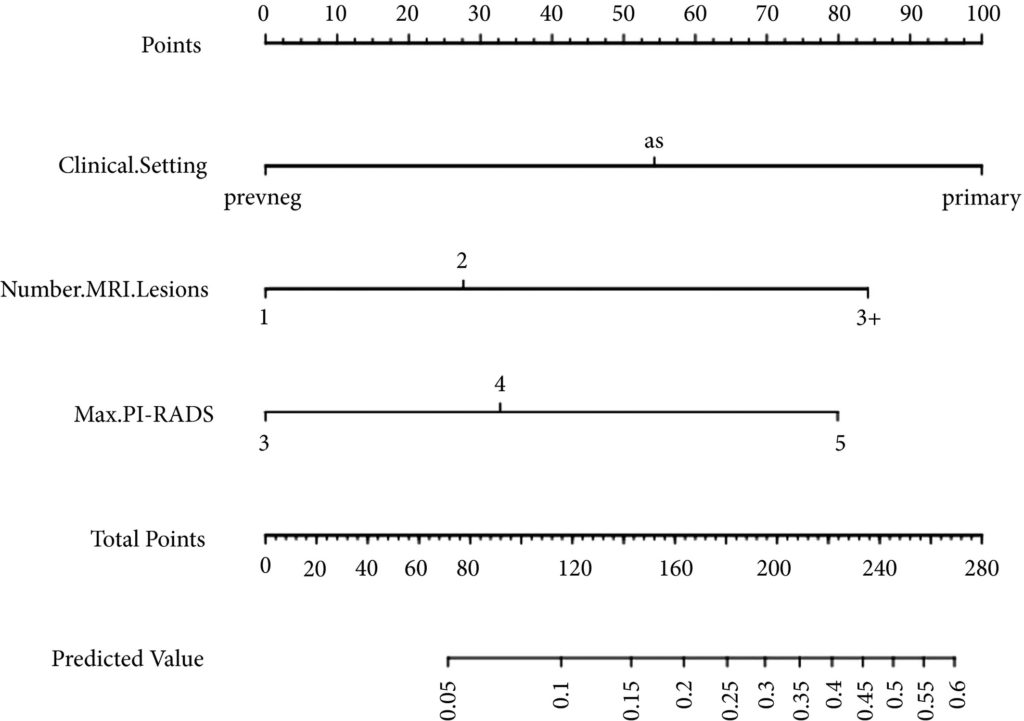
A consecutive sample of men undergoing TRUS/MRI‐fusion‐guided biopsy with the UroNav® system (Invivo International, Best, The Netherlands) who also underwent concurrent systematic biopsy was included. By comparing the grade of cancer diagnosed on targeted and systematic biopsy cores, we identified cases where clinically significant disease (Gleason score ≥3+4) was only found on systematic and not targeted cores. Multivariable logistic regression analyses were used to identify predictive factors for finding significant cancer on systematic cores only. We then used these data to develop a nomogram and evaluated its utility using decision curve analysis.

Fig 1. Nomogram for predicting the diagnosis of clinically significant on systematic biopsy only and missed on targeted biopsy.

Results

Of the 398 men undergoing TRUS/MRI‐fusion‐guided biopsy in our study, there were 46 (11.6%) cases in which clinically significant cancer was missed on targeted biopsy and detected on systematic biopsy. The clinical setting, number of MRI lesions identified, and the highest Prostate Imaging‐Reporting and Data System (PI‐RADS) score of the lesions, were all found to be predictors of this. Our model had a good discriminative ability (concordance index = 0.70). The results from our decision curve analysis show that this model provides a higher net clinical benefit than either biopsying all men or omitting biopsy in all patients when the threshold probability is <30%.

Conclusion

We found that omitting concurrent systematic biopsy in men undergoing TRUS/MRI‐fusion‐guided biopsy would miss significant disease in more than one in 10 patients. We propose a prediction model with good discriminative ability that can be used to improve patient selection for performing concurrent systematic biopsy in order to minimise the number of missed significant cancers. It is important that our model is validated in external cohorts before being employed in routine clinical practice.

Editorial: Can systematic biopsy be safely avoided at the time of MRI/ultrasonography fusion biopsy?

In clinical practice, the need for maximising prostate cancer detection is often balanced against the theoretical risks of infection, bleeding, and pain associated with taking additional cores. In this novel study, Sathianathen et al. [1] provide a tool for measuring the oncological benefit of including concurrent systematic biopsy (SB) at the time of MRI‐guided targeted biopsy (TB). There were several key findings: (i) Amongst patients undergoing MRI‐guided biopsy (all biopsy settings), 11.6% were found to have significant cancers detected by SB alone; (ii) Amongst patients who had clinically significant cancers detected by SB alone, 52.2% were sampled within sextants outside the targeted regions of interest; (iii) According to the proposed nomogram, patients with prior negative biopsies, fewer MRI lesions, and lower Prostate Imaging‐Reporting and Data System (PI‐RADS) scores were at the lowest risk of missing significant cancer when SB was omitted.

Based on the present study, biopsy setting appears to be a key factor for deciding whether to omit SB. In the subset of patients undergoing primary biopsy, the authors found that 18.5% of cancers were detected by SB alone. These results are consistent with those of the MRI‐FIRST trial, which showed 14% of cancers were detected by SB only, 20% by TB only, and 66% by combining both techniques [2]. MRI‐FIRST concluded that in the primary biopsy setting, there was no difference between SB and TB in detection of clinically significant prostate cancer, although combining both techniques provided the highest detection rate.

Prior negative biopsy cohorts are generally at lower risk of harbouring significant cancer, as many cancers have already been ‘selected out’ by initial biopsies. In this setting, TB plays an important role in sampling tumour foci in difficult‐to‐reach regions of the prostate (e.g., anterior and apical) [3]. According to the authors’ nomogram, prior negative biopsy patients were least likely to benefit from concurrent SB. While the authors suggest a paradigm of selectively omitting SB, some authors have proposed omitting both TB and SB altogether in select patients. A previously reported multi‐institutional nomogram can be used to predict benign pathology after MRI‐guided biopsy, which can help reduce the number of unnecessary biopsies after MRI in the prior negative biopsy setting [4]. This clinical tool was further externally validated and optimised by Bjurlin et al. [5].

The ‘active surveillance (AS)’ setting typically refers to a confirmatory MRI‐guided biopsy in men with Grade Group 1 prostate cancer prior to enrollment in AS. Recently, the presence of cribriform morphology in Grade Group 2 patients was confirmed to be a key poor prognostic feature that would exclude patients from AS [6]. The present study, however, did not account for different Gleason pattern 4 morphologies in their analysis, as ‘significant cancer’ was defined by Grade Group alone. Studies by independent groups have found that TB combined with SB was more accurate than either modality alone for detecting cribriform at the time of MRI‐guided biopsy [78]. Therefore, concurrent SB is required to properly sample cribriform cancers in patients who are considering AS.

In this study, Sathianathen et al. [1] provide clinicians with a clinical tool for quantifying the added oncological value of concurrent SB. However, concurrent SB is probably prudent for most patients, particularly for those considering AS or focal therapy for which accurate determination of whole gland grade, cancer volume, and cribriform status are essential. As reducing the number of cores has not yet been shown to reduce biopsy‐related complications, are we willing to suboptimise cancer sampling without proven compensation?

by Matthew Truong

References

  1. Sathianathen, NJWarlick, CAWeight, CJ et al. A clinical prediction tool to determine the need for concurrent systematic sampling at the time of magnetic resonance imaging‐guided biopsy. BJU 2019123612– 7
  2. Salami, SSBen‐Levi, EYaskiv, O et al. In patients with a previous negative prostate biopsy and a suspicious lesion on magnetic resonance imaging, is a 12‐core biopsy still necessary in addition to a targeted biopsy? BJU Int 2015115562– 70
  3. Truong, MWang, BGordetsky, JB et al. Multi‐institutional nomogram predicting benign prostate pathology on magnetic resonance/ultrasound fusion biopsy in men with a prior negative 12‐core systematic biopsy. Cancer 2018124278– 85
  4. Bjurlin, MARenson, ARais‐Bahrami, S et al. Predicting benign prostate pathology on magnetic resonance imaging/ultrasound fusion biopsy in men with a prior negative 12‐core systematic biopsy: external validation of a prognostic nomogram. Eur Urol Focus 2018. [Epub ahead of print] https://doi.org/10.1016/j.euf.2018.05.005
  5. Kweldam, CFKümmerlin, IPNieboer, D et al. Presence of invasive cribriform or intraductal growth at biopsy outperforms percentage grade 4 in predicting outcome of Gleason score 3+4=7 prostate cancer. Mod Pathol 2017301126– 32
  6. Truong, MFeng, CHollenberg, G et al. A comprehensive analysis of cribriform morphology on magnetic resonance imaging/ultrasound fusion biopsy correlated with radical prostatectomy specimens. J Urol 2018199106– 13
  7. Prendeville, SGertner, MMaganti, M et al. Role of magnetic resonance imaging targeted biopsy in detection of prostate cancer harboring adverse pathological features of intraductal carcinoma and invasive cribriform carcinoma. J Urol 2018200104– 13


Article of the Week: Comparison of transperineal mpMRI/fusPbx and sysPbx

Every Week the Editor-in-Chief selects an Article of the Week from the current issue of BJUI. The abstract is reproduced below and you can click on the button to read the full article, which is freely available to all readers for at least 30 days from the time of this post.

In addition to the article itself, there is an accompanying editorial written by a prominent member of the urological community. This blog is intended to provoke comment and discussion and we invite you to use the comment tools at the bottom of each post to join the conversation.

If you only have time to read one article this week, it should be this one.

Prospective comparison of transperineal magnetic resonance imaging/ultrasonography fusion biopsy and transrectal systematic biopsy in biopsy-naïve patients


Angelika Borkowetz*, Boris Hadaschik†‡, Ivan Platzek§, Marieta Toma, Georgi TosevTheresa Renner*, Roman Herout*, Martin Baunacke*, Michael Laniado §, Gustavo Baretton, Jan Philipp Radtke, Claudia Kesch, Markus Hohenfellner† , Michael Froehner*, Heinz-Peter Schlemmer**, Manfred Wirth* and Stefan Zastrow*


*Department of Urology, Technische Universitat Dresden, Dresden, Germany, Department of Urology, University Hospital Heidelberg, Heidelberg, Germany, Department of Urology, University Hospital Essen, Essen, Germany, §Department of Radiology and Interventional Radiology, Technische Universitat Dresden, Dresden, Germany, Department of Pathology, Technische Universitat Dresden, Dresden, Germany, and **Department of Radiology, German Cancer Research Centre (DKFZ), Heidelberg, Germany


Read the full article

Abstract

Objectives

To evaluate the value of multiparametric magnetic resonance imaging (mpMRI) in the detection of significant prostate cancer (PCa) and to compare transperineal MRI/ultrasonography fusion biopsy (fusPbx) with conventional transrectal systematic biopsy (sysPbx) in biopsy-naïve patients.

Patients and Methods

This multicentre, prospective trial investigated biopsy-naïve patients with suspicion of PCa undergoing transperineal fusPbx in combination with transrectal sysPbx (comPbx). The primary outcome was the detection of significant PCa, defined as Gleason pattern 4 or 5. We analysed the results after a study period of 2 years.

Results

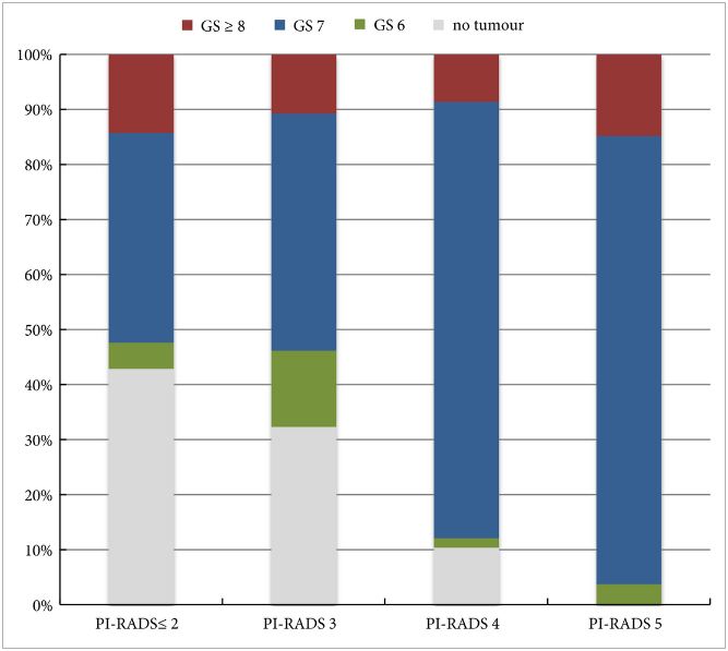
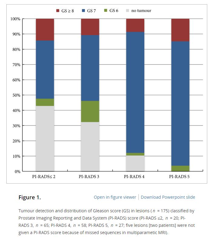
The study included 214 patients. The median (range) number of targeted and systematic cores was 6 (2–15) and 12 (6–18), respectively. The overall PCa detection rate of comPbx was 52%. FusPbx detected more PCa than sysPbx (47% vs 43%; P = 0.15). The detection rate of significant PCa was 38% for fusPbx and 35% for sysPbx (P = 0.296). The rate of missed significant PCa was 14% in fusPbx and 21% in sysPbx. ComPbx detected significantly more significant PCa than fusPbx and sysPbx alone (44% vs 38% vs 35%; P < 0.005). In patients presenting with Prostate Imaging Reporting and Data System (PI-RADS) 4 and 5 lesions there was a higher detection rate of significant PCa than in patients presenting with PI-RADS ≤3 lesions in comPbx (61% vs 14%; P < 0.005).

Conclusions

For biopsy-naïve men with tumour-suspicious lesions in mpMRI, the combined approach outperformed both fusPbx and sysPbx in the detection of overall PCa and significant PCa. Thus, biopsy-naïve patients may benefit from sysPbx in combination with mpMRI targeted fusPbx.

Read more articles of the week

Editorial: The new frontier of prostate biopsy: determining the role of image-guidance in moving the needle

One of the most pressing topics in urological oncology concerns the role of MRI/ultrasonography (US)-fusion guided biopsies in detecting prostate cancer. The literature on this emerging technology is permeated by questions regarding when it should be used, how it should be performed, and which patients stand to benefit. The stakes are high to figure this out, as real patients will continue to suffer from missteps made in diagnosis and treatment of prostate cancer whilst we seek to improve our detection methods.

In this issue of BJUI, Borkowetz et al. [1] compare prostate cancer detection rates between MRI/US-fusion targeted and conventional systematic biopsies. They prospectively enrolled a cohort of biopsy-naïve men who had an elevated PSA level and/or an abnormal DRE. All men received both a transperineal MRI/US-fusion biopsy and a systematic TRUS-guided biopsy. They found that combining both approaches led to improved detection rates for clinically significant and overall prostate cancer compared to either method alone.

A strength of this study [1] is its focus on biopsy-naïve men, for which data on the comparison of biopsy techniques is relatively limited. Siddiqui et al. [2] were instrumental in demonstrating that targeted MRI/US-fusion biopsy, compared to the systematic TRUS biopsy, was associated with improved diagnostic accuracy for higher-risk tumours and decreased detection of low-risk disease. However, the large majority of men in the study had received a prior biopsy, making it difficult to generalise the results to biopsy-naïve men. Focusing on biopsy-naïve men is of great importance – men referred for a biopsy after an elevated PSA level or abnormal DRE increasingly face conflicting opinions about the next best step in diagnostic evaluation, especially given the recent influx of imaging and biomarker tests that aim to guide this decision point.

One group previously compared MRI-guided and systematic TRUS biopsy in a large group of biopsy-naïve of men and found the MRI-guided in-bore technique to be superior in detecting significant cancers and avoiding insignificant cancers [3]. However, that study was limited by both its definition of ‘significant cancer’ (it included Gleason 3 + 3 disease) and the variance in the definition of ‘low-risk’ cancer between MRI-guided and TRUS biopsies. By contrast, the present authors uniformly defined significant cancer as Gleason ≥3 + 4. This is just one example highlighting the widespread disagreement over the parameters used to measure the efficacy of targeted-biopsy techniques in cancer detection. Continued incorporation of standardised scales such as the Prostate Imaging Reporting and Data System (PI-RADS), which was also used in the accompanying article, will help to mitigate some of this disagreement in future studies. Of course, these standardised scales are still subject to inter- and intra-observer variability, which may decrease with further clarification of the grading systems, as well as appropriate reader training [4].

Whilst Borkowetz et al. [1] helped to fill important gaps in the literature, their study was not without limitations. They included PI-RADS 2 scores for targeted biopsies although many urologists would consider these lesions insignificant and not worth targeting. Furthermore, comparing a transperineal MRI/US-fusion with a transrectal systematic approach makes it difficult to separate the true effect of the targeted approach from that of the anatomical approach. Using both methods in each patient also precludes any comparison of complication rates between the biopsy approaches, an important clinical endpoint for both the urologists administering the biopsies and the patients enduring the complications.

Methodology aside, the new frontier of prostate biopsy technique still relies on a basic triad of efficacy: (i) improved accuracy of cancer detection, (ii) reduced complication rates, and (iii) manageable cost and practicality of widespread implementation. For the first tenet, the recently published PROstate MRI Imaging Study (PROMIS) trial cemented the ability of multi-parametric MRI to detect clinically significant prostate cancer with greatly improved sensitivity and negative predictive value over TRUS [5]. The ongoing randomised PRostate Evaluation for Clinically Important Disease: Sampling Using Image-guidance Or Not? (PRECISION) trial will hopefully add to the step taken by Borkowetz et al. [1], by comparing MRI-targeted and systematic TRUS biopsies on the outcomes of accuracy in cancer detection, adverse events, patient health-related quality of life, and cost [6]. Continued investigation in all of these areas will be crucial to guiding how we move the needle in prostate cancer diagnostics.

Sean A. Fletcher, Sebastian Berg and Quoc-Dien Trinh


Division of Urological Surgery, Center for Surgery and Public Health, Brigham and Womens Hospital, Harvard Medical School, Boston, MA, USA


References


1 Borkowetz AHadaschik BPlatzek I et al. Prospective comparison of transperineal magnetic resonance imaging/ultrasonography fusion biopsy and transrectal systematic biopsy in biopsy-naive patients. BJU Int 2018;121: 5360


2 Siddiqui MM, Rais-Bahrami STurkbey B et al. Comparison of MR/ultrasound fusion-guided biopsy with ultrasound-guided biopsy for the diagnosis of prostate cancer. JAMA2015; 313: 3907


3 Pokorny MRde Rooij MDuncan E et al. Prospective study of diagnostic accuracy comparing prostate cancer detection by transrectal ultrasound-guided biopsy versus magnetic resonance (MR) imaging with subsequent MR-guided biopsy in men without previous prostate biopsies. Eur Urol 2014; 66: 229


5 Ahmed HUEl-Shater Bosaily A,Brown LC et al. Diagnostic accuracy of multi-parametric MRI and TRUS biopsy in prostate cancer (PROMIS): a paired validating confirmatory study. Lancet 2017; 389:81522


6 ClinicalTrials.gov. PRostate Evaluation for Clinically Important Disease: Sampling Using Image-guidance or Not? (PRECISION), Identification No. NCT02380027 (2017). Available at: https://clinicaltrials.gov/ct2/show/NCT02380027. Accessed October 2017.


Article of the Month: Combined mpMRI Fusion and Systematic Biopsies Predict the Final Tumour Grading after RP

Every Month the Editor-in-Chief selects an Article of the Month from the current issue of BJUI. The abstract is reproduced below and you can click on the button to read the full article, which is freely available to all readers for at least 30 days from the time of this post.

In addition to the article itself, there is an accompanying editorial written by a prominent member of the urological community. This blog is intended to provoke comment and discussion and we invite you to use the comment tools at the bottom of each post to join the conversation.

Finally, the third post under the Article of the Week heading on the homepage will consist of additional material or media. This week we feature a video from Angelika Borkowetz, discussing her paper.

If you only have time to read one article this week, it should be this one.

Direct comparison of multiparametric magnetic resonance imaging (MRI) results with final histopathology in patients with proven prostate cancer in MRI/ultrasonography-fusion biopsy

Angelika Borkowetz*, Ivan Platzek, Marieta Toma, Theresa Renner*, Roman Herout*, Martin Baunacke*, Michael Laniado, Gustavo Baretton, Michael Froehner*, Stefan Zastrow* and Manfred Wirth*


*Department of Urology, Department of Radiology and Interventional Radiology, and
Department of Pathology, Technische Universitat Dresden, Dresden, Germany


Read the full article

Objective

To compare multiparametric magnetic resonance imaging (mpMRI) of the prostate and histological findings of both targeted MRI/ultrasonography-fusion prostate biopsy (PBx) and systematic PBx with final histology of the radical prostatectomy (RP) specimen.

Patients and Methods

A total of 105 patients with prostate cancer (PCa) histopathologically proven using a combination of fusion Pbx and systematic PBx, who underwent RP, were investigated. All patients had been examined using mpMRI, applying the European Society of Urogenital Radiology criteria. Histological findings from the RP specimen were compared with those from the PBx. Whole-mount RP specimen and mpMRI results were directly compared by a uro-pathologist and a uro-radiologist in step-section analysis.

AugAOTM1

Results

In the 105 patients with histopathologically proven PCa by combination of fusion PBx and systematic PBx, the detection rate of PCa was 90% (94/105) in fusion PBx alone and 68% (72/105) in systematic PBx alone (P = 0.001). The combination PBx detected 23 (22%) Gleason score (GS) 6, 69 (66%) GS 7 and 13 (12%) GS ≥8 tumours. Fusion PBx alone detected 25 (26%) GS 6, 57 (61%) GS 7 and 12 (13%) GS ≥8 tumours. Systematic PBx alone detected 17 (24%) GS 6, 49 (68%) GS 7 and 6 (8%) GS ≥8 tumours. Fusion PBx alone would have missed 11 tumours (4% [4/105] of GS 6, 6% [6/105] of GS 7 and 1% [1/105] of GS ≥8 tumours). Systematic PBx alone would have missed 33 tumours (10% [10/105] of GS 6, 20% [21/105] of GS 7 and 2% [2/105] of GS ≥8 tumours). The rates of concordance with regard to GS between the PBx and RP specimen were 63% (n = 65), 54% (n = 56) and 75% (n = 78) in fusion, systematic and combination PBx (fusion and systematic PBx combined), respectively. Upgrading of the GS between PBx and RP specimen occurred in 33% (n = 34), 44% (n = 46) and 18% (n = 19) in fusion, systematic and combination PBx, respectively. γ-correlation for detection of any cancer was 0.76 for combination PBx, 0.68 for fusion PBx alone and 0.23 for systematic PBx alone. In all, 84% (n = 88) of index tumours were identified by mpMRI; 86% (n = 91) of index lesions on the mpMRI were proven in the RP specimen.

Conclusions

Fusion PBx of tumour-suspicious lesions on mpMRI was associated with a higher detection rate of more aggressive PCa and a better tumour prediction in final histopathology than systematic PBx alone; however, combination PBx had the best concordance for the prediction of GS. Furthermore, the additional findings of systematic PBx reflect the multifocality of PCa, therefore, the combination of both biopsy methods would still represent the best approach for the prediction of the final tumour grading in PCa.

Read more articles of the week

Editorial: Role of systematic biopsy in the era of mpMRI and US fusion guidance

The success of multiparametric MRI (mpMRI) and MRI/ultrasound (US) fusion-guided biopsies in improving the detection of prostate cancer in patients with occult disease (elevated PSA level with prior negative biopsies) and optimising the detection of clinically significant cancer has been reported by centres that have served as early adopters of these techniques [1, 2]. Technological advances in MRI and associated imaging protocols, as well as increased clinical experience with MRI interpretation have led to increased prospective detection and characterisation of clinically significant prostate cancer. This, in conjunction with increasing experience with MRI/US fusion-guided prostate biopsy techniques, has led to the re-evaluation of the contributory role and utility of systematic template US-guided prostate biopsies in the diagnosis of prostate cancer. It is an attractive proposition to forego the systematic biopsy when performing MRI-directed fusion biopsy, as this would minimise the duration, morbidity, and overall cost of the biopsy procedure and post-biopsy pathology processing. However, before adopting this approach, it is important to first consider the potential possibility of missing clinically significant cancer diagnoses when relying on the targeted biopsy cores in isolation.

In this issue of BJUI, Borkowetz et al. [3] report their results of biopsy histological yields on systematic biopsies compared with MRI/US fusion biopsies in their series of patients who underwent radical prostatectomy (RP). These results corroborate previously reported comparisons of fusion biopsy of suspicious lesions on MRI performed concurrently with systematic biopsy, consistently showing an improved detection of both overall prostate cancer foci and, more importantly, an improved detection of clinically significant higher grade cancer foci [1, 2]. It is important to note that the overall detection rate and detection rate for clinically significant prostate cancer was highest when fusion and systematic biopsies were evaluated in conjunction with each other. Another important factor to consider when evaluating the utility and value of these biopsy techniques is the concordance of the pathology of the biopsy specimen with the final pathology of the RP specimen, the ‘gold standard’. The concordance of Gleason grade assigned on targeted fusion-biopsy cores and RP outperformed that of systematic biopsy cores and RP. This, in essence, suggests that targeted biopsy can perform as well, and likely better, than the systematic biopsy approach of sampling the prostate with a systematic-sextant approach, which has been the long standing standard of care for the diagnosis of prostate cancer. Again, it is important to note that the greatest concordance in this study was achieved when the results of the fusion and systematic biopsy cores were combined.

The question now arises regarding the ‘cost’ for the incremental improvement in cancer detection provided by the combination of both MRI-directed fusion biopsy and the systematic biopsy approach. The improved negative predictive value parallels the increased sensitivity for cancer detection by having a larger sampling of the prostate by augmenting the number of biopsy cores sampled and submitted for histopathological evaluation. The area under the curve for detection of clinically significant cancer reported by Borkowetz et al. [3] was not improved by adding systematic biopsies to the targeted biopsies. However, this experience described a mixed population of patients, most of whom had undergone prior prostate biopsy with benign pathology. This creates an enriched population who likely harbours prostate cancers that are more occult to the systematic biopsy approach, thus improving the diagnostic yield of MRI-directed biopsies even further. This is concordant with the work presented by Mendhiratta et al. [4], where systematic biopsies added little to the diagnosis of clinically significant prostate cancer in a population of men undergoing MRI/US fusion-guided biopsy after prior cancer-negative biopsy sessions.

Alternatively, current datasets for biopsy naïve patients have not shown the same degree of convincingly improved detection with targeted biopsies over systematic biopsies. In fact, Delongchamps et al. [5] recently reported a slightly lower rate of overall cancer detection with fusion-guided targeted biopsies vs systematic biopsy cores; however, the difference in detection of clinically significant prostate cancer was not statistically significant. Further study of the role of targeted biopsy in the biopsy naïve patient population is warranted, as there is suggestion that cancer detection efficiency per needle core is significantly improved with MRI-directed biopsies over systematic biopsies [6]. Alternatively, in patients with prior negative systematic biopsies and continued clinical suspicion for prostate cancer, a repeat biopsy session with targeted cores alone may be appropriate, particularly as these patients have previously undergone standard-of-care, extended sextant biopsy.

Read the full article
Jason A. Pietryga* and Soroush Rais-Bahrami*,
*Department of Radiology, and Department of Urology, University of Alabama at Birmingham, Birmingham, AL, USA


References


Video: Combined mpMRI Fusion and Systematic Biopsies Predict the Final Tumour Grading after RP

Direct comparison of multiparametric magnetic resonance imaging (MRI) results with final histopathology in patients with proven prostate cancer in MRI/ultrasonography-fusion biopsy

Angelika Borkowetz*, Ivan Platzek, Marieta Toma, Theresa Renner*, Roman Herout*, Martin Baunacke*, Michael Laniado, Gustavo Baretton, Michael Froehner*, Stefan Zastrow* and Manfred Wirth*


*Department of Urology, Department of Radiology and Interventional Radiology, and
Department of Pathology, Technische Universitat Dresden, Dresden, Germany


Read the full article

Objective

To compare multiparametric magnetic resonance imaging (mpMRI) of the prostate and histological findings of both targeted MRI/ultrasonography-fusion prostate biopsy (PBx) and systematic PBx with final histology of the radical prostatectomy (RP) specimen.

Patients and Methods

A total of 105 patients with prostate cancer (PCa) histopathologically proven using a combination of fusion Pbx and systematic PBx, who underwent RP, were investigated. All patients had been examined using mpMRI, applying the European Society of Urogenital Radiology criteria. Histological findings from the RP specimen were compared with those from the PBx. Whole-mount RP specimen and mpMRI results were directly compared by a uro-pathologist and a uro-radiologist in step-section analysis.

AugAOTM1

Results

In the 105 patients with histopathologically proven PCa by combination of fusion PBx and systematic PBx, the detection rate of PCa was 90% (94/105) in fusion PBx alone and 68% (72/105) in systematic PBx alone (P = 0.001). The combination PBx detected 23 (22%) Gleason score (GS) 6, 69 (66%) GS 7 and 13 (12%) GS ≥8 tumours. Fusion PBx alone detected 25 (26%) GS 6, 57 (61%) GS 7 and 12 (13%) GS ≥8 tumours. Systematic PBx alone detected 17 (24%) GS 6, 49 (68%) GS 7 and 6 (8%) GS ≥8 tumours. Fusion PBx alone would have missed 11 tumours (4% [4/105] of GS 6, 6% [6/105] of GS 7 and 1% [1/105] of GS ≥8 tumours). Systematic PBx alone would have missed 33 tumours (10% [10/105] of GS 6, 20% [21/105] of GS 7 and 2% [2/105] of GS ≥8 tumours). The rates of concordance with regard to GS between the PBx and RP specimen were 63% (n = 65), 54% (n = 56) and 75% (n = 78) in fusion, systematic and combination PBx (fusion and systematic PBx combined), respectively. Upgrading of the GS between PBx and RP specimen occurred in 33% (n = 34), 44% (n = 46) and 18% (n = 19) in fusion, systematic and combination PBx, respectively. γ-correlation for detection of any cancer was 0.76 for combination PBx, 0.68 for fusion PBx alone and 0.23 for systematic PBx alone. In all, 84% (n = 88) of index tumours were identified by mpMRI; 86% (n = 91) of index lesions on the mpMRI were proven in the RP specimen.

Conclusions

Fusion PBx of tumour-suspicious lesions on mpMRI was associated with a higher detection rate of more aggressive PCa and a better tumour prediction in final histopathology than systematic PBx alone; however, combination PBx had the best concordance for the prediction of GS. Furthermore, the additional findings of systematic PBx reflect the multifocality of PCa, therefore, the combination of both biopsy methods would still represent the best approach for the prediction of the final tumour grading in PCa.

Read more articles of the week
© 2024 BJU International. All Rights Reserved.