Tag Archive for: adenocarcinoma

Posts

Is Gleason 6 really cancer?

The recently published Viewpoint of the National Cancer Institute working group on “Overdiagnosis and Overtreatment in Cancer” by Esserman and colleagues [1] raises continued discussion as to whether some lesions currently classified as carcinomas should have the designation of “cancer” removed, based on low rates of progression, death, and other adverse outcomes. Pertinent to those interested in urology, a central example in the article is prostatic adenocarcinoma.

One simple answer to this question is that to a small extent, a subgroup of prostatic lesions has already been reclassified as not cancer: In current practice, needle biopsy or radical prostatectomy specimens with an overall Gleason score (GS) of 5 or less are now quite rare in current practice. This shift is due in part to modern updates to the Gleason grading system [2], under which many tumors now reach thresholds for GS6 or above. However, at least some lesions previously considered adenocarcinoma with a low overall GS would now be categorized as atypical adenomatous hyperplasia or adenosis in the era of immunohistochemistry for markers of prostatic basal cells. Nonetheless, the current and more controversial debate surrounds whether some (or all?) tumors currently classified as GS6 could be recategorized as not “cancer”.

Arguments against removing the cancer designation from some prostatic adenocarcinomas:

A major difficulty from the pathologic standpoint in adopting a non-cancer nomenclature for some tumors (such as GS6 adenocarcinomas) is that the Gleason pattern 3 component of a GS 3+3=6 tumor (small, round prostatic glands that lack a basal cell layer and infiltrate between benign glands) is for all intents and purposes identical to the Gleason pattern 3 component of a GS 3+4=7 or higher prostate cancer. These similarities are not limited exclusively to the microscopic appearance but also include a number of immunohistochemical and molecular features, as summarized in a recent article addressing this question [3]. Therefore, no pathologic features are as yet defined that ideally predict whether Gleason pattern 3 glands in a biopsy specimen represent a pure GS6 tumor or a component of higher-grade tumor in which the high-grade component is not represented. Not surprisingly, it is not unusual for tumors with GS6 on needle biopsy to be upgraded to GS7 at radical prostatectomy [3], particularly when a high tumor volume is present in the needle biopsy.
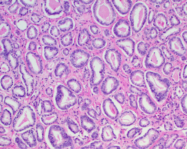
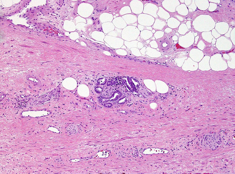
Gleason pattern 3 glands from a GS7 tumor, identical to those of a GS6 tumor.

To compare to other cancers with low risk of aggressive behavior, basal cell carcinoma and squamous cell carcinoma of the skin similarly show locally infiltrative properties, supporting their classification as carcinomas by a classical pathologic definition. Despite that the word “carcinoma” continues to be used for these tumors, most patients are not concerned that they have a life-threatening disease and these lesions are even excluded from the American Cancer Society statistics regarding cancers [4]. In the same way, Gleason pattern 3 glands exhibit infiltrative growth by extending between benign glands, invading nerves, and sometimes extending outside of the prostate. This difference in mindset regarding some types of “cancers” could be considered supportive evidence for the assertion in the recent Melbourne Consensus Statement that uncoupling prostate cancer diagnosis from intervention may be more appropriate than removing its “cancer” nomenclature.


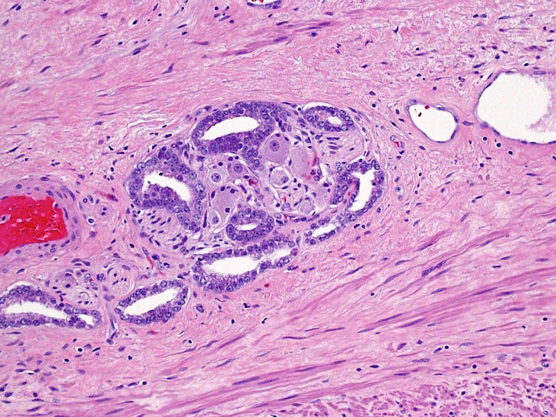
This small GS6 adenocarcinoma was an incidental finding in a radical cystoprostatectomy specimen for bladder cancer but surprisingly extended into periprostatic fat via this focus of perineural invasion.

Supporting removal of the cancer designation from some prostatic adenocarcinomas:

A valid argument of the NCI Viewpoint is that a neoplasm should have a substantive rate of progression and patient death if it is to be considered a cancer. Likewise, others have questioned whether low-volume GS6 tumors fulfill other molecular and pathogenetic hallmarks of cancer, such as unlimited replicative potential and other features [5].

In general, benign and malignant neoplasms can be regarded as having some prototypical gross and microscopic pathologic characteristics, such as a circumscribed vs infiltrative growth and homogeneous vs pleomorphic cell population. However, differentiating benign from malignant lesions also relies heavily on parameters specific to the organ involved. Clear cell renal cell carcinoma, another genitourinary tract tumor, often does not possess these prototypical features of malignancy. Tumors often form a well-circumscribed mass without an “invasive” growth pattern and they often are composed of a uniform population of cells. However, based on known behavior of these tumors, their status as a malignancy is not in doubt. Conversely, renal oncocytoma is a benign neoplasm that shares some of these general features (a round mass composed of a homogeneous population of renal tubular cells). Occasionally oncocytomas appear infiltrative by extending into the perinephric fat or renal vein, yet their status as benign is also not the subject of debate. If some prostate cancers do not have a substantial likelihood of resulting in progression and death, they may not meet an important criterion for a diagnosis of cancer, despite that other features, such as infiltration of tissues, invasion of nerves, and loss of the basal cell layer are characteristic of a malignant neoplasm.

Since a diagnosis of GS6 by needle biopsy is not always predictive of a radical prostatectomy overall GS6, a major challenge to such an approach would be to determine where such a cutoff could be drawn between “cancer” and “not cancer” [5]. If based on tumor volume, it would be difficult to conceptualize that a small amount of GS6 glands would be regarded as a benign lesion, whereas a large amount of identical glands would represent a malignant lesion. Alternatively, the presence of Gleason pattern 4 could used as the point of differentiation (GS7 or above). In the endometrium, a disorganized proliferation of crowded glands with some cytologic features of cancer is regarded as complex atypical hyperplasia. Diagnosis of adenocarcinoma is then reserved for proliferations with a confluent growth of these glands, similar to the threshold for recognizing a component of cribriform glands as Gleason pattern 4. A limitation to such an approach, however, is that a substantial fraction of patients with a needle biopsy GS6 are upgraded to GS7 at radical prostatectomy, as discussed above. Likewise, the ability to treat and monitor GS6 adenocarcinoma nonsurgically is not quite analogous to that of endometrial hyperplasia.

Higher magnification of image 2 shows Gleason pattern 3 glands invading a nerve with ganglion cells.

Other points of discussion

The NCI Viewpoint also suggests that high-grade prostatic intraepithelial neoplasia (HGPIN) no longer be considered cancer or even neoplasia.  A comparison to ductal carcinoma in situ (DCIS) of the breast for this argument is somewhat flawed, as HGPIN neither contains the word “carcinoma” nor is justification for treatment in and of itself. Its status as a risk factor for a future cancer even remains debated. The proposal to remove “neoplasia” from HGPIN is also a confusing one, particularly as cervical cancer is noted as an example of the successful application of screening, in which “cervical intraepithelial neoplasia” is the preferred term for precancerous lesions. The authors suggest the designation “indolent lesions of epithelial origin” (IDLE) for cancers in this category to convey their low likelihood of aggressive behavior. However, would recognizing the status of these lesions as at least premalignant neoplasms be more appropriate?

Likely a typographical error in the Viewpoint is that the authors also cite reclassification of urothelial papilloma as papillary urothelial neoplasm of low malignant potential [1]. Since urothelial papilloma has never been considered a malignant neoplasm, the authors likely meant reclassifying “grade 1 urothelial carcinoma” to papillary urothelial neoplasm of low malignant potential.

References
[1]        Esserman LJ, Thompson IM, Reid B. Overdiagnosis and Overtreatment in Cancer: An Opportunity for Improvement. JAMA. 2013 Jul 29:

[2]        Epstein JI, Allsbrook WC, Jr., Amin MB, Egevad LL. The 2005 International Society of Urological Pathology (ISUP) Consensus Conference on Gleason Grading of Prostatic Carcinoma. Am J Surg Pathol. 2005 Sep: 29:1228-42

[3]        Carter HB, Partin AW, Walsh PC, et al. Gleason score 6 adenocarcinoma: should it be labeled as cancer? J Clin Oncol. 2012 Dec 10: 30:4294-6

[4]        Siegel R, Naishadham D, Jemal A. Cancer statistics, 2013. CA Cancer J Clin. 2013 Jan: 63:11-30

[5]        Ahmed HU, Arya M, Freeman A, Emberton M. Do low-grade and low-volume prostate cancers bear the hallmarks of malignancy? Lancet Oncol. 2012 Nov: 13:e509-17


Sean Williamson is Senior Staff Pathologist in the Department of Pathology and Laboratory Medicine, Henry Ford Health System, Detroit MI, USA. @Williamson_SR

Concurrence of squamous cell carcinoma, sarcomatoid carcinoma and adenocarcinoma in relapsed prostate cancer originated from adenocarcinoma

Squamous cell carcinoma (SqCC) and sarcomatoid carcinoma (SC) are rare subtypes of prostate cancer. We report a rare case with concurrence of SqCC, SC and adenocarcinoma in a relapsed tumour originated from adenocarcinoma.

Authors: Ruiying Diao1,2, Kin Lam Fok3, Zhongfu Zhang1,2, Li Zhao2,4, Lisha Mou1,2,5, Shuolei Sun1,2, Lijun Zhou1,2 and Zhiming Cai2,6
1Department of Urology, Peking University Shenzhen Hospital, Shenzhen, China
2Guangdong Key Laboratory of Male Reproductive Medicine and Genetics, Peking University Shenzhen Hospital, Shenzhen, China
3Epithelial Cell Biology Research Center, Department of Physiology, School of Biomedical Sciences, Faculty of Medicine, Chinese University of Hong Kong, Hong Kong, China
4Department of Laboratory Medicine, Peking University Shenzhen Hospital, Shenzhen, China
5Institute of Urology, Shenzhen PKU-HKUST Medical Center, Shenzhen, China
6Department of Urology, Second People’s Hospital of Shenzhen, Shenzhen, China

Corresponding Author: Zhiming, Cai. Guangdong Key Laboratory of Male Reproductive Medicine and Genetics, Peking University Shenzhen Hospital, and Department of Urology, Second People’s Hospital of Shenzhen, Shenzhen, China. E-mail: [email protected]


Abstract
Squamous cell carcinoma (SqCC) and sarcomatoid carcinoma (SC) are rare subtypes of prostate cancer. We report a rare case with concurrence of SqCC, SC and adenocarcinoma in a relapsed tumour originated from adenocarcinoma. A 62-year-old man diagnosed with prostate adenocarcinoma (Gleason score 5+4=9)  received androgen blockade (AB) treatment and 89Sr radiotherapy. An increase in total prostate-specific antigen level was noted 13 months after treatment. Pathological analyses on biopsies from transurethral resection of the prostate revealed the concomitance of SqCC and SC with adenocarcinoma (Gleason score 5+5=10). Expression of the epithelial markers E-cadherin and β-catenin were significantly down-regulated, while the mesenchymal marker vimentin was up-regulated in both SqCC and SC. The expression of androgen receptor (AR) was down-regulated in SqCC but elevated in SC. The altered epithelial and mesenchymal markers and the heterogeneous AR expression in the relapsed tumour suggest that the concurrence of unusual subtypes may arise from the epithelial-to-mesenchymal transition and/or the differential function of AR on prostatic epithelial and stromal cells. The present study raises concerns about antiandrogen therapy regimen for prostate cancer.

Introduction
Prostate cancer is the most common malignant tumour in men > 70 years old and the morbidity of prostate cancer has been markedly increasing in recent years [1, 2]. Prostatic squamous cell carcinoma (SqCC) and sarcomatoid (SC) are rare subtypes of prostate cancer that account for 0.5–1% and < 0.1% of all prostate tumours, respectively [3-5]. The prognosis of patients with SqCC and SC is usually poor. Understanding the origin and initiation of SqCC and SC may benefit the development of an effective therapeutic regimen. SqCC and SC have mainly been observed after endocrine therapy or radiotherapy [4]. It has been postulated that the selection pressure from endocrine therapy is one of the driving forces for clonal selection in SqCC, but the exact mechanism underlying the occurrence of SqCC and SC de novo is not yet understood. The present study reports a rare pathological differentiation from initial adenocarcinoma to the concomitance of three kinds of pathological subtypes, adenocarcinoma, SqCC and SC. The expression of androgen receptor (AR), epithelial markers E-cadherin and β-catenin, and the mesenchymal marker vimentin in specimens before and after therapy were examined to evaluate their possible involvement in the transformation of pathological phenotypes.

Case Report
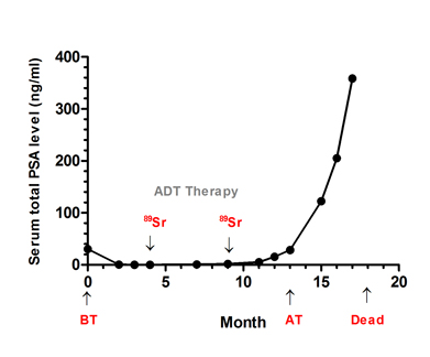
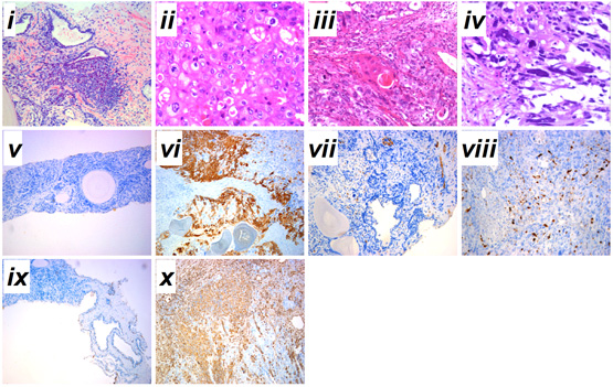
A 62-year-old patient was admitted to our hospital on 3 February 2010 with a 36-month history of progressive LUTS. Before these symptoms, there had been no previous history of urinary tract disease. His PSA level during admission was 30.5 ng/mL (Fig. 1A). The initial pathological analysis from needle biopsy led to a diagnosis of moderately differentiated adenocarcinoma with small acinar infiltration and proliferation mainly centered in the peri-acinar region (Gleason score 5+4=9, FIG. 1B, i). A bone scan revealed widespread metastases. The patient was treated with androgen deprivation therapy for 10 months as the initial regimen. During this period, 89Sr radionuclide therapy against prostate cancer was used twice, once at 4 months and once at 9 months, and the patient’s total PSA level was initially suppressed but was then elevated at a later stage (FIG. 1A). TURP was undertaken for pathological analysis at 13 months because of the increase in total PSA level (FIG. 1A). Specimens were fixed intact in 4% formalin and then sectioned transversally at regular intervals for random sampling (10 points). Pathological diagnosis showed concurrent adenocarcinoma, SqCC and SC (Gleason score 5+5=10). A 55% area of the TURP specimen showed the pathological structure of adenocarcinoma with abundant clear cytoplasm and enlarged nucleolus (FIG. 1B, ii). An area of ~25% of the TURP specimen showed the differentiation of SqCC with individual cell keratinization (cytokeratin high molecular weight; FIG. 1B, vi), intercellular bridges, and/or keratin pearl formation (FIG. 1B, iii). An area of nearly 20% of the TURP specimen showed bizarre atypia with giant cells and a tumour-induced osteoclastic phenotype with S-100 positive (a marker for osteosarcoma) in SC (FIG. 1B, iv). From the 13th month to time of death, serum total PSA increased rapidly to nearly 400ng/mL (FIG. 1A).
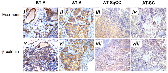
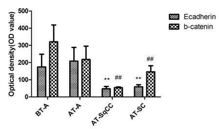
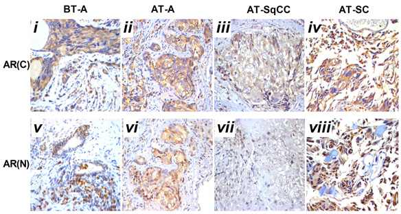
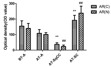
Compared with samples from the needle biopsy, the intensities of AR (FIG. 2B), and E-cadherin and β-catenin (FIG. 3B) immunoreactivity were all significantly lowered in SqCC. Similar to the pattern in the SqCC, the expression of E-cadherin and β-catenin in specimens after therapy was also lower in SC compared with those before therapy. The expression of the mesenchymal marker vimentin was increased after therapy (FIG. 1B, x), but the expression of AR was elevated in SC (FIG. 2B, iv, viii). Moreover, the AR C-terminal ligand-binding domain was mainly located in the cytoplasm of osteosarcomatous cells (FIG. 2B, iv), while the AR N-terminal transcription activation-binding domain was mainly located in the nucleus of osteosarcomatous components (FIG. 2B, viii).

12-036FIG.1A12-036FIG.1B


Fig. 1 Serum total PSA levels and diagnosis of prostatic cancer before and after therapy. A, Serum total PSA levels from admission to death. B, (i) prostatic adenocarcinoma from prostate needle biopsy before therapy; (ii) prostatic adenocarcinoma from TURP after therapy; (iii) prostatic sqCC from TURP after therapy; (iv) SC after the TURP after therapy; immunohistochemical staining for (v–vi) cytokeratin high molecular weight, (vii-viii) S-100 and (ix-x) vimentin expressions before and after therapy. Haematoxylin-eosin stain, x200. Admission time was set as 0 month. ADT, androgen deprivation therapy; 89Sr, 89Sr radiation; BT, before therapy; AT, after therapy. i-iv, scale bar=50 μm; v-x, scale bar=200 μm.

12-036FIG.2A12-036FIG.2B


Fig. 2 Immunohistochemical staining for AR(C) and AR(N) in the adenocarcinoma, SqCC, and SC specimens before and after therapy. A, Immunohistochemical staining for AR(C) and AR(N) in the adenocarcinoma, SqCC, and SC specimens before and after therapy (i–viii). B, Statistical analysis for immunohistochemical staining of AR.
Note: AR(C), AR C-terminal ligand-binding domain; AR(N): AR N-terminal transcription activation-binding domain. Magnification, X200. BT, before therapy; AT, after therapy, **, compared with BT-adenocarcinoma group (BT-A, AR[C]), P<0.05; ##, compared with BT-adenocarcinoma group (BT-A, AR[N]), P<0.05. i-x, scale bar=50 μm.

12-036FIG.3A12-036FIG.3B


Fig. 3 Immunohistochemical staining for E-cadherin and β-catenin expressions before and after therapy . A, Immunohistochemical staining for E-cadherin and β-catenin expression before and after therapy (i-viii). B, Statistical analysis for immunohistochemical staining of E-cadherin and β-catenin. Magnification, X200. BT, before therapy; AT, after therapy; **, compared with BT-adenocarcinoma group (BT-A, E-cadherin), P<0.05; ##, compared with BT-adenocarcinoma group (BT-A, β-catenin), P

Discussion
Sarcomatoid carcinoma and SqCC are unusual histological prostatic tumours with a low incidence rate. About half of them can arise from patients with initial acinar adenocarcinoma after endocrine therapy or radiotherapy [4], but the mechanism underlying the occurrence of histological variants of prostate carcinoma remains unknown. In the present case, the concurrence of SqCC and SC was observed in a relapsed tumour originated from adenocarcinoma. The characteristics of the unusual concurrence of multiple carcinoma may provide some clues as to the pathogenesis of histological variants of prostate cancer.
Serum PSA level is the main indicator for estimating the prognosis of prostate cancer [6]. It has been reported that PSA can regulate and transactivate AR expression in prostate cancer cells [7]. During the initial phase of antiandrogen therapy, serum PSA concentration decreased, then rapidly increased at a later stage (FIG. 1A), indicating insensitivity to the hormone therapy [8]. Pathological analysis of the specimens from the needle biopsies suggested that primary multifocal adenocarcinoma tumour occurred both in the right and left lobes. Even with androgen deprivation therapy and radionuclide therapy, the total PSA level of the patient rose gradually from the 13th month (FIG. 1A), therefore, TURP was undertaken for more precise pathological analysis. Intra-operative localization in the TURP specimen indicated that extensive multifocal carrion-like tissues were found in the the middle, left and right lobes of the prostate. Samples from all the three lobes were found to be tumours, based on histological features and immunohistochemical evidence of epithelial and sarcomatoid-like differentiation, including vimentin (FIG. 1B, x) and PSA (FIG. 1A).
Histological analysis of the relapsed tumour found adenocarcinoma, SqCC and SC and revealed a lower AR expression in the area of SqCC, but a significantly higher AR expression in the SC region. Furthermore, the AR C-terminal domain was mainly localized in the cytoplasm of giant cells and tumour-induced osteoclastic cells in the SC, while the AR N-terminal transcription activation-binding domain was mainly in the nucleus of the above cell types. AR has been shown to exert dual functions in prostate cancer proliferation and metastasis. On the one hand, it acts as a suppressor in prostate epithelium, on the other hand, it promotes proliferation in stroma [9, 10]; therefore, the differential expression of AR in relapsed tumour may contribute to the differential response and insensitivity to antiandrogen therapy in the patient [11].
A number of studies have shown that adenocarcinoma can undergo the epithelial-to-mesenchymal transition (EMT) in order to migrate and invade other tissues [12, 13]. EMT is also considered to be a de-differentiation process, which is associated with the loss of epithelial markers and gain of mesenchymal markers [14]. In the present case, mixed populations of E-cadherin- (FIG. 3A, ii, iii) and β-catenin- (FIG. 3A, vi, vii) negative and positive cells were detected in the region between the junction of adenocarcinoma and SqCC. Moreover, down-regulation of E-cadherin (FIG. 3A, iii, iv) and β-catenin (FIG. 3A, vii, viii) and up-regulation of the mesenchymal marker vimentin (FIG.1.Bx) was found in the areas of both SqCC and SC, suggesting that the relapse of three subtypes of prostate cancer may be partially originated from a stem-like cell from the EMT of initial adenocarcinoma; however, the signal that triggers the EMT process in this case remains unknown. Interestingly, it has been suggested that androgen can trigger EMT in prostate tumour epithelial cells, and the effect is inversely correlated with expression levels of AR [15]. While we proposed that EMT may contribute to SqCC, SC and possibly other undifferentiated histological variants of the prostate cancer, the association between AR and EMT suggest that the occurrence of SqCC and SC from the initial adenocarcinoma may be a consequence of the antiandrogen treatment.

Conclusion
The alteration in epithelial and mesenchymal markers and differential AR expression may underlie the concurrence of multiple carcinoma in this case of prostate cancer. The insensitivity of the patient to antiandrogen treatment with a rapid increase in PSA level and the observed differential expression of AR in SqCC and SC raise doubts about the treatment regime, which warrants future investigation.

Acknowledgement
This work was supported by grants from the national High Technology Research and Development Program of China (863 Program, 2006AA02A302 and 2009AA022707) and Bank of Clinical Data of Major Diseases and Biological Specimens of Shenzhen (CXC201005260001A). The authors wish to thank Prof Hsiao Chang Chan (The Chinese University of Hong Kong, Department of Physiology, Epithelial Cell Biology Research Center, China) for her critical comments on the manuscript.

References
1 Jung KW, Park S, Kong HJ, et al. Cancer statistics in Korea: incidence, mortality and survival in 2006-2007. Journal of Korean medical science. 2010 Aug: 25:1113-21
2 Haberland J, Bertz J, Wolf U, Ziese T, Kurth BM. German cancer statistics 2004. BMC cancer. 2010: 10:52
3 Munoz F, Franco P, Ciammella P, et al. Squamous cell carcinoma of the prostate: long-term survival after combined chemo-radiation. Radiat Oncol. 2007: 2:15
4 Mazzucchelli R, Lopez-Beltran A, Cheng L, Scarpelli M, Kirkali Z, Montironi R. Rare and unusual histological variants of prostatic carcinoma: clinical significance. BJU international. 2008 Nov: 102:1369-74
5 Humphrey PA. Histological variants of prostatic carcinoma and their significance. Histopathology. 2012 Jan: 60:59-74
6 Borley N, Feneley MR. Prostate cancer: diagnosis and staging. Asian journal of andrology. 2009 Jan: 11:74-80
7 Saxena P, Trerotola M, Wang T, et al. PSA regulates androgen receptor expression in prostate cancer cells. The Prostate. 2011 Sep 28:
8 Bruckheimer EM, Kyprianou N. Apoptosis in prostate carcinogenesis. A growth regulator and a therapeutic target. Cell and tissue research. 2000 Jul: 301:153-62
9 Niu Y, Altuwaijri S, Yeh S, et al. Targeting the stromal androgen receptor in primary prostate tumors at earlier stages. Proceedings of the National Academy of Sciences of the United States of America. 2008 Aug 26: 105:12188-93
10 Niu Y, Altuwaijri S, Lai KP, et al. Androgen receptor is a tumor suppressor and proliferator in prostate cancer. Proceedings of the National Academy of Sciences of the United States of America. 2008 Aug 26: 105:12182-7
11 Nantermet PV, Xu J, Yu Y, et al. Identification of genetic pathways activated by the androgen receptor during the induction of proliferation in the ventral prostate gland. The Journal of biological chemistry. 2004 Jan 9: 279:1310-22
12 Yuen HF, Chua CW, Chan YP, Wong YC, Wang X, Chan KW. Significance of TWIST and E-cadherin expression in the metastatic progression of prostatic cancer. Histopathology. 2007 Apr: 50:648-58
13 Acevedo VD, Gangula RD, Freeman KW, et al. Inducible FGFR-1 activation leads to irreversible prostate adenocarcinoma and an epithelial-to-mesenchymal transition. Cancer cell. 2007 Dec: 12:559-71
14 Li Q, Mattingly RR. Restoration of E-cadherin cell-cell junctions requires both expression of E-cadherin and suppression of ERK MAP kinase activation in Ras-transformed breast epithelial cells. Neoplasia. 2008 Dec: 10:1444-58
15 Zhu ML, Kyprianou N. Role of androgens and the androgen receptor in epithelial-mesenchymal transition and invasion of prostate cancer cells. FASEB journal : official publication of the Federation of American Societies for Experimental Biology. 2010 Mar: 24:769-77

Date added to bjui.org: 19/03/2013

DOI: 10.1002/BJUIw-2012-036-web

Metastatic colonic adenocarcinoma presenting with gross painless haematuria

Urethral metastasis from distal primary sites is rare, accounting for less than 0.02% of all urological malignancies. Metastasis from a colorectal primary is even rarer. 


Authors: Moran, Diarmaid; O’Connor, Kevin; Kavanagh, Dara; Kelly, Peter; Fitzpatrick, John; O’Malley, Kiaran. Mater Misericordiae, Department of Urology, Eccles Street, Dublin, Ireland.

Corresponding Author: Diarmaid Moran, Mater Misericordiae, Department of Urology, Eccles Street, Dublin, Ireland.  E- mail: [email protected]


Case Report
In May 2007, a 45 year old man underwent an open sigmoid colectomy, loop ileostomy and partial resection of the posterior bladder wall for a pT4 adenocarcinoma of the sigmoid colon. He initially presented with recurrent, treatment-resistant urinary tract infections. Diagnostic flexible cystoscopy revealed an inflammatory mass at the posterior bladder wall. No obvious fistula was seen at that time. Colonoscopy revealed a large suspicious looking sigmoid mass. Histology from both the diagnostic trans-urethral resection and colonoscopic biopsy demonstrated adenocarcinoma, consistent with a bowel primary. A staging CT of his thorax, abdomen and pelvis demonstrated a complex pelvic mass, suspicious for malignancy at the sigmoid colon / bladder region. There was no evidence of distal metastatic disease.  A sigmoid colectomy with en bloc resection of a portion of bladder was performed. A primary end-to-end colorectal anastomosis was constructed with proximal defunctioning ileostomy. Post operative histology revealed a 7cm moderately differentiated colonic adenocarcioma with transmural bladder invasion to the bladder mucosa. Thirteen lymph nodes retrieved were free of tumour. Colonic, soft tissue and bladder mucosal margins were clear. There were no features of microsatellite instability. Post operatively he received adjuvant chemotherapy using the FOLFOX regimen (Folinic acid, Fluorouracil (5-FU), Oxaliplatin). Following completion of this treatment he underwent reversal of his loop ileostomy. Follow up with the colorectal, urology and oncology teams was completed according to institutional guidelines. He had an annual colonoscopy along with bi-annual CT of thorax, abdomen and pelvis and measurement of CEA and CA-125 tumour markers.  All of the above investigations were within normal limits with no evidence of disease recurrence or metastasis when last seen routinely in December 2009.
However the patient presented emergently four months later complaining of gross painless haematuria. Attempted cystoscopy revealed an abnormal, exophytic lesion in his proximal penile urethra (Fig. 1).


Figure 1. Urethroscopic view of metastasis at the bulbar urethra


This lesion obscured the urethral lumen but the flexible cystoscope was able to pass into the bladder at the 10 0’ clock position. Completion cystoscopy revealed no additional bladder neoplasm. Biopsies of this urethral lesion confirmed adenocarcinoma with villo-glandular morphology, consistent with colorectal metastasis. A CT scan revealed liver metastasis, which correlated with positive PET scan assessment. His PET scan also showed a ‘hot spot’ at the junction of his penile and bulbar urethra (Fig. 2).


Figure 2a. Coronal PET CT slices demonstrating ‘hotspot’ at the bulbar urethra


Figure 2b. Axial PET CT slices demonstrating ‘hotspot’ at the bulbar urethra


Following discussion at our institution’s multi-disciplinary team meeting the management of this patient’s urethral metastasis involved local urethroscopic excision followed by local radiotherapy. The patient was carefully counseled regarding the potential complications (urethral stricturing, recurrence) and limitations of this course of treatment. In view of the fact that he had hepatic metastases, formal urethral excision with reconstruction was deemed not suitable. He is currently undergoing chemotherapy for distal disease control. He remains well and is voiding per urethra with a good subjective flow rate.


Discussion
 
Urethral metastasis from distal primary sites is rare, accounting for less than 0.02% of all urological malignancies. Metastasis from renal cell carcinoma [1], melanoma [2], prostate adenocarcinoma [3] and lung [8] have all been described. Metastasis from a colorectal primary is even rarer. To the best of our knowledge there have been only ten previous cases reported in the medical literature [4-11].
Various hypotheses on the mechanism of metastasis have been proposed, though there remains controversy as to which is most likely. The mode of spread could potentially arise from direct infiltration, retrograde lymphatic or a retrograde haematogenous route. Some authors have suggested that recurrence may occur secondary to changes in pelvic lymphatic flow arising from operative intervention [5]. This theory is supported by the finding that female patients who undergo a cystectomy for bladder cancer are more likely to develop vaginal metatasis when there are positive pelvic nodes [12]. However another possible explanation is that in these cases, vaginal metastasis occurs as a result of having more locally advanced disease and may not be due to disruption of normal lymphatic drainage. The most likely mechanism of spread in this case is the hypothesis proposed by Yoshimura et al [13]. That is, that urethral metastasis arises from direct seeding of colonic cancer cells via the urine.
The presentation of urethral metastasis, which predominantly occurs in the bulbar urethra (in males) is quite varied. In male patients, mixed lower urinary tract symptoms (LUTS) and acute urinary retention are the most commonly reported symptoms. In only two other reported cases was frank painless haematuria the predominant symptom. Occasionally a penile or perineal mass is palpable which suggests a more locally advanced stage. In female patients voiding difficulty, dysuria and bloody discharge appear to predominate.
Given the rare nature of urethral metastasis arising from a colorectal primary various authors have suggested that routine urethroscopy +/- cytological brushings as part of the follow up care of the patient is not warranted [4,10].  All patients with a history of locally advanced colonic / rectal adenocarcinoma presenting with new-onset LUTS, haematuria or acute urinary retention should have a full work-up of both their upper and lower urinary tracts. In addition we suggest that even in the absence of urinary symptoms all patients should have bi-annual urinary dipstick to check for microscopic haematuria. Those with microscopic haematuria may then undergo further invasive evaluation. Annual urine cytology could also be sent for laboratory analysis although its diagnostic yield may be limited.
Management of urethral metastasis arising from a gastrointestinal primary should be undertaken via a multi-disciplinary approach. In those with a solitary urethral lesion, local surgical excision is the treatment modality of choice. Urethral reconstruction or urinary diversion may be undertaken at the time of excision or at a later stage. The timing of a secondary procedure may depend on the extent of the lesion, the extent of urethral tissue excised and available expertise to reconstruct the urethra. Although urethral metastasis usually infers a poor prognosis in most cases, an excellent long term survival of seven years post urethrectomy has been previously described [5]. As our patient had extensive distal metastasis (liver) needing additional chemotherapeutic intervention the consensus following multi-disiplinary review was that formal urethral excision and reconstruction was not appropriate. We elected to treat his urethral lesion with local urethroscopic excision followed by radiotherapy. While there is a paucity of data in the medical literature relating to survival advantage offered by this course of treatment, it can provide symptomatic relief of lower urinary tract symptoms. This stopped his haematuria and he is currently voiding normally. He remains well six months post diagnosis of his urethral metastasis.


Conclusion
 
Urethral metastasis arising from a colorectal primary adenocarcinoma is rare. Treatment must be tailored according to disease stage. The addition of routine urinary dipstick analysis for the presence of microscopic haematuria as part of the follow up protocol for patients with bladder involvement from a colorectal primary may facilitate earlier detection of local recurrence and improve outcomes. This requires evaluation in larger cohorts.


References
1. Fukata S, Inoue K, Moriki T, Shuin T. A solitary metastasis of renal cell carcinoma to the urethra. J Urol. 2000 Apr;163(4):1245-6.
2. Gassara M, Delongchamps NB, Legrand G, Vieillefond A, Saighi D, Debré B, Conquy S, Zerbib M. Primary metastatic urethral melanoma: a case study. Prog Urol. 2010 Jan;20(1):80-2
3. Iverson AP, Blackard CE, Schulberg VA. Carcinoma of the prostate with  urethral metastases., J  Urol. 1972 Dec;108(6):901-4.
4. Chitale SV, Burgess NA, Sethia KK, et al: Management of urethral metastasis from colorectal carcinomas. ANZ J Surg 74:925-927, 2004.
5. Okayena T, Inoue y, Ogawa A., Solitary urethral recurrence of sigmoid colon carcinoma. Urol Int. 1991;47:105-7
6. Kupfer HW, Theunissen P, Delaere KP: Urethral metastasis from a rectal carcinoma. Acta Urol Belg 63: 31-32, 1995
7. Selikowitz SM, Olsson CA: metastatic urethral obstruction. Arch Surg 107: 906-908, 1973
8. Roberts TW, Melicow MM: Pathology and natural history of urethral tumours in females: review of 65 cases. Urology 10: 583-589, 1978
9. Straagier J, Van-Poppel H, Mertens V, et al: Adenocarcinoma of the rectum with a solitary metastasis to the urethra in a female. Eur J Surg Oncol 20: 696-697, 1994
10. Chang HY, Chuang CK, Ng KF, Liao SK., Urethral metastasis from a colon carcinoma Urology 69: 3; 575, 2007
11. H. Pastor Navarro, M.J. Donate Moreno, J. Martinez Ruiz, P.Carrion Lopez, C. Martinez Sanchiz, J.A. Virseda Rodriguez   Actas Urol Esp. 2010; 34(3):304-305
12. Chin JL, Wolf RM, Huben RP, Pontes JE., Vaginal recurrence after cystectomy for bladder cancer. J.Urol. 1985; 134; 58-61)
13. Koji Yoshimura, Yoshiaki Isogawa, Hiroshi Yoshida, Norio Kawase, Yoji Taki., Int. J. Urol., 1999, 6, 479-482

 
Date added to bjui.org: 12/07/2011 


DOI: 10.1002/BJUIw-2011-027-web


© 2024 BJU International. All Rights Reserved.