Tag Archive for: artificial intelligence

Posts

BJUI journal prizes

Every year the BJUI awards three prizes to trainee urologists who have played a significant role in contributing to the work published in the journal. The prizes go towards travel costs enabling the trainees to visit international conferences. In 2020, due to the coronavirus pandemic leading to the cancellation of many of these conferences, the usual prize-giving ceremonies have not taken place so here we are introducing you to the prize winners and their work. We hope they will be able to spend their prize money in 2021.

Global prize

This is awarded to authors who are trainees based anywhere in the world other than the Americas and Europe. Usually presented at the USANZ annual meeting. In 2020 the prize was awarded to Sho Uehara for his work on artificial intelligence in prostate cancer diagnosis.

Sho Uehara MD Ph.D Tokyo, Japan
Assistant professor, Department of Urology
Tokyo Medical and Dental University

Email: [email protected]


Sho Uehara received a Ph.D. from the graduate school of Tokyo Medical and Dental University, Tokyo, Japan, in 2018.  He is now working as a urologist and an assistant professor at the university hospital. His research interests include prostate cancer diagnostics, and utilization of machine learning for them.

Membership of academic societies:

JUA (The Japanese Urological Association), EAU (European Association of Urology) and AUA (American Urological Association)


Coffey-Krane prize

The Coffey-Krane prize is awarded to an author who is a trainee based in The Americas. Normally presented at the AUA annual conference. Dr Nathan Wong received this year’s award for his work on using machine learning to predict biochemical cancer recurrence following prostatectomy.
Dr Nathan Wong
Associate Professor
Westchester Medical Center and New York Medical College

Dr Nathan Wong is an assistant professor and associate program director in the Department of Urology at Westchester Medical Center and New York Medical College. He specializes in urologic oncology and robotics surgery. His main interests are in technology, clinical trials and surgical education. He completed a Society of Urologic Oncology fellowship at Memorial Sloan Kettering Cancer Center in New York City and urology residency at McMaster University in Hamilton, Ontario in Canada. 


John Blandy prize

This prize is for authors who are trainees based in Europe. Presented at the BAUS annual conference; the winner gives a presentation. This year the prize went to Nicholas Raison for his work on a RCT on cognitive training in robotic surgery.
Nicholas Raison is Vattikuti fellow at the MRC Centre for Transplantation and Mucosal Cell Biology, King’s College London and a Urology Specialist Registrar in the London Deanery.

Article of the week: Deep learning computer vision algorithm for detecting kidney stone composition

Every week, the Editor-in-Chief selects an Article of the Week from the current issue of BJUI. The abstract is reproduced below and you can click on the button to read the full article, which is freely available to all readers for at least 30 days from the time of this post.

If you only have time to read one article this week, we recommend this one. 

Deep learning computer vision algorithm for detecting kidney stone composition

Kristian M. Black*, Hei Law, Ali Aldoukhi*, Jia Deng and Khurshid R. Ghani*

*Department of Urology, University of Michigan, Ann Arbor, MI, and Department of Computer Science, Princeton University, Princeton, NJ, USA

Read the full article

Abstract

Objectives

To assess the recall of a deep learning (DL) method to automatically detect kidney stones composition from digital photographs of stones.

Materials and Methods

A total of 63 human kidney stones of varied compositions were obtained from a stone laboratory including calcium oxalate monohydrate (COM), uric acid (UA), magnesium ammonium phosphate hexahydrate (MAPH/struvite), calcium hydrogen phosphate dihydrate (CHPD/brushite), and cystine stones. At least two images of the stones, both surface and inner core, were captured on a digital camera for all stones. A deep convolutional neural network (CNN), ResNet‐101 (ResNet, Microsoft), was applied as a multi‐class classification model, to each image. This model was assessed using leave‐one‐out cross‐validation with the primary outcome being network prediction recall.

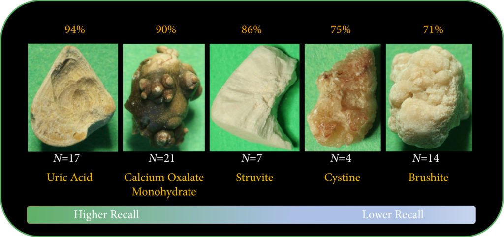
Fig.2. Representative samples for each stone composition prior to cropping. A total of 63 stones were used in this study including: 17 UA, 21 COM, seven struvite, four cystine, and 14 brushite stones consisting of a total of 127 images. Automatic stone composition recall was highest for UA stones at 94%.

Results

The composition prediction recall for each composition was as follows: UA 94% ( = 17), COM 90% ( = 21), MAPH/struvite 86% ( = 7), cystine 75% ( = 4), CHPD/brushite 71% ( = 14). The overall weighted recall of the CNNs composition analysis was 85% for the entire cohort. Specificity and precision for each stone type were as follows: UA (97.83%, 94.12%), COM (97.62%, 95%), struvite (91.84%, 71.43%), cystine (98.31%, 75%), and brushite (96.43%, 75%).

Conclusion

Deep CNNs can be used to identify kidney stone composition from digital photographs with good recall. Future work is needed to see if DL can be used for detecting stone composition during digital endoscopy. This technology may enable integrated endoscopic and laser systems that automatically provide laser settings based on stone composition recognition with the goal to improve surgical efficiency.

Read more Articles of the week

Editorial: Can artificial intelligence optimize case selection for hemi‐gland ablation?

The victory of ‘AlphaGo’ over humans in Go, one of the most complex games with more than 10170 board configurations, has yielded tremendous attention worldwide [1]. The later version, ‘AlphaGo Zero’, has brought artificial intelligence (AI) to the next level by demonstrating an absolute superiority, winning 100‐0 against the champion‐defeating AlphaGo [2]. It is exciting, and perhaps shocking, to realize what AI can achieve.

In this issue of BJUI, the study by Zhou et al. [3] is the first to utilize AI to optimize case selection for hemi‐gland ablation. In this study, classification and regression tree (CART) analysis, which is a form of supervised machine‐learning algorithm, was used to identify laterality of prostate cancer. In the conventional approach, case selection was based on biopsy results and MRI findings. For the CART model, in addition to the common clinical variables (i.e. age, PSA, prostate volume, biopsy and MRI results), biopsy coordinate‐derived spatial features were also used as model inputs. The model output was the probability of unilateral clinically significant prostate cancer considered suitable for hemi‐gland ablation. Whole‐mount prostatectomy specimens were used as the standard of reference. The CART model correctly identified laterality in 80% of the cases, compared to 73% with the conventional approach. The positive predictive value of the CART model was 83%, compared to 53% with the conventional approach. The superiority of the CART model has been demonstrated, and the area under curve was 0.82.

Artifical intelligence has been widely adopted in the field of Urology [4]. For prostate cancer detection in particular, our group evaluated the diagnostic performances of four machine‐learning models based on clinical variables in a biopsy cohort of 1625 men [5]. The machine‐learning models achieved excellent performances in detecting clinically significant prostate cancer, with an accuracy of up to 95.3%. Algohary et al. [6] constructed three machine‐learning models to identify the presence of clinically significant prostate cancer based on MRI radiomic features in patients who underwent active surveillance. When compared with the Prostate Imaging–Reporting and Data System (PI‐RADS) scoring system, the machine‐learning models were able to improve overall accuracy by 30–80%.

Fehr et al. [7] developed an automated system to classify Gleason scores based on MRI images. The automated system could distinguish between Gleason scores of 6 and 7 or above cancers with an accuracy of up to 93%. The differentiation between Gleason score 3+4 and 4+3 disease also yielded an accuracy of up to 93%. Importantly, the performance of AI and machine‐learning models is highly dependent on the quality and accuracy of the data being input. In terms of prostate cancer detection, either mapping biopsy or whole‐mount prostatectomy specimens should be considered to represent the ‘ground truth’.

There are a number of challenges in implementing AI in clinical practice. First, decision‐making in healthcare requires logical deduction and explanation. The data processing in AI, however, is often described as a ‘black box’. Taking AlphaGo as an example, some ‘moves’ were considered incomprehensible even by world‐class players. Second, although results from AI are promising, there is in general a lack of regulations and standards to assess its safety, efficacy and validity. Liability issues can be problematic in case of medical mishaps. Third, doctors are human. Conflict of interest does exist, and how we can utilize AI in a complementary rather than a competitive manner is a challenging obstacle to overcome.

Nevertheless, AI has huge potential in improving healthcare. Collaborative effort is needed globally to develop and optimize AI systems, and to increase its acceptability and practicality upon implementation. Future studies answering clinically important questions using appropriate standards of reference will be of paramount importance in paving the way for the AI era in urology.

by Jeremy Yuen‐Chun Teoh, Edmund Chiong and Chi‐Fai Ng

References

  1. Silver DHuang AMaddison CJ et al. Mastering the game of Go with deep neural networks and tree search. Nature 2016529484– 9
  2. Silver DSchrittwieser JSimonyan K et al. Mastering the game of Go without human knowledge. Nature 2017550354– 9
  3. Zhou SRPriester AMJayadevan R et al. Using spatial tracking with magnetic resonance imaging/ultrasound‐guided biopsy to identify unilateral prostate cancer. BJU Int 2020125399– 406
  4. Chen JRemulla DNguyen JH et al. Current status of artificial intelligence applications in urology and their potential to influence clinical practiceBJU Int 2019124567– 77
  5. Wang GTeoh JYChoi KSDiagnosis of prostate cancer in a Chinese population by using machine learning methods. Conf Proc IEEE Eng Med Biol Soc 201820181– 4
  6. Algohary AViswanath SShiradkar R et al. Radiomic features on MRI enable risk categorization of prostate cancer patients on active surveillance: Preliminary findings. J Magn Reson Imaging 201848818– 28
  7. Fehr DVeeraraghavan HWibmer A et al. Automatic classification of prostate cancer Gleason scores from multiparametric magnetic resonance images. Proc Natl Acad Sci USA 2015112E6265– 73


Residents’ podcast: Artificial intelligence applications in urology

Maria Uloko is a Urology Resident at the University of Minnesota Hospital. In this podcast she is joined by Dr Christopher Weight, an Associate Professor in the Department of Urology at the University of Minnesota. They are discussing a recent BJUI Article of the month:

Current status of artificial intelligence applications in urology and their potential to influence clinical practice

Read the full article

Abstract

Objective

To investigate the applications of artificial intelligence (AI) in diagnosis, treatment and outcome prediction in urologic diseases and evaluate its advantages over traditional models and methods.

Materials and methods

A literature search was performed after PROSPERO registration (CRD42018103701) and in compliance with Preferred Reported Items for Systematic Reviews and Meta‐Analyses (PRISMA) methods. Articles between 1994 and 2018 using the search terms “urology”, “artificial intelligence”, “machine learning” were included and categorized by the application of AI in urology. Review articles, editorial comments, articles with no full‐text access, and nonurologic studies were excluded.

Results

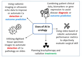
Initial search yielded 231 articles, but after excluding duplicates and following full‐text review and examination of article references, only 111 articles were included in the final analysis. AI applications in urology include: utilizing radiomic imaging or ultrasonic echo data to improve or automate cancer detection or outcome prediction, utilizing digitized tissue specimen images to automate detection of cancer on pathology slides, and combining patient clinical data, biomarkers, or gene expression to assist disease diagnosis or outcome prediction. Some studies employed AI to plan brachytherapy and radiation treatments while others used video-based or robotic automated performance metrics to objectively evaluate surgical skill. Compared to conventional statistical analysis, 71.8% of studies concluded that AI is superior in diagnosis and outcome prediction.

Conclusion

AI has been widely adopted in urology. Compared to conventional statistics AI approaches are more accurate in prediction and more explorative for analyzing large data cohorts. With an increasing library of patient data accessible to clinicians, AI may help facilitate evidence‐based and individualized patient care.

More podcasts

BJUI Podcasts now available on iTunes, subscribe here https://itunes.apple.com/gb/podcast/bju-international/id1309570262


Dr Weight specializes in the surgical treatment of urologic cancers including prostate, bladder, kidney, adrenal, testis and penile cancer. He performs open, endoscopic, laparoscopic, robotic (da Vinci) and retroperineoscopic surgery.

Dr Weight completed his residency training at Cleveland Clinic where he received several awards including the George and Grace Crile Traveling Fellowship Award, the Society of Laparoendoscopic Surgeons Resident Achievement Award and the ASCO Genitourinary Cancer Symposium Merit Award. Dr. Weight then completed a fellowship in Urologic Oncology at Mayo Clinic, where he also completed a Masters degree in Clinical and Translational Research from Mayo Graduate School and was awarded the Mayo Fellows Association Humanitarian Award.

Dr Weight believes that medical research is a key component to offering excellent patient care. His research is focused on improving patient outcomes and the use of artificial intelligence in different urologic applications. He is an author of more than 45 peer-reviewed publications and book chapters and has been invited to speak at regional, national and international conferences. 

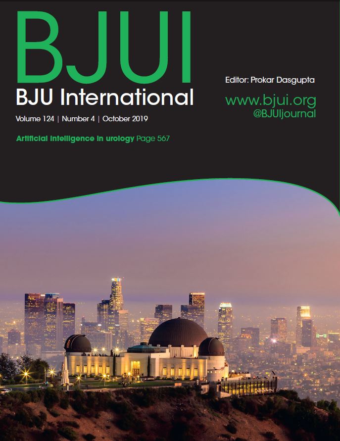
October 2019 – About the cover

The Article of the Month for October was written by researchers primarily from Los Angeles, California, USA: Current status of artificial intelligence applications in urology and their potential to influence clinical practice

The cover image shows the Griffith Observatory, with the surrounding view of LA. The observatory is “Southern California’s gateway to the cosmos”. It is named after its creator and funder Griffith J. Griffith who wanted a free public observatory – it is now a world-leader in public astronomy and has received over 80 million visitors in its nearly 90-year history. It is also one of the best vantage points from which to view the Hollywood sign.


Article of the month: Current status of artificial intelligence applications in urology and their potential to influence clinical practice

Every month, the Editor-in-Chief selects an Article of the Month from the current issue of BJUI. The abstract is reproduced below and you can click on the button to read the full article, which is freely available to all readers for at least 30 days from the time of this post.

In addition to the article itself, there is an editorial  and a visual abstract produced by prominent members of the urological community. These are intended to provoke comment and discussion and we invite you to use the comment tools at the bottom of each post to join the conversation. 

If you only have time to read one article this month, it should be this one.

Current status of artificial intelligence applications in urology and their potential to influence clinical practice

Jian Chen*, Daphne Remulla*, Jessica H. Nguyen*, D. Aastha, Yan Liu, Prokar Dasgupta and Andrew J. Hung*

*Catherine & Joseph Aresty Department of Urology, Center for Robotic Simulation & Education, University of Southern California Institute of Urology, Computer Science Department, Viterbi School of Engineering, University of Southern California, Los Angeles, CA, USA, and Division of Transplantation Immunology and Mucosal Biology, Faculty of Life Sciences and Medicine, Kings College London, London, UK

Read the full article

Abstract

Objective

To investigate the applications of artificial intelligence (AI) in diagnosis, treatment and outcome prediction in urologic diseases and evaluate its advantages over traditional models and methods.

Materials and methods

A literature search was performed after PROSPERO registration (CRD42018103701) and in compliance with Preferred Reported Items for Systematic Reviews and Meta‐Analyses (PRISMA) methods. Articles between 1994 and 2018 using the search terms “urology”, “artificial intelligence”, “machine learning” were included and categorized by the application of AI in urology. Review articles, editorial comments, articles with no full‐text access, and non-urologic studies were excluded.

Results

Initial search yielded 231 articles, but after excluding duplicates and following full‐text review and examination of article references, only 111 articles were included in the final analysis. AI applications in urology include: utilizing radiomic imaging or ultrasonic echo data to improve or automate cancer detection or outcome prediction, utilizing digitized tissue specimen images to automate detection of cancer on pathology slides, and combining patient clinical data, biomarkers, or gene expression to assist disease diagnosis or outcome prediction. Some studies employed AI to plan brachytherapy and radiation treatments while others used video based or robotic automated performance metrics to objectively evaluate surgical skill. Compared to conventional statistical analysis, 71.8% of studies concluded that AI is superior in diagnosis and outcome prediction.

Conclusion

AI has been widely adopted in urology. Compared to conventional statistics AI approaches are more accurate in prediction and more explorative for analyzing large data cohorts. With an increasing library of patient data accessible to clinicians, AI may help facilitate evidence‐based and individualized patient care.

Read more Articles of the week


Editorial: Machines in urology: a brief odyssey of the future

Artificial intelligence (AI) will bring in a new wave of changes in the medical field, likely altering how we practice medicine. In a timely contribution, Chen et al. [1] outline the current landscape of AI and provide us with a glimpse of the future, in which sophisticated computers and algorithms play a front-and-centre role in the daily hospital routine.

Widespread adoption of electronic medical records (EMRs), an ever-increasing amount of radiographic imaging, and the ubiquity of genome sequencing, among other factors, have created an impossibly large body of medical data. This poses obvious challenges for clinicians to remain abreast of new discoveries, but also presents new opportunities for scientific discovery. AI is the inevitable and much-needed tool with which to harness the ‘big data’ of medicine.

Currently, the most immediate and important application of AI appears to be in the field of diagnostics and radiology. In prostate cancer, for example, machine learning algorithms (MLAs) are not only able to automate radiographic detection of prostate cancer but have also been shown to improve diagnostic accuracy compared to standard clinical scoring schemes. MLAs can use clinicopathological data to predict clinically significant prostate cancer and disease recurrence
with a high degree of accuracy. The same has been shown for other urological malignancies, including urothelial cancer and RCC. Implementation of MLAs will lead to improved accuracy and reproducibility, reducing human bias and variability. We also predict that as natural language processing becomes more sophisticated, the troves of nonstructured data that exist in EMRs will be harnessed to deliver improved and more personalized patient care. Patient data and clinical outcomes can be analysed in short time, drawing from a deep body of knowledge, and leading to rapid insights that can guide medical decision-making.

Current AI technology, however, remains experimental and we are still far from the widespread implementation of AI within clinical medicine. A valid criticism of today’s AI is that it functions in the setting of a ‘black box’; the rules that govern the clinical decision-making of an algorithm are often poorly understood or unknowable. We cannot become operators of machines for which we know not how they work, to do so would be to practice medicine blindly.

Another barrier to incorporating AI into common practice is the level of noise in healthcare data. MLAs will use whatever data that are fed to the algorithm, thus running the risk of producing predicative models that include nonsensical variables gleaned from the noise. This concept is similar to multiple hypothesis-testing, where if you feed enough random information into a model, a pattern might emerge. Furthermore, none of the studies described by Chen et al. have been externally validated on large, representative datasets of diverse patients. MLAs trained on a narrow patient population run the risk of creating predictions that
are not generalizable. This problem has already been popularized within genome analysis, where one study found that 81% of all genome-wide studies were taken from individuals of European ancestry [2]. It is easy to imagine situations where risk score calculators or biomarkers are validated using non-representative datasets, leading to less accurate and even inappropriate treatment decisions for underrepresented patient populations. At best, MLAs that are not validated using stringent principles can lead to erroneous disease models. At worst, they can bias the delivery of healthcare to patients, leading to worse patient outcomes and exacerbation of healthcare disparities.

Chen et al. write of the possibility of AI in urology today. What about the future? Imagine a world in which computers with a robotic interface see patients in clinics, design and carry out complex medical treatment plans, and perform surgery without the aid of a human hand. This future may not be far off [3]. Or, even stranger, consider a world in which generalizable AI exists. Estimates of the dawn of this technology range, however the most optimistic projections put the timeline on the order of 20–30 years. Not far behind could be the ‘singularity’, a moment when technological advancement occurs at such an exponential rate that improbable scientific discoveries happen almost instantaneously, setting off a feed-forward cycle leading to an inconceivable superintelligence.

The future is, of course, hard to predict. Nevertheless, AI and the ensuing technology will certainly transform the practice of urology, albeit not without significant challenges and growing pains along the way. The urologist of the future may look very different indeed.

by Stephen W. Reese, Emily Ji, Aliya Sahraoui and Quoc-Dien Trinh


References

  1. Chen J, Remulla D, Nguyen JH et al. Current status of artificial intelligence applications in Urology and its potential to influence clinical practice. BJU Int 2019; 124: 567–77
  2. Popejoy AB, Fullerton SM. Genomics is failing on diversity. Nature 2016; 538: 161–4
  3. Grace K, Salvatier J, Dafoe A, Zhang B, Evans O. When Will AI Exceed Human Performance? Evidence from AI Experts, 2017


Video: Current status of artificial intelligence applications in urology

Current status of artificial intelligence applications in urology and their potential to influence clinical practice

Read the full article

Abstract

Objective

To investigate the applications of artificial intelligence (AI) in diagnosis, treatment and outcome prediction in urologic diseases and evaluate its advantages over traditional models and methods.

Materials and methods

A literature search was performed after PROSPERO registration (CRD42018103701) and in compliance with Preferred Reported Items for Systematic Reviews and Meta‐Analyses (PRISMA) methods. Articles between 1994 and 2018 using the search terms “urology”, “artificial intelligence”, “machine learning” were included and categorized by the application of AI in urology. Review articles, editorial comments, articles with no full‐text access, and non-urologic studies were excluded.

Results

Initial search yielded 231 articles, but after excluding duplicates and following full‐text review and examination of article references, only 111 articles were included in the final analysis. AI applications in urology include: utilizing radiomic imaging or ultrasonic echo data to improve or automate cancer detection or outcome prediction, utilizing digitized tissue specimen images to automate detection of cancer on pathology slides, and combining patient clinical data, biomarkers, or gene expression to assist disease diagnosis or outcome prediction. Some studies employed AI to plan brachytherapy and radiation treatments while others used video based or robotic automated performance metrics to objectively evaluate surgical skill. Compared to conventional statistical analysis, 71.8% of studies concluded that AI is superior in diagnosis and outcome prediction.

Conclusion

AI has been widely adopted in urology. Compared to conventional statistics AI approaches are more accurate in prediction and more explorative for analyzing large data cohorts. With an increasing library of patient data accessible to clinicians, AI may help facilitate evidence‐based and individualized patient care.

View more videos

Visual abstract: Current status of artificial intelligence applications in urology and their potential to influence clinical practice

See more infographics

BAUS/BJUI/USANZ Joint Session AUA 2019

British Association of Urological Surgeons/BJU International/Urological Society of Australia and New Zealand (BAUS/BJUI/USANZ) Joint Session AUA 2019

Sunday, May 5th 2:00 – 5.00 PM. McCormick Place Convention Center South Building – Room S102 BC


Registries /Smart Data /Complications – CHAIR: Duncan Summerton


1400-1420 Alan Partin

A contemporary look at biomarkers for diagnosis of Prostate Cancer

1420-1440 Chris Harding (BJUI sponsored BAUS lecture)

The Mesh Story – lessons learned and future plans

1440-1500 Nick Watkin

PROMs in Urology

1500-1520 Stephen Mark

Big Data and Urology – a pilot trial in New Zealand

1520-1540 Afternoon tea
 

Education /Training /Innovation – CHAIR: Prokar Dasgupta


1540-1600 Andrew Hung (BJUI sponsored lecture)

The emerging role of Artificial Intelligence in Surgical Science

1600-1620 Jonathan Kam

Zero learning curve Percutaneous Nephrolithotomy Access – Prone endoscopic combined intrarenal surgery and multimedia training aid to teach urology trainees

1620-1640 Madhu Koya (BJUI sponsored USANZ lecture)

Cx bladder reduces flexible cystoscopy in haematura and superficial TCC

1640-1700 Kamran Ahmad

Innovation in healthcare systems

1700-1705 BJUI Coffey-Krane Award for trainees based in The Americas presented by Prokar Dasgupta
1700-1900 BJUI Reception


© 2024 BJU International. All Rights Reserved.