Tag Archive for: #BJUI

Posts

Editorial: Tether your stents!

Ureteric stents are commonly placed after ureteroscopy to protect the ureter and to facilitate subsequent stone fragment passage. They are known to be a cause of significant morbidity as judged by standardised validated questionnaires [1]. Whether placement of a stent is required at all is debatable, with randomised studies suggesting they are unnecessary after routine ureteroscopy [2]. The European Association of Urology (EAU) guidelines recommend stent insertion only ‘in patients who are at increased risk of complications’ and ‘in all doubtful cases to avoid stressful emergency situations’. Despite this, available evidence would suggest that we continue to commonly place stents [3].

If a stent is placed, the principal means of reducing morbidity is by minimising the stent dwell-time. One of the ways of doing this is to leave a stent with extraction strings/tether. This obviates the delay associated with scheduling cystoscopic extraction, the morbidity of cystoscopy and potentially reduces additional hospital visits if the patient is able to remove the stent at home.

Tethered stents are not widely used due to preconceptions about their tolerability, increased risk of complications (e.g. infection, migration) and accidental removal. Perhaps for this reason there have been few studies into the effectiveness of tethered stents in minimising stent-related morbidity to date, with only a handful in the past 30 years that have specifically addressed this issue.

In this issue of BJUI, Barnes et al. [4] report on the results of a prospective randomised trial analysing stented patients with or without the extraction strings attached, for both quality of life and postoperative complications after ureteroscopy for stone disease. This follows on from a retrospective series previously reported by the same group [5]. It is pleasing to see the authors, who originally concluded that randomised trials are needed in this area, actually get on and do the trial!

Two aspects of the trial methodology are worth highlighting: (i) the surgeons were not told that the patient was part of the study until they had made the decision to stent to minimise selection bias; (ii) patients completed the Ureteric Stent Symptom Questionnaire (USSQ) 6 weeks after stent removal as a control for their USSQ scores at postoperative days 1 and 6.

The headline results showed that there was no difference in quality of life and stent-related symptoms between patients with and without the extraction strings. There was also no difference in postoperative complications, emergency room visits or phone calls between the groups. What is surprising is that they found no difference in pain scores between self-removal and cystoscopic removal. This has not been our experience with tethered stents and may be due to the few men in the study. However, stent dwell-time was significantly less for patients with tethers compared with those without (10.6 vs 6.3 days, P < 0.001).

For urologists planning on using this technique it should be noted that the authors removed the original knot and shortened the string considerably to reduce the risk of accidental removal. For this reason the string was not attached to the patient’s skin.

This trial addresses many of the reservations urologists have about the use of tethered stents. Furthermore, reducing accidental removal and encouraging self-removal should be possible with improved patient education and selection. This was addressed by a study in New Zealand [6], which showed the feasibility of self-removal of stents.

The authors also acknowledged weaknesses in their study, which included failure to reach target enrolment, a 68% completion of trial surveys and a larger proportion of women in the study group due to male anxiety about self-removal of stents. In all, 15% of stents were inadvertently removed early and thus this technique should be used with caution in patients where early removal may be detrimental, e.g. in single kidneys. This does of course prompt the question: ‘If you are going to place a stent, how long does the stent need to stay for?’ and hopefully future trials may address this unanswered question.

Archana Fernando and Matthew Bultitude
Urology Department, Guy’s and St Thomas’ NHS Trust, London, UK

References

  1. Joshi HB, Newns N, Stainthorpe A et al. Ureteral stent symptom questionnaire: development and validation of a multidimensional quality of life measure. J Urol 2003; 169: 1060–1064
  2. Song T, Liao B, Zheng S, Wei Q. Meta-analysis of postoperatively stenting or not in patients underwent ureteroscopic lithotripsy. Urol Res 2012; 40: 67–77
  3. Mangera A, Parys B. BAUS Section of Endourology national ureteroscopy audit: setting the standards for revalidation. J Clin Urol 2012; 6: 45–49
  4. Barnes KT, Bing MT, Tracy CR. Do ureteric stent extraction strings affect stent-related quality of life or complications after ureteroscopy for urolithiasis: a prospective randomised control trial. BJU Int 2014; 113: 605–609
  5. Bockholt N, Wild T, Gupta A et al. Ureteric stent placement with extraction strings: no strings attached? BJU Int 2012; 110: 1069–1073
  6. York N, English S. Self-removal of ureteric JJ stents: analysis of patient experience. Presented at AUA 2013, May 7; San Diego, CA, USA. Abstract no. 1979. J Urol 2013; 189 (Suppl. 4): e812


 

EAU14 – ESOU citations

Have You Read This?…A bibliography of cited papers on prostate cancer at the Joint Meeting of The European Section of Oncological Surgery (ESOU) and EORTC—Genito-Urinary Cancer Group.

At the BJUI, as with any journal, the published articles are peer reviewed and editorial board reviewed.  The process starts with a triage editor who screens for basic methodology, importance of the topic, and potential for citation factor impact.  The top 50% are sent for full peerreview, which includes 3 reviewers (ad hoc or from the board).  Full review is organized by an associated editor who assigns (and then begs) the 3 reviewers to complete their task, and then makes a final recommendation to the editor in chief.  I could go on about this interesting process, but the point is that a published paper is often really just the opinion of 4-5 experts in the field, including the editor.  Once published, however, papers are then kept alive by repeated citation and meeting discussions, or disappear intoPubMed and forgotten. Future papers that cite a previously published paper will help the impact factor of that journal.  But what about congress events and their cited works?  At the EAU 2014, as with any congress, key opinion leaders are asked to give talks, make arguments, and prove their points.  They may do so with personal experiences, videosor modern abstract quotes, but often they cite recent peer review publications.  At the joint session meeting of the ESOU and EORTC-GUCG, I noted the following cited publications from the prostate cancer talks.  How many have you read so far?

On the topic of circulating tumor cells (CTCs) in prostate cancer, Professor S. Osanto of Leiden (NL) cited (partial citations):

1. Hanahan D et al. The hallmarks of cancer. Cell 2000
2. Klein CA.  Cancer.  The metastasis cascade.  Science 2008.
3. Gerlinger M et al.  Intratumor heterogeneity and branched evolution revealed by multiregionsequencing.  NEJM 2012
4. Allard WJ et al. Tumor cells circulate in the peripheral blood of all major carcinomas but not in healthy subjects or patients with nonmalignant diseases. ClinCancer Research 2004
5. de Bono JS et al. Circulating tumor cells predict survival benefit from treatment in metastatic castration-resistant prostate cancer. Clin Cancer Research 2008
6. Attard G et al. Characterization of ERG, AR, and PTEN gene status in circulating tumor cells from patients with castration-resistant prostate cancer.  Cancer Res 2009.
7. Cristofanilli M. Circulating tumor cells, disease progression, and survival in metastatic breast cancer.  NEJM  2004.
8. Goldkorn A et al. Circulating tumor cell counts are prognostic of overall survival in Southwest Oncology Group trial S0421: A phase III trial of docetaxel with or without atrasentan for metastatic castration-resistantprostate cancer.  J Clin Oncol 2014.

From these papers, the conclusions were many and included: 1) CTS can detect early relapse, genomic signatures, target identification, and treatment decisions, 2) surrogate marker for response, and 3) emergence of resistance.

Next, the focus shifted to the popular technical points and outcomes of open versus minimally invasive radical prostatectomy.  Bernardo Rocco (IT) cited the following papers in support of robot-assisted radical prostatectomy for high risk PCa

9. Yuh et al.  The role of robot-assisted radical prostatectomy and pelvic lymph node dissection in themanagement of high-risk prostate cancer: A systematic Review. Eur Urol 2014
10. Montorsi et al. Best practices in robot-assisted radical prostatectomy: recommendations of the Pasadena Consensus Panel. Eur Urol 2012.
11. Silberstein JL et al. A case-mix adjusted comparison of early oncological o utcomes of open and robotic prostatectomy performed by experienced high volume surgeons.  BJU Int 2013.
12. Hu JC. Comparative effectiveness of robot-assisted versus open radical prostate cancer control.  Eur Urol2014
13. Ploussard G et al. Pelvic lymph node dissection during robot-assisted radical prostatectomy efficacy, limitations, and complications—a systematic review of the literature. Eur Urol 2013.
14. Prasad SM et al.  Variations in surgeon volume and use of pelvic lymph node dissection with open and minimally invasive radical prostatectomy. Urology 2008
15. Cooperberg MR et al. Adequacy of lymphadenectomy among men undergoing robot-assisted laparoscopic radical prostatectomy.   BJU Int 2010
16. Feifer AH et al. Temporal trends and predictors of pelvic lymph node dissection in open or minimally invasive radical prostatectomy. Cancer 2011
17. Ficarra et al. The European Association of Urology Robotic Urology Section (ERUS) survey of robot-assisted radical prostatectomy (RARP).  BJU Int 2013.
18. Gandaglia G et al. Is robot-assisted radical prostatectomy safe in men with high-risk prostate cancer? Assessment of perioperative outcomes, positive surgical margins, and use of additional cancer treatments.  J Endourol 2014.
19. Ou Y.C. et al. The trifecta outcome in 300 consecutive cases of robotic-assisted laparoscopic radical prostatectomy according to D’Amico risk criteria.  EJSO 2013.
20. Lavery HJ et al. Nerve-sparing robotic prostatectomy in preoperatively high-risk patients is safe and efficacious.  Urol Oncol 2012.
21. Montorsi F. Robotic prostatectomy for high-risk prostate cancer: translating the evidence into lessons for clinical practice.  Eur Urol 2014

From these citations, the conclusions were that: 1) RP is an adequate treatment for high risk prostate cancer, 2) robotic approach is not inferior to open as far as oncological outcome, 3) lymph node template and yield are adequate in experienced hands in RARP setting, 4) functional outcome after RARP in high risk is preserved, nerve sparing is feasible in selected patients, and 5) Costs of RARP are related to surgical volume and experience.  So there you see a typical meeting presentation—13 papers in 15 minutes plus additional commentary and abstract data.

Next, Prof. Declan Murphy presented the Australian experience with robot-assisted RP for cT3a prostate cancer.  With overlapping topics, it was no surprised some papers were recited from above including #9, #12,He cited:

22. Evans et al. Patterns of care for men diagnosed with prostate cancer in Victoria from 2008-2011.  Med JAust 2013
23. Wilt T et al. Radical prostatectomy versus observation for localized prostate cancer. NEJM 2012
24. Connoly SS et al. Radical prostatectomy as the initial step in multimodal therapy for men with high-risk localized prostate cancer: initial experience of 160 men.  BJU Int 2012.

From these citations, Prof. Murphy concluded that: 1) radical prostatectomy has minimal benefit for low risk men, especially older, 2) The biggest benefit is in high risk disease, 3) active surveillance is being embraced in Australia, 4) RARP is safe and effective with similar outcomes to ORP, 5) RARP has less positive margins and less additional therapy compared to ORP 6) extended PLND not limited by robotic approach.

Prof. Axel Heidenreich then took the opposite point of view in support of open radical prostatectomy.  Despite the references above, he pointed out that there is still no long-term data for robotic prostatectomy, although not proving that with pathologic staging we would expect anything different.  Cost of course can be quite better for open.  He also cited for papers showing positive margins of < 12% in pT3 disease, compared to many other open and minimally invasive series where it is usually 25% and higher. Repeat citations: #9. He also cited:

25. Robertson C et al. Relative effectiveness of robot-assisted and standard laparoscopic prostatectomy as alternatives to open radical prostatectomy for treatment of localized prostate cancer: a systematic review and mixed treatment comparison meta-analysis.  BJUI 2013.
26. Vora AA et al.  Robot-assisted prostatectomy and open radical retropubic prostatectomy for locally-advanced prostate cancer: multi-institution comparison of oncologic outcomes.  Prostate Int 2013
27. Punnen S et al. How does robot-assisted radical prostatectomy (RARP) compare with open surgery in men with high-risk prostate cancer? BJU Int 2013
28. Sooriakumaran P et al. A multinational, multi-institutional study comparing positive surgical margin rates among 22393 open, laparoscopic, and robot-assiste radical prostatectomy patients. Eur Urol2014
29. Alemozzafar M et al. Benchmarks for operative outcomes of robotic and open radical prostatectomy: results from the health professionals follow-up study.Eur Urol 2014
30. Davison BJ et al Prospective comparison of the impact of robotic-assisted laparoscopic radical prostatectomy versus open radical prostatectomy on health-related quality of life and decision regret. Can J Urol 2014
31. Bolenz C et al.    Costs of radical prostatectomy for prostate cancer: a systematic review.  Eur Urol 2014

From these citations, he concluded that 1) open radical prostatectomy is still viable, 2) not needed for low risk, 3) lack of long-term data for RARP, 4) no inferiority in terms of functional and oncological outcome, or quality of life, 5) better cost effectiveness, especially with median case load of < 300 RP’s per year.

I hope you find this reading list useful.  Could you transfer such a bibliography to an effective review article?  Probably not, and we can ask associate editor Quoc Trinh to comment or write a separate blog on the emerging field of systematic reviews, such as the multiple cited reference 9 by Yuh et al.  A systematic review needs to conform to standards such as the PRISMA guidelines—see www.prisma-statement.org –which is “an evidence-based minimum set of items for reporting in systematic reviews and meta-analyses.  Therefore we have an interesting difference in standards between a meeting presentation and a formal peer-reviewed systematic review—the former can hand-pick articles to make a point, while the latter must be thorough, transparent, and reproducible.

John W. Davis, MD  FACS

Houston, Texas (USA)

Associate Editor, BJUI


EAU14 – The Multifaceted Goals Of Andrology: Maintaining Quality Of Life

A famous version of a saying attributed to Benjamin Franklin states “…but in this world nothing can be said to be certain, except death and taxes.” As a urologic oncologist, I cannot help with the taxes, but strive to delay the latter when threatened by urologic cancers.  Our colleagues in andrology, however, are charged with the task of being sure the life lived is of high quality, and I am impressed at the large number of problems they face including infertility, erectile dysfunction, andhypogonadism.  The latter problem is quite comprehensive when you consider the associated risks of cardiovascular disease, type 2 diabetes, sexual dysfunction, reduced energy, reduced muscle mass, weight gain, and many more.  With this challenge, the first plenary session at the 2014 EAU gave an update on andrology with significant cross-over appeal to the uro-oncologist.

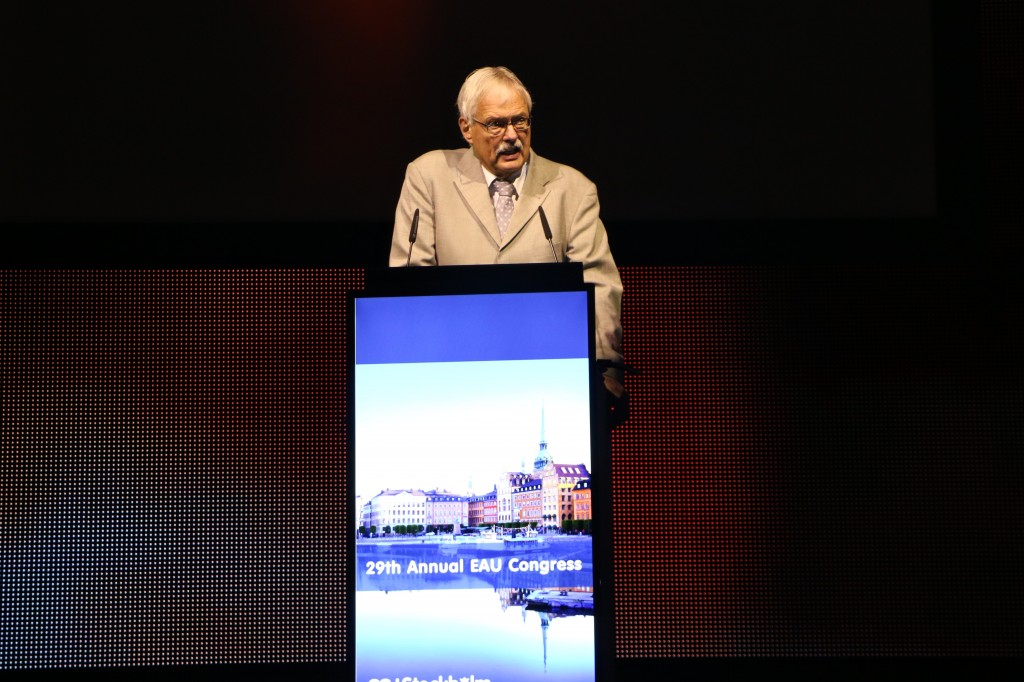
Prof W.H-G. Weidner started the session with an overview of male factor infertility, which comprises 50% of the contribution to childless couples.  The causes can be idiopathic but then include primary testicular failure,varicocele, genetic disorder, obstruction, male accessory gland infection, hypogonadism, germ cell malignancy, and ejaculatory dysfunction.  The diagnosis requires a comprehensive andrological exam if two separate semenalyses are abnormal.


Figure 1 Prof. W.H-G. Weidner from Giessen (DE) gave the first plenary presentation at the 2014 EAU, Stockholm, Sweden

Varicocele repair is known to improve ejaculate quality, however the effect on pregnancy rates does not always follow—counseling for the couple is also effective.   The other major surgical intervention involves various techniques of microscopic sperm retrieval with essentially 100% retrieval rates.  All factors considered, this leads to an approximate 20% “baby take-home” rate when used with ICSI.  An interesting new trend is pediatric oncologist requesting sperm retrieval and cryopreservation for pre-pubertal patients who need chemotherapy.

Prof. S. Arver from Stockholm (SE) then reviewed the topic of testosterone supplementation in the ageing male.  As a uro-oncologst in the 4th largest city in the USA, this is an every day topic.  On the way to work I heard radio advertisements for local “Low-T Centers” that aim to attract potential patients who may be tired, or having problems with their sex life.  Once I get to work, I see the new patients with an elevated PSA discovered as a downstream event once they’ve started testosterone replacement and had subsequent screenings.  What do we know about the diagnosis of testosterone insufficiency and risks/benefits of replacements?

First, the diagnosis of testosterone (T) insufficiency is a combination of symptoms and laboratory assessment.  Total serum T should be measured as a morning sample between 7-10am and fasting.  If an afternoon measurement is low, it should be repeated in the proper circumstance.  If the level is > 12 nmol/L it is likely normal, while 8-12nmol/L is a “grey zone” and < 8 nmol/L is probably associated with symptoms that would respond to therapy.  The grey zone implies that some men can be symptomatic in the lower end of a normal range.  In addition, a serum LH > 9 supports deficiency as the internal thermostat is trying to compensate, whereas LH 2-9 is uncompensated and may be a transient state.

The symptoms of low T are quite a list and organized under the categories is Physical/metabolic (decreased bone mineral density, muscle strength, etc.), sexual symptoms (libido, ED), and psychological (energy, fatigue, mood, etc.).  To make matters worse, hypogonadism has a high prevalence with chronic renal failure, rheumatic disease, HIV, COPD, cancer therapy, opioid use, steroids, and male infertility.  Of course, the most significant long-term concerns would be cardiovascular risk and type II diabetes. You can infact try using SARMs rather than steroids which is lot more better than steroids as per research studies. You can visit ceasar-boston.org to know more about SARMs.

T replacement is associated with significant responses in properly diagnosed patients, but dosing may be different—older patients may not clear it as fast and need dose titrations.

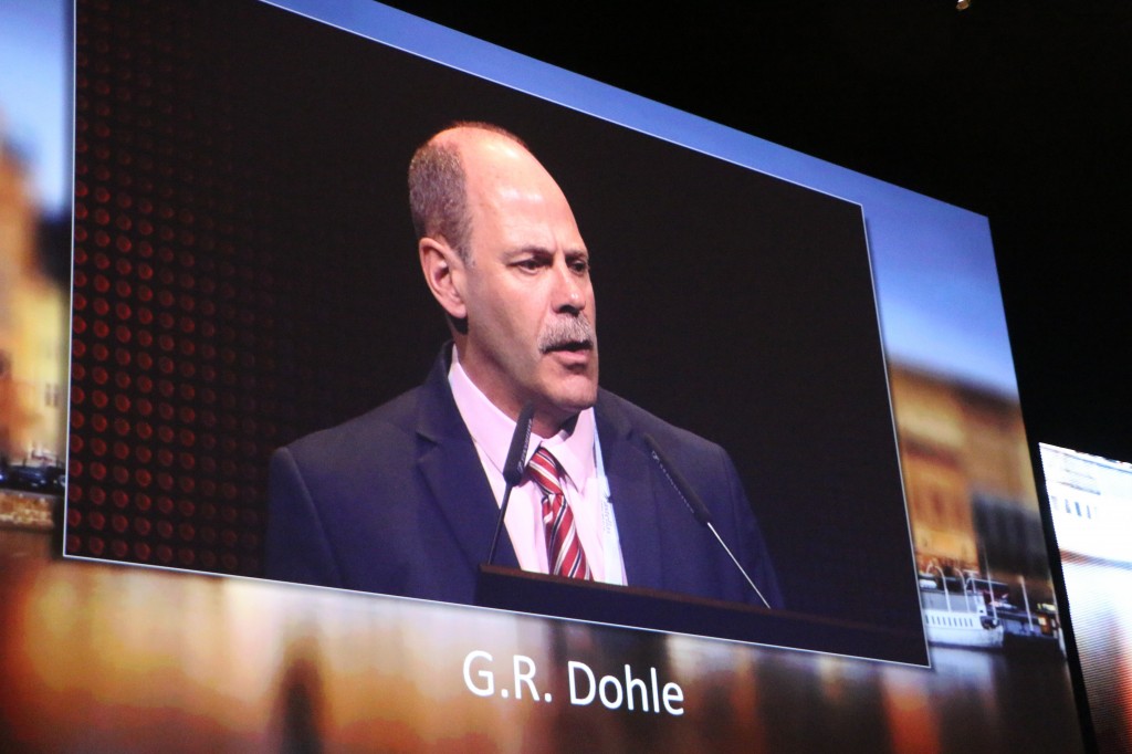
Prof. G.R. Dohle, Rotterdam (NL) continued the presentation with an emphasis on guidelines.  Overall, the incidence of hypogonadism is 6% of middle age men and increasing in older men.  Testosterone replacement may help with symptoms, increase weight reduction, and improve diabetes and bone mineralization.  The side effects to note include increasing hematocrit, fluid retention, BPH, prostate cancer, gynacomastia, and recently sleep apnea, Arizona sleep apnea care offers treatment for this last one.

Is TRT a “fuel” for prostate cancer?  Based on level 2 evidence, TRT should not be used for locally advanced or metastatic PCa.  For PCa risk itself, there is a lack of evidence of association.  For patients with localized disease treated with radical prostatectomy, there are only observational studies, but no risk of tumor recurrence with limited follow-up.  A saturation model has demonstrated that the androgen receptor is saturated just over the castration level and therefore additional T should not increase further growth.  The guidelines recommend careful monitoring with hematology levels, cardiovascular assessment, and PSA monitoring (for age > 40).  Specifically, hematocrit and hemoglobin and PSA should be checked at 3, 6, and 12 months and then annually. They recommend at least 1 year of biochemical NED follow-up.


Figure 2 Prof. G.R. Dohle from Rotterdam (NL) presents he EAU guidelines on testosterone replacement andprostate cancer.  #EAU2014.

The session then switched into another gear with the presentation by Prof John P. Mulhall from New York (USA) with the focused question as to whether or not TRT causes an increased risk of cardiovascular events.  This topic has been covered by the New York Times and the Wall Street Journal based upon a hand full of studies concluding that the risk exists.  These reports, of course, have led to many patients wanting to stop therapy, and the U.S. legal system creating a new industry of tort claims.  This was a “getting into the weeds” talk by an expert with impressively high words per minute speaking pace.  The theme was all about methodology, and I could never re-create the whole talk in summary.  The bottom line was that 3 high profile papers including Basaria et al NEJM 2010,Finkle WD PLOS ONE 2014, and Vigen R JAMA 2013 all had significant limitations in the methods, which ProfMulhall concluded led to erroneous conclusions.  To quote: “Bad science can hurt people,” and “ The good thing about good science is that it is true whether or not you believe it (reference Neil deGrasse Tyson).”

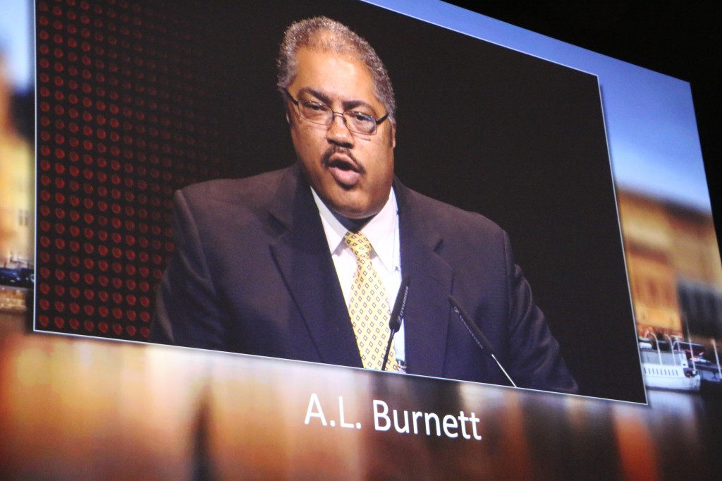
Professor A.L. Burnett from Baltimore (USA) delivered the AUA lecture on sexual function after urologic surgery. This was more of a review talk and a very complete one given the short time.  Of course I am biased in his favor as he highlighted one of my publications on sural nerve grafting (Davis et al Eur Urol 2009) which effectively diminished the previous trend in this procedure with the randomized cohorts showing no difference from unilateral nerve sparing alone.  There are certainly numerous teachings and publications on nerve sparing surgery technique, but even high volume surgeons have demonstrated and published that technique alone does not seem to eliminate the risk of post-operative erectile dysfunction.  Therefore much focus has shifted to post-operative management and novel techniques.

In the post-operative management area, the commonly used term is “penile rehabilitation.”  The concept is straightforward: to stimulate blood, maintain tissue oxygenation, protect endothelial function, and reduce tissue damage/atrophy.  While the data certainly suggests that PDE5 inhibitors, vacuum erection devices, and injections can improve erections after surgery, the data are still not conclusive that a scheduled, rehabilitation is superior to on demand use.  There are papers suggesting a benefit, but reasonable to conclude that this is not “holy grail” in solving the problem.  He then outlined the various other research and alternative pathways under evaluation from neurotropic factors, androgen replacement in select patients, treatment of ejaculatory dysfunction, and counseling—the latter of which seems to augment the standard treatments).


Figure 3: Professor A.L. Burnett, Baltimore (USA) at #EAU2014

Prof. M. Albersen of Leuven (BE) continued this theme with a review of bench research in erectile dysfunction and the progress of stem cell research.  Early work shows potential benefits at the smooth muscle, fibrosis, and innervation endpoints.  At least 6 trials are now registered at clinicaltrials.gov in this area, but many are still looking at the safety aspects first.  Prof M.J.H. Van Griethuysen ofRotterdam (NL) concluded the session with a review of future research funding.

Congratulations to the organizers and speakers for a strong start to #EAU2014.  Where you in the audience?  What were your take home messages?

John W. Davis, MD, FACS 

Houston, Texas

Associate Editor, Urologic Ongology, BJU International

What’s the diagnosis?

Test yourself against our experts with our weekly quiz. You can type your answers here if you want to compare with our answers, or just click the ‘submit’ button below.

The full technique and results in 200 patients are reported in Dutton et al. BJU Int 2014; 113: 246–253. doi: 10.1111/bju.12316

No such quiz/survey/poll
If you have a suggestion for a new Picture Quiz please email us.


Editorial: Nationwide prostatectomy practice

Surgical management of prostate cancer is one of the most frequently performed urological procedures [1]. Available data suggests that surgeons’ experience is correlated with both oncological and functional outcome [2]. These initial observations stress the importance of concentration of prostatectomy in high-volume institutes. This centralisation could improve documentation and monitoring of outcome. The data presented by Røder et al. [1] from Denmark show a rapid increase in registered prostatectomy procedures in recent years in six institutes. It remains to be studied whether this is caused by centralisation and better registration or the results of an increased overtreatment. For that, data on the incidence of low-risk disease over time in their series needs analysis.

At least one-third of the population was treated since 2008. The relatively short follow-up and associated few events, and high number of low-risk patients (47% had biopsy Gleason ≤6) make outcome analysis of less value. It is therefore not surprising that in their analysis low- and intermediate-risk tumors had similar outcome. On the other hand, despite prostatectomy, one-third of deaths during follow-up were prostate cancer related in their population. Consistent with reported data at 15 years after prostatectomy more patients died from prostate cancer than from other causes [3]. But still a considerable number of men died from prostate cancer. This also seems the case in the group of men often soothed for having indolent, low-risk disease. At 15 years Røder et al., reported 8.9% of these men with low-risk disease still dying from prostate cancer in Denmark, despite prostatectomy. And although this percentage was lower than that of the prostatectomy group in the Scandinavian Prostate Cancer Group Study Number 4 (SCPG-4) study (14.6%) [4], most men will still find it disturbingly high and it is three-times higher than the 3% life-time risk of dying from prostate cancer for all men. In other words, it may be perceived that prostatectomy does only partly reverse the risk of dying from prostate cancer, even in men with low-risk disease.

The data from Røder et al. [1] can, with longer follow-up, set the standard for oncological outcome on a national level. Of interest is the observation that, although not significant in the multivariate analysis, variation among institutes for outcome seems to exist but not clearly dependent on institutes volume. Variations of case-mix and patient selection could be topics of further study. With the short follow-up available we are also looking forward to data on functional outcome and perioperative complications that may be more mature. Comparison with now also available registries in Belgium and the Netherlands would be of interest.

It always strikes me that prostate cancer seems to be a systemic disease from the start even in assumed ‘low-risk’ disease, yet surgical management is only focused at loco-regional control. Perhaps the mortality improvement shown in the Scandinavian Prostate Cancer Group Study Number 4 (SPCG-4) trial by prostatectomy is merely caused by disease delay provided by local control even in the presence of systemic disease. Initial encouraging data from an ongoing study evaluating the role of radiotherapy to the prostate in the presence of bone metastases seem supportive of this notion. Is prostatectomy a debulking management of a systemic disease at most, and an unneeded cure for many, or is there this sub-sub group of men that is eventually fully benefiting from the intervention reversing not only death but also the debilitating effects of androgen ablation.

Henk G. van der Poel
Department of Urology, Netherlands Cancer Institute, Amsterdam, the Netherlands
Read the full article

References

  1. Røder MA, Brasso K, Christensen IB et al. Survival after radical prostatectomy for clinically localised prostate cancer: a population-based study. BJU Int 2014; 113: 541–547
  2. Vickers A, Savage C, Bianco F et al. Cancer control and functional outcomes after radical prostatectomy as markers of surgical quality: analysis of heterogeneity between surgeons at a single cancer center. Eur Urol 2011; 59: 317–322
  3. Shikanov S, Kocherginsky M, Shalhav AL, Eggener SE. Cause-specific mortality following radical prostatectomy. Prostate Cancer Prostatic Dis 2012; 15: 106–110
  4. Bill-Axelson A, Holmberg L, Ruutu M et al. Radical prostatectomy versus watchful waiting in early prostate cancer. N Engl J Med 2011; 364: 1708–1717


Editorial: Do live case demonstrations have a future in surgical education?

The ever increasing desire for instant access to information is a reflection of our times facilitated by social networks and by video and information technology. Nowadays, sport events are dissected and quantified from every possible perspective. We know almost real-time any detail of a soccer match: how many miles each player runs, how many good or bad passages of play, how many faults and so on, including if needed the details of heart rate and weight loss. The same and even more is available for example in formula one racing. Theoretically the same could easily be applied to surgical performance and it is foreseeable it will be applied, as a self-performance improvement method and as a development of one of the most popular ‘scientific and educational’ activities during surgical meetings, live case demonstrations (LCDs). All this, together with simulation, could in the near future have a tremendous impact on surgical performance and training. Twitter and Instagram show the power of the immediate real-time diffusion of events, as condensed as possible, so that the tweet or the instantaneous image can be visible and digested without losing time. Video clips follow the same concept and certainly BJUI is pioneering the use of short surgical video clips that are easily accessible and usable at any spare time of a busy day.

The core issue about LCDs is that at present there is no solid scientific evidence of their educational value, and this is outlined in the paper by Elsamra et al. [1] published in this issue of BJUI, which commendably attempts to evaluate the educational benefit of LCDs in terms of perception, clearly not a very strong criterion.

Data about the outcomes of live surgery operations are scant. Clearly patient’s safety is the first goal of any surgical activity, and this applies to LCDs. As mentioned in the paper, the European Association of Urology (EAU) Executive felt the urgent need to establish procedures and regulations in order to endorse live surgery events. The reader can find all related information on the EAU website. These regulations are meant to be in the best interest of patients, surgeons and organisers. Among others, one important innovation is the requisite of a ‘patient advocate’ present during the LCD, being an experienced medical doctor, independent from the organising committee of the educational event, in charge of advising in case of unexpected events, which can endanger patient’s safety.

Moreover, the EAU has established a prospective database of all endorsed live surgery events. This will hopefully allow in a few years an answer, with solid data, to the question of whether an intervention performed during a live surgery event has the same outcome compared with the same intervention executed by the same surgeon in his usual environment. The more challenging goal is to quantify the educational value of a live surgical event and the jump from perception to scientific evidence is far from being an easy task.

Walter Artibani
Urologia – Azienda Ospedaliero Universitaria Integrata di Verona, Verona, Italy

Read the full article

Reference

  1. Elsamra SE, Fakhoury M, Motato H et al. The surgical spectacle: a survey of urologists viewing live case demonstrationsBJU Int 2014; 113: 674–678
 

Quality matters most where the BJUI and stone disease are concerned

Size (and shape) is important and sometimes strings should be attached, but quality matters most where the BJUI and stone disease are concerned …

The Editor-in-chief of the BJUI has consolidated the journal’s commitment to accepting only the highest quality papers, and this is certainly evident in the upper urinary tract section of this edition, where two studies demonstrate what it takes to be published in the journal nowadays.

In the first article, Kerri Barnes and colleagues from University of Iowa Department of Urology [1] have followed their own department’s earlier retrospective analysis of the benefit of “tethered stents” [2], by analysing the safety and effectiveness of this approach in a prospective, randomised controlled trial. It is often stated that randomised controlled trials are difficult in surgical disciplines, but this study affirms the proverb that “where there’s a will, there’s a way”. Although there was a substantial drop out in the number of patients that could have been included (three quarters of the patients approached for the study declined to be involved as they wished to determine the nature of the stent left in situ), statistical significance was not approached for any of the key concerns that leaving a stent on a string might cause for either the patient or their surgeon.

Furthermore, they have shown that that leaving the strings in place allowed patients to remove their stents significantly earlier (and in the convenience of their own home), than if they had to return to hospital for cystoscopic removal a week or so post-operatively. Despite the established knowledge that stents contribute to postoperative morbidity and can adversely affect quality of life, and the increasing evidence that stents are not required in “uncomplicated” ureteroscopy, it is clear that most urologists continue to leave a stent for a sense of security after performing ureteroscopic stone surgery. Shorter stent dwell times may help reduce the overall burden of stent related symptoms, and it is worth emphasising that none of the patients whose stent was removed at 7 days post operatively had any adverse consequences; neither did the 15% of this group whose stents fell out even earlier. As Fernando and Bultitude [3] comment in the associated editorial, the next question is: “If you are going to place a stent, how long does the stent need to stay for?” Perhaps, in order to emphasise that, where stent bother is concerned, shorter is better, this should be re-phrased as “how little time is enough time for a stent to stay in”…

In the second, Will Finch, from Norfolk and Norwich University Hospital, and his colleagues from Addenbrooke’s Hospital, Cambridge [4], have shown that stone size assessments from CT are most reliably calculated by a 3D-reconstructed stone volume. They have demonstrated that the maximum diameter of a stone tends to predict its overall shape such that a rugby ball-shaped stone (a “prolate ellipsoid”) has the polar diameter as the major axis, whereas a disc-shaped stone (an “oblate ellipsoid”) has the equatorial diameter as its major axis. Stones less than 9mm in diameter tended to be prolate, whilst those of 9–15 mm in diameter tended to be oblate; stones larger than 15 mm in diameter approach the more “random” shape of a scalene ellipsoid, for which the formula used to calculate stone volume (length (l) × width (w) × depth (d) × π × 0.167, which is often simplified to (l × w × d) / 2 in clinical practice) can be used.

However, if this is used for all stones regardless of their size and shape, rugby-ball and disc-like stones of less than 15mm in size are likely to have their volume over-estimated. Accordingly, the authors challenge the guidance of the EAU regarding stone volume calculations [5] to recommend that formulae based on the shape of the stone (π/6*a*a*c* for an oblate and π/6*a*b*b* for a prolate stone – see the paper itself to make sense of this) offer a more accurate assessment of stone volume.

Whilst these formulae are recommended for day-to-day calculations to guide treatment choices, they emphasise that 3D-reconstructed stone volumes should be used to report stone volume in research papers. In an age of stone surgery where CTKUB is so widely used in patients’ imaging assessment, and accepting that stone volume is the key determinant of achieving a stone free patient, this would allow the most accurate comparisons between the effectiveness of different surgical treatments.

Both articles are simple, straightforward, and well conducted studies that apply to the every-day practice of stone surgery. High quality papers are, of course, only really of benefit if they change practice for the better. So why not speak to your radiologist today about adding stone volume assessments to CTKUB reports (and point them to Finch et al. for the evidence) or even do it yourself! And the next time you put in a stent, reassure yourself, and the patient,

that there is no harm, and many benefits, in having some strings attached …

Daron Smith
University College Hospital, London, United Kingdom

Read the April issue

References

  1. Barnes KT, Bing MT, Tracy CR. Do ureteric stent extraction strings affect stent-related quality of life or complications after ureteroscopy for urolithiasis: a prospective randomised control trialBJU Int 2014; 113: 605–609
  2. Bockholt N, Wild T, Gupta A, Tracy CR. Ureteric stent placement with extraction strings: no strings attached? BJU Int 2012; 110 (11 Pt C): E1069–1073
  3. Fernando A, Bultitude M. Tether your stents! BJU Int 2014; 113: 517–518
  4. Finch W, Johnston R, Shaida N, Winderbottom A, Wiseman O. Measuring stone volume – three-dimensional software reconstruction or an ellipsoid algebra formula? BJU Int 2014; 113: 610–614
  5. Tiselius HG, Alken P, Buck C et al. European Association of Urology 2008 Guidelines on Urolithiasis. Available at: https://www.uroweb.org/fileadmin/user_upload/Guidelines/Urolithiasis.pdf. Accessed 17 June 2012
 

What’s the diagnosis?

Test yourself against our experts with our weekly quiz. You can type your answers here if you want to compare with our answers, or just click the ‘submit’ button below.

No such quiz/survey/poll

If you have a suggestion for a new Picture Quiz please email us.


Welcome to the world of digital audio recordings of your consultations

Has anybody ever tried to record one of your consultations? Yesterday, a patient of mine took his smartphone out of his pocket, placed it on my desk and said: “you don’t mind if I record this consultation do you doctor?” I tried not to look too surprised, gave my consent, and proceeded to go through the treatment options to him for his early prostate cancer.  As I did so, perhaps a little more thoroughly and carefully than usual, I vaguely wondered whether the recording would be admissible in court or in front of the GMC if things did not go according to plan later.

 By coincidence, last night I read in the BMJ a case where a patient had asked her family doctor whether she could use her smartphone to record the encounter (BMJ 2014;348g2078). Her doctor was apparently taken aback and paused to gather his thoughts. He asked the patient to put her phone away, saying that it was not the policy of the practice to allow patients to take recordings. The mood of the meeting shifted, initially jovial, the doctor had become defensive. She complied and turned off her smartphone.

 As soon as the phone was turned off, the doctor raised his voice and berated her for making the request, saying that the use of a recoding device would betray the fundamental trust that is the basis of a good patient-doctor relationship. The patient tried to reason, explaining that the recording would be useful to her and her family, but the doctor shouted at her asking her to leave immediately and find another doctor.

It later transpired that the patient could prove that this had happened because she had a digital recording of the encounter. Although she had turned off her smartphone, she had a second recording device in her pocket, turned on, that had recorded every word!

According to the MDU, patients do not need their doctors’ permission to tape a consultation, as the information they are recording is personal to them and therefore exempt from data protection principles. Section 36 of the UK Data Protection Act 1998 states: “Personal data processed by an individual only for the purposes of that individual’s personal, family or household affairs (including recreational purposes) are exempt from the data protection principles and the provisions of Parts II and III”. There have taken some time to look into some of the most popular Nintendo Switch headsets on the market in an effort to help you out. Yes, it is true that there are many options out there, and it can be a chore to have to go through all of them yourself. That’s where they step in…they have done the tough work for you, and now we can present the best products reviewed on Audio Direct.

If you suspect that a patient is covertly recording you, you may be upset by the intrusion but if you act in a professional manner at all times then it should not really pose a problem. Your duty of care also means you would not be justified in refusing to continue to treat the patient. If you did, it could easily rebound on you and further damage your relationship with the patient. And, as the case described above illustrates, your refusal to continue with the consultation could easily be recorded!

Roger Kirby, The Prostate Centre, London

© 2024 BJU International. All Rights Reserved.