Tag Archive for: Editorial

Posts

Beyond our wildest dreams

In this podcast Prokar Dasgupta summarises the success of the BJUI over 2013. For more on podcasts, including how to record your own, go to Podcasts Made Simple.


If anyone had suggested to me in January 2013 that our full article downloads would increase by 15% and the Melbourne Consensus Statement on PSA testing would be viewed over 5000 times @ BJUI.org, I would have stared at them in disbelief. The launch of our web portal in addition to an innovative paper journal, has achieved just that. And much more. We remain one of the Big Three in urology with a Klout score greater than any of our colleagues. These are impossible to achieve via papyrus alone.

The common theme amongst all the fantastic innovation that our Associate Editors have introduced is the highest quality of original articles that we have attracted and published this year. I wanted to take this opportunity to highlight them and thank all our authors for sending us their best manuscripts.

The updated Partin tables (2006–11) remains our most cited paper published in 2013 [1]. It is sheer coincidence that I selected it as our first article of the month in January. It has allowed surgeons to avoid lymphadenectomy during radical prostatectomy in non-palpable Gleason 3+4 disease as the risk of a positive lymph node is <2%. The accompanying 3 minute video on the BJUI Tube channel is an excellent summary for the busy urologist.

I had to appease a number of oncologists when Cooperberg and colleagues showed that radiation for prostate cancer was about 2.5 times more expensive than radical prostatectomy in a comprehensive lifetime cost-utility analysis [2]. Peace was rapidly established at the annual meeting of the British Uro-Oncology group (BUG) where I participated in a balloon debate on the subject this autumn.

The thematic variations continue. It seems that 12 weeks of Tadalafil is effective in ejaculatory and orgasmic dysfunction in patients with ED [3]. Sexual medicine remains an exciting section of the BJUI and I am grateful to the andrologists on our editorial board for diligently reviewing the large number of papers that we receive from investigators in this field.

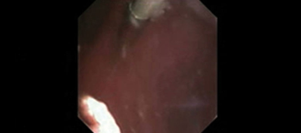
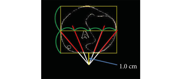
And finally we had two practice changing randomised trials in stone disease. Plasma vaporisation performed better than balloon dilatation for creating PCNL tracts [4]. For the curious, there is a video demonstrating the method if you wish to learn it.

The Portland trial has a simple message that you just can’t ignore; a single dose of NSAID before ureteric stent removal prevents severe pain afterwards. This is going to become standard of care if it has not already [5].

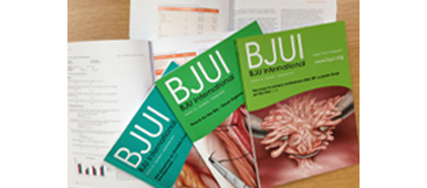
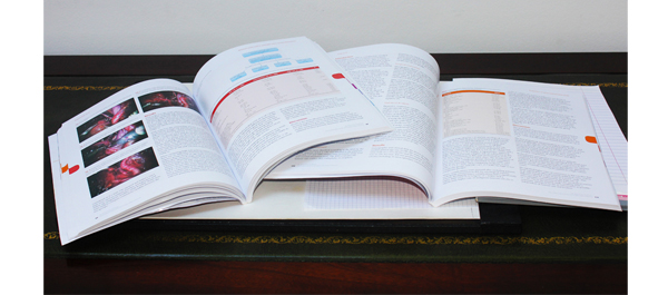
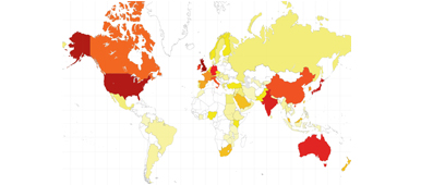
Many of our readers will wonder why we continue with a paper journal when the web has been so successful? The map here shows our global reach, which includes a number of subscribers who prefer to, or by necessity, read the print journal (∼30%). Moreover in a BJUI Online Poll, 75% of our readers reported taking the paper journal out of its plastic sheath and reading it, with over 50% doing so within a week. The transition will thus take longer and while the web remains our main portal, the beautifully designed paper BJUI will still land on your doorstep.

Prokar Dasgupta
Editor in Chief, BJUI

Guy’s Hospital, King’s Health Partners

References

  1. Eifler JB, Feng Z, Lin BM et al. An updated prostate cancer staging nomogram (Partin tables) based on cases from 2006 to 2011. BJU Int 2013; 111: 22–29
  2. Cooperberg MR, Ramakrishna NR, Duff SB et al. Primary treatments for clinically localised prostate cancer: a comprehensive lifetime cost-utility analysis. BJU Int 2013; 111: 437–450
  3. Paduch DA, Bolyakov A, Polzer PK, Watts SD. Effects of 12 weeks of tadalafil treatment on ejaculatory and orgasmic dysfunction and sexual satisfaction in patients with mild to severe erectile dysfunction: integrated analysis of 17 placebo-controlled studies. BJU Int 2013; 111: 334–343
  4. Chiang PH, Su HH. Randomized and prospective trial comparing tract creation using plasma vaporization with balloon dilatation in percutaneous nephrolithotomy. BJU Int 2013; 112: 89–93
  5. Tadros NN, Bland L, Legg E, Olyaei A, Conlin MJ. A single dose of a non-steroidal anti-inflammatory drug (NSAID) prevents severe pain after ureteric stent removal: a prospective, randomised, double-blind, placebo-controlled trial. BJU Int 2013; 111: 101–105
Original publication of this editorial can be found at: BJU Int 2013; 112: 1051–1052. doi: 10.1111/bju.12524


Editorial: Does HAL assistance improve outcomes in patients who receive postoperative intravesical therapy?

There is growing evidence that hexaminolevulinate (HAL) fluorescence cystoscopy increases detection of bladder cancer at the time of transurethral resection of bladder tumours (TURBT) and that this results in lower recurrence rates [1, 2]. One limitation in many prior studies was the lack of standardisation about the use of immediate postoperative chemotherapy, which has been shown to reduce recurrence in patients with non-muscle-invasive bladder cancer [3]. This raises the question of whether the benefit of HAL in reducing recurrences would be eliminated if patients did in fact receive postoperative intravesical chemotherapy, which would help eradicate any missed residual tumour.

There have been several studies that attempt to bring clarity to this issue. A study by Geavlete et al. [4] randomised 362 patients suspected of having bladder cancer to HAL vs white-light (WL) TURBT with a single postoperative mitomycin C instillation given in all cases. The authors found that the recurrence rate at 3 months was lower in the HAL group (7.2% vs 15.8%) due to fewer ‘other site’ recurrences when compared with the WL group. There continued to be an advantage for the HAL group with lower 1- and 2-year recurrence rates compared with the WL group (21.6% vs 32.5% and 31.2% vs 45.6%, respectively). The study did not stratify patients specifically to those with low-grade non-invasive tumours but patients with single tumors had a trend toward less recurrence (23.3% vs 35.3%, P = 0.064).

Grossman et al. [2] published the long-term follow-up for 551 patients enrolled in a prospective, randomised study of HAL vs WL for Ta or T1 urothelial bladder cancer with similar rates of intravesical therapy in the two groups (46% and 45%, respectively). They found that the median time to recurrence was 9.4 months in the WL group and 16.4 months in the HAL group (P = 0.04) but they did not report specifically on patients who received postoperative intravesical therapy. A meta-analysis of raw data from prospective studies on 1345 patients with suspected bladder cancer evaluating HAL-assisted cystoscopy vs WL found that both patients with low- and high-risk disease had statistically significant lower recurrence rates [1]. This meta-analysis was unable to stratify based on use of postoperative intravesical therapy.

O’Brien et al. [5] performed a randomised prospective study of HAL-assisted vs conventional WL TURBT, with all patients scheduled to get a single treatment of postoperative intravesical mitomycin C. There were 86 and 82 patients with cancer in the HAL and WL groups who completed the 12 months follow-up, respectively. In this study, 63% and 77% of the HAL and WL patients received mitomycin C, respectively. There was an increased detection of carcinoma in situ (CIS) in the HAL group (26% vs 14%) but no significant difference in recurrence at 3 and 12 months. When stratifying by low-grade tumours, the 3-month recurrence rates for HAL and WL were 19% vs 9% and at 12 months were 16% vs 22%, so that no significant differences were noted but the study was not powered to evaluate this subgrouping.

What can be concluded then about whether HAL assistance improves outcomes in patients who receive postoperative intravesical therapy? It appears the results are inconclusive and this is not surprising. The risk reduction of postoperative intravesical chemotherapy is primarily limited to patients with a single low-grade papillary tumour and one would need to treat 8.5 patients with peri-TUR chemotherapy to prevent one recurrence [3]. The benefits of peri-TUR chemotherapy in patients at intermediate- and high-risk are not well established [6]. Most of the studies of HAL-assisted TUR have not treated with postoperative intravesical therapy systematically. The studies that have tried to uniformly give postoperative therapy have not been sufficiently powered to evaluate those patients most likely to benefit, namely low-grade non-invasive cancer. As such, one cannot determine whether the benefit of potentially detecting additional low-grade tumours by HAL in patients with low-risk disease could be matched by postoperative intravesical therapy and such a study would require a very large number of low-grade solitary papillary tumours. However, it would minimise the benefits of HAL to focus on the benefit in the lowest risk patients. A meta-analysis of randomised trials found that HAL reduced the risk of recurrence independent of level of risk, such that there was reduced recurrence in patients with CIS, T1 and high-grade disease [1]. These are patients for which immediate postoperative intravesical therapy has shown minimal benefit and for which the benefit of HAL cannot be explained away. Furthermore a small but meaningful number of low-risk patients can be found to have intermediate- or high-risk disease, which would change their subsequent management [4]. As such if one had to choose between approaches rather than apply both, the use of HAL would appear to result in a greater benefit in managing patients with bladder cancer.

Yair Lotan
Department of Urology, UT Southwestern Medical Center at Dallas, Dallas, TX, USA

Read the full article

References

  1. Burger M, Grossman HB, Droller M et al. Photodynamic diagnosis of non-muscle-invasive bladder cancer with hexaminolevulinate cystoscopy: a meta-analysis of detection and recurrence based on raw data. Eur Urol 2013; 64: 846-54
  2. Grossman HB, Stenzl A, Fradet Y et al. Long-term decrease in bladder cancer recurrence with hexaminolevulinate enabled fluorescence cystoscopy. J Urol 2012; 188: 58–62
  3. Sylvester RJ, Oosterlinck W, van der Meijden AP. A single immediate postoperative instillation of chemotherapy decreases the risk of recurrence in patients with stage Ta T1 bladder cancer: a meta-analysis of published results of randomized clinical trials. J Urol 2004; 171: 2186–2190
  4. Geavlete B, Multescu R, Georgescu D, Jecu M, Stanescu F, Geavlete P. Treatment changes and long-term recurrence rates after hexaminolevulinate (HAL) fluorescence cystoscopy: does it really make a difference in patients with non-muscle-invasive bladder cancer (NMIBC)? BJU Int 2012; 109: 549–556
  5. O’Brien T, Ray E, Chatterton K, Khan S, Chandra A, Thomas K. Prospective randomized trial of hexylaminolevulinate photodynamic-assisted transurethral resection of bladder tumour (TURBT) plus single-shot intravesical mitomycin C vs conventional white-light TURBT plus mitomycin C in newly presenting non-muscle invasive bladder cancer. BJU Int 2013; 112:1096–1104
  6. Kamat AM, Lotan YR. Perioperative intravesical therapy after transurethral resection for bladder cancer. J Urol 2010; 183: 19–20

Editorial: Better late than early for long-term survival in patients with recurrence after renal carcinoma

In this paper, Brookman-May et al. [1] used a large multi-institutional database of over 13 000 patients from 23 centres in both Europe and the USA to examine the prognostic indicators of cancer-specific survival (CSS) in patients who had recurrence after primary surgery for RCC. Their analysis was based on a subset of 1712 patients who had recurrence during a median follow-up period of 50 months. All patients had undergone either radical nephrectomy or nephron-sparing surgery, with no evidence of metastasis at the time of surgery.

The authors have previously shown, in a related study based on a subset of 5000 patients from the same database, that lymphovascular invasion, Fuhrman grade 3–4, and pT stage > pT1 at the time of diagnosis were significantly associated with the development of late recurrence (defined as after >5 years) [2]. In this paper, the primary objective was to look at the effect of time to tumour recurrence (TTR) on CSS. In addition, clinical and histopathological comparisons were made between patients with early (<5 years) and late recurrence (>5 years).

Patients often want to know whether if they are recurrence-free after a period of time, their subsequent risk of dying from recurrence is reduced; this paper goes some way towards answering this question and showing that those with later recurrence had improved survival times. Specifically, the authors found that TTR was an independent predictor of CSS; i.e. if patients recurred early they had a worse CSS than those recurring late. This is similar to results from another group who reported that recurrent disease, particularly before 12 months, was associated with a poorer prognosis [3]. In the first 4 years of follow-up, a shorter TTR independently predicted lower CSS after recurrence [1]. When divided into those with early recurrence, Group A (N = 1402), and those with late recurrence, Group B (N = 310), patients in Group A were more likely to be male, of advanced age, have a greater tumour diameter and stage, have Fuhrman grade 3–4, with lymphovascular invasion and positive lymph node disease, than those in Group B. Patients in Group A had a 3-year CSS of 30% compared with those in Group B whose CSS was better at 41%. Age and gender were also independent predictors of CSS.

These results can help to guide the aftercare management of patients after primary surgery. Currently, primary surgery is the only recommended option for patients with localized RCC, although results from several phase III clinical trials looking at the role of adjuvant therapy, such as the SORCE, PROTECT and S-TRAC trials, are still awaited [4]. Furthermore, it is not known which group of patients are suitable for adjuvant chemotherapy, which is reflected in the subtly differing eligibility criteria for recruitment to the various trials [4]. The authors of the present study pointed out that a method of risk stratification may be useful to allow equal representation of early and late recurrence patients in treatment arms for clinical trials. Potentially, understanding the predictors of early recurrence may help to identify patients for whom adjuvant therapy may be beneficial.

Only 12% of patients with localized RCC in the present cohort developed recurrence after surgery [1]. This rate is lower than that found in the literature, where 20–30% recurrence rates of localized RCC have been reported [2, 5, 6]. Brookman-May et al. speculate that this lower rate is attributable to both an increase in early detection as well as improved surgical management in recent years. Furthermore, they acknowledge that the database is heterogeneous and that the study therefore has all the inherent limitations of a retrospective study.

The present paper clearly shows that the earlier the recurrence after surgery the lower the survival rate, but a clear strategy for the surveillance of localized RCC after primary surgery is currently lacking. Most follow-up protocols exercise a blanket ‘one for all’ policy with follow-up spaced at regular intervals to ensure patients who recur are detected early. Such a policy may not be intensive enough to detect early recurrence in some patients and may be excessive for the majority of patients where the risk of recurrence is low. Risk stratification of patients, by understanding the predictors of CSS after surgery, may help to tailor surveillance protocols to the individual and identify those for whom adjuvant therapy may be beneficial.

Kathie Wong and Ben Challacombe
The Urology Centre, Guy’s Hospital, Guy’s and St Thomas’ NHS Foundation Trust, London, UK

Read the full article

References

  1. Brookman-May S, May M, Shariat S et al. Time to recurrence is a significant predictor of cancer-specific survival after recurrence in patients with recurrent renal cell carcinoma – results from a comprehensive multi centre database (CORONA/SATURN Project). BJU Int 2013; 112: 909–916
  2. Brookman-May S, May M, Shariat SF et al. Features associated with recurrence beyond 5 years after nephrectomy and nephron-sparing surgery for renal cell carcinoma: development and internal validation of a risk model (PRELANE score) to predict late recurrence based on a large multicenter database (CORONA/SATURN Project). Eur Urol 2012; 64: 472–477
  3. Rodriguez-Covarrubias F, Gomez-Alvarado MO, Sotomayor M et al. Time to recurrence after nephrectomy as a predictor of cancer-specific survival in localized clear-cell renal cell carcinoma. Urol Int 2011; 86: 47–52
  4. Kim SP, Crispen PL, Thompson RH et al. Assessment of the pathologic inclusion criteria from contemporary adjuvant clinical trials for predicting disease progression after nephrectomy for renal cell carcinoma. Cancer 2012; 118: 4412–4420
  5. Hollingsworth JM, Miller DC, Daignault S, Hollenbeck BK. Five-year survival after surgical treatment for kidney cancer: a population-based competing risk analysis. Cancer 2007; 109: 1763–1768
  6. Breda A, Konijeti R, Lam JS. Patterns of recurrence and surveillance strategies for renal cell carcinoma following surgical resection. Expert Rev Anticancer Ther 2007; 7: 847–862

Editorial: Totally X-ray-free percutaneous nephrolithotomy: caveat emptor

In the accompanying paper, Yan et al. [1] present the outcomes of their study on percutaneous nephrolithotomy (PCNL) guided only by ultrasonography (US).

This is the largest series (705 patients) to date on PCNL purely under US control and reports stone-free and complication rates that are consistent with those commonly reported for PCNL guided by X-ray or by a combination of X-ray and US.

Since its introduction more than three decades ago, PCNL has traditionally been performed under fluoroscopic control by the majority of urologists, even though US guidance has now gained wide acceptance as a means of achieving renal access. Now, the most important international guidelines suggest that US be used in addition to fluoroscopy [2]. US guidance has the following advantages: it minimizes radiation exposure, allows the detection of viscera that can sometimes lie in the trajectory of the puncturing needle and avoids contrast-related complications. Furthermore, US provides imaging of the collecting system in three-dimensional orientations and helps to distinguish between anterior and posterior calyces with great accuracy. Nevertheless, the innovative concept proposed by Yan et al. [1], with their impressive series, concerns the whole procedure (puncture, creation of renal access and final look to rule out eventual residual fragments), and not only the safe accomplishment of the puncture solely under US guidance.

Caution should be taken in interpreting their results. This is a purely retrospective study which guarantees only a low level of evidence (3B). In addition, even though major complications arising during the creation of access were not reported in the paper, doubts remain about the safety of using only US guidance in monitoring the dilatation process by either balloon or coaxial dilators. The following questions still need to be addressed. How can the progression of dilators be monitored to avoid excessive inadvertent medial advances with the accompanying high risk of collecting system perforation? How can false passage of a working guidewire be detected early by US? What about obese patients in whom the effectiveness of US is generally impaired?

To balance the risks and benefits of guidance solely by US, a middle ground could be represented by US guidance aided by ureteroscopic monitoring of the dilatation process using the so-called ‘Endovision technique’ [3], as is possible during endoscopic combined intrarenal surgery (ECIRS) (Fig. 1). In view of the risks, it should be stressed that, even though PCNL guided solely by US is an attractive option, biplanar C-arm fluoroscopy should always be present in the operating room.

It is well known that radiation hazard is directly proportional to cumulative radiation exposure time, so US guidance provides an obvious advantage in terms of absence of radiation for patient and operating room staff, but is the extent of this advantage really known? It is important to underline that the amount of radiation exposure during PCNL is not particularly great, measuring on average 0.56 mSv for the patient and 0.28 mSv for the urologist [4]. By contrast, unenhanced CT involves a significant radiation exposure of 8.6 mSv [5], which is of course particularly relevant for patients with stones, who are often quite young and likely to experience recurrence. According to the ‘as low as reasonably achievable’ (or ALARA) principle, replacing CT scans with US in the follow-up would have a much greater impact on reducing radiation exposure in adult patients (in the present series patients undergo two CT scans after surgery, at 48 h and 4 weeks, and one preoperative CT scan!) than would renouncing the safety guaranteed by X-ray monitoring during endourology.

Finally, it is of paramount importance to stress that, in the current climate in which malpractice litigation related to endourology continues to rise [6], it is still advisable that PCNL guided solely by US should be performed only in trials for which approval of the local institutional review board has been obtained.

To conclude, Yan et al. [1] propose an alternative approach to PCNL that involves solely US guidance, but some doubts remain. Only further well designed, prospective, comparative and possibly randomized studies will allow us to draw definitive conclusions.

Guido Giusti
Head of Stone Center & European Training, Center in Endourology, Humanitas Clinical and Research Center, Milan, Italy

Read the full article

References

  1. Yan S, Xiang F, Yongsheng S. Percutaneous nephrolithotomy guided solely by ultrasonography: a 5-year study of >700 cases. BJU Int 2013; 112: 965–971
  2. Türk C, Knoll T, Petrik A et al. 2013 EAU Guidelines on Urolithiasis
  3. Scoffone CM, Cracco CM et al. Endoscopic Combined intrarenal surgery in galdakao-modified supine valdivia position: a new standard for percutaneous nephrolithotomy? Eur Urol 2008; 54: 1393–1403
  4. Kumari G, Kumar P, Wadhwa P, Aron M, Gupta NP, Dogra PN. Radiation exposure to the patient and operating room personnel during percutaneous nephrolithotomy. Int Urol Nephrol 2006; 38: 207–210
  5. Katz SI, Saluja S, Brink JA, SForman HP. Radiation dose associate with unenhanced CT for suspected renal colic: impact of repetitive studies. AJR Am J Roentgenol 2006; 186: 1120–1124
  6. Duty B, Okhunov Z, Okeke Z, Smith A. Medical malpractice in endourology: analysis of closed cases from the State of New York. J Urol 2012; 187: 528–532

Editorial: How many cores are needed to detect nearly all prostate cancers?

Virtual prostate biopsy and biopsy simulation: lessons to be learned

Prostate biopsies, transrectal or transperineal, still constitute the pillars of prostate cancer detection today [1]. With the lack of reliable imaging tools (new MRI techniques are promising but still investigational [2]); random biopsies offer the sole adequate cancer detection option [3]. However, random biopsies are far from efficient in detecting all tumours and even less efficient in detecting all significant cancer ‘spots’. To improve sensitivities and specificities, increasing the biopsy core numbers, targeting more lateral aspects and encouraging repeat biopsies have been recommended [4]. Recently, HistoScanning™ [5] and template biopsies [6] have been introduced to further improve biopsy quality and efficiency. The latest innovations include the fusion of MRI pictures with the TRUS image to offer optimal targeting of suspicious areas [7]. And yet, these efforts are far from solving the main problem. How can we perform a biopsy and be confident to detect most of the cancers, i.e. significant malignant areas.

The present study [1] does, what should have been done a long time ago, namely to create a reliable and reproducible biopsy simulation model to allow the investigation of various biopsy schemes, core lengths and numbers. Based on a series of 109 radical prostatectomy specimens, a three-dimensional (3D) prostate and prostate cancer model was created using novel 3D slicer software and various prostate biopsy schemes were simulated. Using this method, the detection rate for tumours with a tumour volume (TV) of ≥0.5 mL plateaued at 77% (69 of 90) using a 12 core (3 × 4) scheme, standard 17-mm biopsy cutting length without anteriorly directed biopsy (ADBx) cores. Twenty of 21 (95%) tumours with a TV of ≥0.5 mL not detected by this scheme originated in the anterior peripheral zone or transition zone [1].

Confirming our earlier data with the Vienna nomograms [8], increasing the biopsy cutting length and depth/number of ADBx cores (14–18 cores) improved the detection rate for tumours with a TV of ≥0.5 mL in the 12-core scheme [1]. The best biopsy scheme used a 22-mm cutting length and a 12-core scheme with additional volume-adjusted ADBx cores. Using this combination, 100% of ≥0.5 mL tumours in prostates <50 mL in volume and 94.7% of ≥0.5 mL tumours in prostates >50 mL in volume were detected.

Certainly, these numbers will not be reproducible in real-time TRUS or transperineal biopsies (detections rates of 95–100% as seen in this simulation model, cannot be achieved without adequate imaging tools, which are not available yet), but they aid significantly in rethinking our biopsy strategy. So, if we summarise the present findings and combine them with published data, the future will demand a TRUS-fusion biopsy technique, involving 14–18 cores (or more if volume increases), involving the anterior zones of the prostate and using a 22-mm cutting length of the biopsy core vs a 15–17 mm core as is used currently. Obviously real-time prospective trials are needed to confirm these findings but nothing indicates that the outcome would be otherwise.

Bob Djavan
Department of Urology, New York University School of Medicine, NYU, New York, NY, USA

Read the full article

References

  1. Kanao K, Eastham JA, Scardino PT, Reuter VE, Fine SW. Can transrectal needle biopsy be optimised to detect nearly all prostate cancer with a volume of ≥0.5 mL? A three-dimensional analysis. BJU Int 2013; 112: 898–904
  2. Delongchamps NB, Peyromaure M, Schull A et al. Pre-biopsy Magnetic Resonance Imaging and prostate cancer detection: comparison of random and MRI-targeted biopsies using three different techniques of MRI-TRUS image registration. J Urol 2013;189: 493–499
  3. Djavan B, Rocco B. Optimising prostate biopsy. BMJ 2011; 344: d8201
  4. Thompson I, Thrasher JB, Aus G et al. Guideline for the management of clinically localized prostate cancer: 2007 update. J Urol 2007; 177: 2106–2131
  5. Simmons LA, Autier P, Zát’ura F et al. Detection, localisation and characterisation of prostate cancer by prostate HistoScanning(™)BJU Int 2012; 110: 28–35
  6. Huo AS, Hossack T, Symons JL et al. Accuracy of primary systematic template guided transperineal biopsy of the prostate for locating prostate cancer: a comparison with radical prostatectomy specimens. J Urol 2012; 187: 2044–2049
  7. Sonn GA, Natarajan S, Margolis DJ et al. Targeted biopsy in the detection of prostate cancer using an office based magnetic resonance ultrasound fusion device. J Urol 2013; 189: 86–92
  8. Djavan B, Margreiter M. Biopsy standards for detection of prostate cancer. World J Urol 2007; 25: 11–17

The impact of the BJUI and what influences it today: does impact factor matter?

Over the last decade, urological researchers have been increasingly interested with, and driven by, the impact factor (IF) of the journal to which they are submitting. This bibliometric tool measures the way in which a journal receives citations of its articles over time. IF is calculated by dividing the number of current citations a journal receives for articles published in the two previous years, by the number of articles published in those same years.

Although IF represents a proxy for the popularity of a journal within its field, several academic and scientific organizations now use the IF to judge the value of a scientist or of a research team using it for national and international academic evaluations. This questionable policy has generated a vicious circle that has driven authors to prefer journals with higher IFs and, consequently, journal editorial boards (and publishers) to plan (soft or strong) strategies to increase this index. As a result a higher IF attracts the best articles in the field and increases the number of subscriptions to a journal. There are a number of potential biases influencing the IF values including self-citation, the number of articles published per year, and the type of articles accepted. We will explain how all of these nuances can play a significant role in calculating the IF.

Some journals subtly suggest that authors and reviewers cite articles published in their own journal within the references of newly submitted papers. This slightly dubious practice can bias the true value of the IF. Reassuringly when looking at the urological journals, the self-citation factor generally seems to play a limited role, as most journals have a percentage <10%. The policy of the BJUI Editorial Board does not support a self-citation practice. The decision to start each BJUI issue with some editorial comments (the Editor’s Choice section) is only to offer to readers the opportunity to have expert comment on the most important papers published within each edition. Indeed, the invited authors are only requested to cite the featured article and no others from the BJUI.

The number of papers published per journal volume and throughout each year is another significant factor influencing the IF value. Table 1 clearly shows the wide variability in the number of papers published from 2010–2011 in the different urologic journals. The new BJUI policy is to significantly reduce the number of published papers/year. Reflecting this decision, the BJUI Editorial Board has agreed to significantly improve the review process with the aim of selecting only the most relevant and original of the submitted manuscripts. A new rapid triage review process should allow us to select only the best 30–40% of submitted manuscripts to send to 3–4 experts for a more focused and precise review process. This mechanism has produced a significantly increased rejection rate in favour, we hope, of a better selection of topics and papers for our readership [2].

Table 1. Items cited in 2012 and items published in 2010–2011 in the most important urological journals. Data from ISI Web of Journal Citation Reports (JCR)
Abbreviated Journal Title Cites in 2012 Items published in 2010–2011 Impact Factor
EUR UROL 4.662 445 10.476
NAT REV UROL 580 121 4.793
PROSTATE 1.395 963 3.843
J UROLOGY 4.864 1316 3.696
J SEX MED 2.638 751 3.513
BJU INT 3.323 1091 3.046
WORLD J UROL 673 233 2.888
UROLOGY 2.843 1173 2.424
CURR OPIN UROL 360 164 2.195


Bibliometric analyses have shown that review articles are cited more frequently than full original research papers. Therefore publishing good quality review articles written by expert opinion leaders in the field represents an excellent strategy to increase a journal’s IF. Although, we recognize the impact of review articles on IF, the current policy of the BJUI remains unchanged with only relatively few review articles included in each issue. As a result we will continue to give maximal attention to the clinical and basic research papers.

Finally the IF is in many ways only an index of the popularity of a journal because it equally weights citations from highly reputed journals alongside citations from more obscure journals [1]. However a journal’s true credit is also based on the prestige of the citing journals and the Eigenfactor scores is currently used to reflect this measure. Table 2 shows that the BJUI is third of all urological journals according to this less popular bibliometric tool. Another contemporary measure of impact, particularly influenced through the internet is the “Klout Score”. This system, which uses social media analytics to rank users according to online social influence via the Klout Score, giving a numerical value between 1 and 100. The BJUI currently has a score of 56, higher than its contemporaries. Therefore we can conclude that the BJUI today is a journal with a good reputation throughout the urologic field.

Table 2. Relationship between prestige (Eigenfactor® Score) and popularity (Impact Factor score) of urological journals. Data from ISI Web of Journal Citation Reports (JCR)
Rank Abbreviated Journal Title Eigenfactor® Score Impact Factor IF rank
 1 J UROLOGY 0.08109 3.696 4
 2 EUR UROL 0.05503 10.476 1
 3 BJU INT 0.04248 3.046 7
 4 UROLOGY 0.03896 2.424 11
 5 J SEX MED 0.01738 3.513 6
 6 PROSTATE 0.01624 3.843 3
 7 J ENDOUROL 0.01571 2.074 15
 8 NEUROUROL URODYNAM 0.00897 2.674 10
 9 WORLD J UROL 0.00750 2.888 8
10 INT J UROL 0.00582 1.734 16


The editorial board of a traditional urological journal like the BJUI must take into consideration both the IF and other scoring systems as indicators of its popularity and prestige. The strategies we employ to give better bibliometric parameters should predominantly reflect an increase in the quality of the papers published as we must remember that the journal is primarily produced for the readership and not just for those who wish to publish in it [3].

Vincenzo Ficarra1, Associate Editor,
Ben Challacombe2, Associate Editor,
Prokar Dasgupta2, Editor in Chief

1Department of Experimental and Clinical Medical Sciences, Urology Unit, University of Udine, Italy. 2King’s Health Partners, London UK

References

  1. Franceschet M. The difference between popularity and prestige in the sciences and in the social sciences: a bibliometric analysis. J Informetr 2010; 4: 55–61
  2. Dasgupta P. The most read surgical journal on the web. BJU Int 2013; 111: 1–3
  3. Schulman CC. What you have always wanted to know about the impact factor and did not dare to ask. Eur Urol 2005; 48: 179–181

Original publication of this editorial can be found at: BJU Int 2013; 112: 873–874, doi: 10.1111/bju.12472

Editorial: Regaining continence after radical prostatectomy: RARP vs. ORP

Functional outcomes represent relevant criteria to evaluate the success of radical prostatectomy (RP) in the treatment of localised and locally advanced prostate cancer. Indeed, while the primary goal of RP remains the complete extirpation of the primary tumour, patients’ satisfaction can be negatively affected by urinary incontinence and/or erectile dysfunction after RP.

In this issue of BJUI, Geraerts et al. [1] evaluated urinary continence recovery and voiding symptoms in a well-conducted, single-centre, prospective non-randomised study comparing two contemporary series of patients who underwent either open retropubic RP (RRP) or robot-assisted RP (RARP) for clinically localised or locally advanced prostate cancer. Patients were assigned to each group according to their or their surgeon’s preference. High-risk prostate cancers were preferably treated with an open access to offer a more accurate extended lymphadenectomy. The study showed that the urinary continence recovery rate was significantly shorter in the RARP group than in the RRP group (16 vs 46 days; P = 0.008). Interestingly, the RA approach remained an independent predictor of time to urinary continence recovery on multivariable Cox regression analysis (P = 0.03; hazard ratio [HR] 1.52, 95% CI 1.03–2.26). Therefore, this study confirmed previously published results. In 2003, Tewari et al. [2] reported a shorter time to recovery of urinary continence in patients who underwent RARP (44 days) than those who received RRP (160 days). In 2008, Kim et al. [3] reported a median time to continence in RARP patients of 1.6 months, significantly lower than the 4.3 months in the RRP patients. Interestingly, Geraerts et al. [1] identified other independent predictors of time to continence, such as patient’s age >65 years (P = 0.02; HR 0.67, 95% CI 0.45–0.96) and the preoperative continence status (P = 0.004; HR 1.69, 95% CI 1.18–2.43). In all, 28% of patients who received RRP and 34% of those who underwent RARP were preoperatively defined as incontinent using a symptom-specific questionnaire [1]. These patients classifiable as ‘Cx’ according to the Survival, Continence and Potency (SCP) classification [4] represent a confounding population in the Geraerts et al. study who should be evaluated separately.

An interesting question is whether the reported difference in time to continence in favour of RARP is also significant from the clinical perspective. Urinary continence in patients who underwent RARP recovered 1 month early than those treated with traditional RRP. The King’s Health questionnaire seems to confirm a positive effect of this outcome on the patient’s quality of life (QoL). Indeed, there were better results in the RARP compared with RRP group at 1 and 3 months after RP. Moreover, at 12 months after RP, patients who underwent RRP were more physically limited (P = 0.01) and took more precautions to avoid urine loss (P = 0.01) than those who received a RARP [1]. These data seem to be in conflict with the reported overlapping 12-month urinary continence rates (96% in RRP and 97% in RARP group). Moreover, looking at the 12-month urinary continence rate, the Geraerts et al. study does not confirm the results of a recent cumulative analysis of available comparative studies showing a better 12-month urinary continence rate after RARP compared with RRP (odds ratio 1.53; P = 0.03) [1].

Interestingly, the 12-month urinary continence rate reported after RRP by Geraerts et al. is significantly higher (96%) than the values reported in the comparative studies included in the meta-analysis (88.7%) and in the most important and recent RRP non-comparative series (60–93%) [5]. This aspect appears to confirm the important role of surgeon experience. Indeed, in this Belgium series most of the open procedures were performed by an expert surgeon with experience of >3000 RRPs, and thus able to reach excellent functional outcomes for urinary continence recovery. In favour of robotic surgeons, we could consider that they were able to reach overlapping results after <200 cases.

In conclusion, the study published by Geraerts et al. [1] showed that modern RP in expert hands is able to achieve excellent results for urinary continence recovery regardless of the approach. However, pure and RA laparoscopy has pushed open surgeons to improve technical and postoperative aspects to achieve comparable outcomes. RARP can offer some advantages over traditional RRP, above all for the time to reach urinary continence. This advantage seems to have generated a better QoL profile in patients who underwent RARP at 12 months after RP.

However, the choice between the two techniques must be taken according to all the most relevant parameters including perioperative, functional (continence and potency) and oncological outcomes. Therefore, we strongly support the publication of clinical series or comparative studies reporting results according to the ‘trifecta’, ‘pentafecta’ or SCP systems [6].

Vincenzo Ficarra°, Alessandro Iannettiand Alexandre Mottrie
OLV Vattikuti Robotic Surgery Institute, Aalst, Belgium, °Department of Experimental and Clinical Medical Sciences – Urology Unit – School of Medicine, University of Udine, 
and *Department of Surgical, Oncologic and Gastrointestinal Sciences, Padua, Italy

Read the full article

References

  1. Geraerts I, Van Poppel H, Devoogdt N, Van Cleynenbreugel B, Joniau S, Van Kampen M. Prospective evaluation of urinary incontinence, voiding symptoms and quality of life after open and robot-assisted radical prostatectomyBJU Int 2013; 112:936–943
  2. Tewari A, Srivasatava A, Menon M, Members of the VIP Team. A prospective comparison of radical retropubic and robot-assisted prostatectomy: experience in one institutionBJU Int 2003; 92: 205–210
  3. Kim SC, Song C, Kim W et al. Factors determining functional outcomes after radical prostatectomy: robot-assisted versus retropubicEur Urol 2011; 60: 413–419
  4. Ficarra V, Sooriakumaran P, Novara G et al. Systematic review of methods for reporting combined outcomes after radical prostatectomy and proposal of a novel system: the survival, continence, and potency (SCP) classificationEur Urol 2012; 61:541–548
  5. Ficarra V, Novara G, Rosen RC et al. Systematic review and meta-analysis of studies reporting urinary continence recovery after robot-assisted radical prostatectomyEur Urol 2012; 62: 405–417
  6. Ficarra V, Borghesi M, Suardi N et al. Long-term evaluation of survival, continence and potency (SCP) outcomes after robot-assisted radical prostatectomy (RARP)BJU Int 2013; 112: 338–345

Editorial: Three robotic surgery training methods: is there a clear winner?

All training adds value. A craft-based specialty such as surgery has always recognised this. The advent of advanced minimally invasive surgical technology and techniques has provided both new challenges and new opportunities for surgical performance and for the delivery of training. Conceptually, we have moved from the Halstedian model of ‘See one, do one, teach one’ [1] to an environment where skills are acquired away from the operating room in simulator, inanimate and in vivo (animal) laboratory training sessions. Increased scrutiny of credentialling and medico-legal aspects of robotic surgery have reinforced the importance of training and have led to a number of papers outlining pathways to facilitate this [2, 3].

In the present paper, Hung et al. evaluate the construct validity of three standardised training methods (inanimate, simulator and in vivo) and also compare the three different platforms for cross-method training value. As others have shown, the latest generation of robotic surgery simulators have high face, content and construct validity [4, 5] and the present paper confirms the value of both inanimate and simulator training for novice surgeons. In addition, the authors confirmed the construct validity of a simple in vivo exercise using the daVinci© surgical system by demonstrating that experts outperformed novices. Using Spearman’s rank correlation coefficient, the authors compared the three training methods under evaluation and concluded that they were strongly correlated for construct validity between exert and novice surgeons. While construct validation of these exercises may be established, are they useful for experts? Until realistic virtual reality surgical simulations are available, only a novice, an inexperienced or an occasional robot-assisted surgeon may benefit from virtual reality exercises.

What are we therefore to conclude from this? For certain, the advent of excellent surgical simulators and structured inanimate exercises has provided tools for novice surgeons to acquire console skills in a safe and structured environment. This will enhance their operating performance and reduce aspects of the learning curve such as operating time; however, the lack of availability of in vivo training opportunities greatly limits the applicability of this method of surgical training. In many countries (including Australia and the UK), this type of training is illegal or not available. The robotic surgery industry has strongly recommended that in vivo training should be undertaken in one of their official training facilities before surgeons are given the credentials to use this technology; however, even in the USA where most of these facilities are located, key leaders within the AUA have called for the awarding of credentials for robotic surgery ‘not to be an industry driven process, but one that is a result of a standardized, competency based, peer evaluation system’ [2]. Notably, the current AUA Standard Operating Practices (guidelines) for the awarding of credentials for robotic surgery list in vivo training as being optional.

Our view is that although all training has value, there is not enough evidence that in vivo training (particularly on an animal with a rudimentary prostate), which requires international travel and considerable expense, adds sufficient value to be mandatory in any credentialling process. In fact, we have dropped the requirement to complete in vivo training from our requirements at major robotic surgery centres in Australia in favour of structured Mini-Fellowship training [6]. Hung et al. have confirmed what we already knew, which is that all training adds value; however it is likely that only simulator and inanimate training adds enough value to be incorporated into standardised training in robotic surgery.

The multi-disciplinary ‘Fundamentals of Robotic Surgery’ (FRS) curriculum being created by Dr Richard Satava and associates is working on psychomotor skills tasks that include inanimate models as well as corresponding virtual reality exercises. Multi-institutional validation of the FRS or similar curricula will allow the establishment of training milestones and proficiency benchmarks. We must continue to strive for further development of robotic and surgical simulation to change the training paradigm so that surgical training does not need to be at the expense, however minor, of increased operating time or adverse patient outcome.

Declan G. Murphy* and Chandru P. Sundaram
*Peter MacCallum Cancer Centre, Division of Cancer Surgery, University of Melbourne, Australian Prostate Cancer Research Centre, Epworth Richmond Hospital, Melbourne, Australia, and Department of Urology, Indiana University, Indianapolis, IN, USA

Read the full article

References

  1. Halsted WS. The training of the surgeon. Bull Johns Hop Hosp 1904; XV: 8
  2. Lee JY, Mucksavage P, Sundaram CP, McDougall EM. Best practices for robotic surgery training and credentialingJ Urol 2011;185: 1191–1197
  3. Zorn KC, Gautam G, Shalhav AL et al. Training, credentialing, proctoring and medicolegal risks of robotic urological surgery: recommendations of the society of urologic robotic surgeonsJ Urol 2009; 182: 1126–1132
  4. Finnegan KT, Meraney AM, Staff I, Shichman SJ. da Vinci Skills Simulator construct validation study: correlation of prior robotic experience with overall score and time score simulator performanceUrology 2012; 80: 330–335
  5. Abboudi H, Khan MS, Aboumarzouk O et al. Current status of validation for robotic surgery simulators – a systematic reviewBJU Int 2013; 111: 194–205
  6. Melbourne Uro-Oncology Training Program. Robotic surgery training. Available at: https://www.declanmurphy.com.au/training. Accessed 28 February 2013

Editorial: Does inflammation reduce the risk of prostate cancer?

Chronic inflammation is thought to play an aetiological role in tumorigenesis in several cancers including bladder, oesophagus and liver [1]. Molecular studies show that it plays a critical role in several stages of the carcinogenic process including tumour initiation, promotion, metastases and response to therapy. However the role in prostate cancer is less clear and to date, clinical studies are inconclusive.

The article in this issue of BJUI by Yli-Hemminki et al. [2] appears to show an inverse association with histological inflammation and the risk of prostate cancer. Using data from the Finnish subgroup of the European Randomised Study of Prostate Cancer Screening (ERSPC) study they examined 293 patients with previous negative biopsies over a 10.5-year period and reported an 18% risk of prostate cancer in men with inflammation on initial biopsy (34 of 101 men) as opposed to 27% in those without inflammation (51 of 192 men). Perhaps somewhat surprisingly, histological inflammation did not appear to be significantly associated with PSA concentration, although it did appear that the free/total PSA ratio was higher in men with inflammation.

One potential confounding factor is that inflammation may also play a role in the pathogenesis of BPH. A large scale study showed that the odds ratio for BPH was 8.0 with a history of prostatitis [3]. Furthermore, the Medical Therapy of Prostatic Symptoms (MTOPS) study showed that men with inflammation had a significantly higher risk of BPH progression and acute urinary retention. These factors may impact on PSA levels and the chance of a subsequent prostate biopsy. However, the authors report that the inverse association of prostate cancer and inflammation did not alter when corrected for prostate volume, PSA level and age.

Significantly, those patients who screened positive at first biopsy were already excluded as were those with a suspicion of prostate cancer, such as a small atypical focus. Despite this, the study group probably represents high-risk patients, as they had all previously met the criteria for the first round of biopsies. This is borne out by the fact that the risk of prostate cancer was significantly increased when the men with inflammation on biopsy were compared with the initially screened negative men (hazard ratio 4.3). Unfortunately, we do not know what the rate of inflammation was in the control arm as they were screened negative and hence no biopsies were taken.

The significance of PSA level or prostatic intraepithelial neoplasia was not specifically investigated, although the reported rates were 32.1% and 7.5% respectively. Overall the incidence of inflammation was reported as 65%, but this included both acute and chronic inflammation. A smaller number of patients were given grade 2/3 chronic inflammation (80 and 20 patients, respectively) and only 14 patients had grade 2/3 acute, indicating most had milder degrees of inflammation. Whether this is a representative sample is not entirely clear. As the authors comment, published rates of histological inflammation do vary significantly from 8 to 99% and this does appear to vary according to detection method.

Several case-control studies and a meta-analysis [4] have shown that there is a significant increase in the relative risk of prostate cancer in men with prostatitis; however, these epidemiological studies all suffer from selection bias, in that men with clinical symptoms are more likely present and to be investigated and followed up by a Urologist. This study [2] only examines the role of histological inflammation and, at least partially, removes the selection bias associated with clinical symptoms, although it could still be argued that men with clinical prostatitis may be more likely to present for screening. This study represents a highly selected group of patients that are high risk for prostate cancer and no firm conclusions can be drawn on the general population. As inflammation is so common in prostate specimens, further high-quality large-scale studies are needed with similar long-term follow-up.

Miles A. Goldstraw and Roger S. Kirby
The Prostate Centre, London, UK

Read the full article

References

  1. Coussens LM, Werb Z. Inflammation and cancer. Nature 2002; 420: 860–867
  2. Yli-Hemminski T, Laurila M, Auvinen A et al. Histological inflammation and risk of subsequent prostate cancer among men with initially elevated serum prostate-specific antigen (PSA) concentration in the Finnish prostate cancer screening trial. BJU Int 2013; 112: 735–741
  3. Alcarez A, Hammerer P, Tubaro A, Schroder FH, Castro R. Is there evidence of a relationship between benign prostatic hyperplasia and prostate cancer? Findings of a literature review. Eur Urol 2009; 55: 864–875
  4. Dennis LK, Lynch CF, Torner JC. Epidemiologic association between prostatitis and prostate cancer. Urology 2002; 60: 78–83

Editorial: Is FDG-PET/CT ready for prime time?

Fluorodeoxyglucose positron-emission tomography (FDG PET)/computed tomography (CT) in bladder cancer

In this month’s issue Mertens et al. [1] present a retrospective analysis of the clinical impact of fluorodeoxyglucose positron-emission tomography (FDG PET)/CT in 96 patients with muscle-invasive bladder cancer. Muscle invasion is present in ≈30% of patients presenting with bladder cancer and is associated with a higher incidence of nodal and metastatic disease than non-muscle-invasive tumours [2]. Accurate staging in this patient group will influence management decisions to proceed to local therapies, to instigate neoadjuvant treatment before local therapy, or to offer palliative chemotherapy where there is imaging evidence and subsequent confirmation of metastatic disease [2].

While there have been a few previous studies investigating FDG PET or FDG PET/CT for staging bladder cancer [3-7], with reported sensitivities and specificities ranging from 60 to 81% and 67 to 94% respectively, to date there are few data describing the impact on clinical management. A recent FDG PET/CT study of 57 patients with bladder cancer [3] reported that management was changed in 68% of cases after PET suggesting that FDG PET/CT has a substantial impact on the management of these patients. However, most patients in that study underwent FDG PET/CT for a suspected recurrence (72%) and the remainder for initial staging (21%) or post-treatment monitoring (chemotherapy or radiotherapy; 7%); 44% of patients had metastatic disease.

In the study reported by Mertens et al. [1], clinical data obtained in 96 patients during the patients’ clinical pathway were reviewed retrospectively. FDG PET/CT staging with standard contrast-enhanced CT was discordant in 22% of cases (21 patients), where PET/CT predominantly upstaged patients, consistent with the previous reports [3, 4]. After PET/CT, the treatment recommendations changed in 13.5% (13 patients) due to disease upstaging. In seven of the 13 patients treatment recommendations altered from local to palliative, due to the presence of metastatic disease, and in the remaining six of the 13 patients, neoadjuvant treatment was recommended in addition to planned local therapy. In another four patients management changed as a consequence of detecting other incidental primary tumours with FDG PET/CT.

However, the final clinical impact of FDG PET/CT may be less. When actual treatment changes were recorded, in only eight of these 13 patients were the recommendations implemented, due to patient co-morbidity or patient wishes in the remainder, e.g. FDG PET/CT changed actual treatment in only 8% in this study (eight of 96 patients). Including the four patients in whom incidental other primary tumours were discovered, the management impact of FDG PET/CT was 12.5%.

There is no doubt that from current published data and supported by this study by Mertens et al. [1] that FDG PET/CT improves staging in bladder cancer due to its higher sensitivity for metastatic disease. However, the actual change in management is relatively low and more prospective data will be required to confirm its clinical and cost effectiveness in terms of outcome, both in a single and multicentre setting.

Vicky Goh* and Gary Cook*
*Division of Imaging Sciences and Biomedical Engineering, King’s College London, Department of Radiology, and Clinical PET Imaging Centre, Guy’s and St Thomas’ Hospitals NHS Foundation Trust, London, UK

Read the full article

References

  1. Mertens L, Fioole-Bruining A, Vegt E, Vogel W, van Rhijn B, Horenblas S. Impact of 18F-fluorodeoxyglucose (FDG)-positron-emission tomography/computed tomography (PET/CT) on management of patients with carcinoma invading bladder muscle. BJU Int 2013; 112: 729–734
  2. Kaufman DS, Shipley WU, Feldman AS. Bladder cancer. Lancet 2009; 374: 239–249
  3. Apolo AB, Riches J, Schoder H et al. Clinical value of fluorine-18 2-fluoro-2-deoxy-d-glucose positron emission tomography/computed tomography in bladder cancer. J Clin Oncol 2010; 28: 3973–3978
  4. Kibel AS, Dehdashti F, Katz MD et al. Prospective study of [18F] Fluorodeoxyglucose positron emission tomography/computed tomography for staging of muscle-invasive bladder carcinoma. J Clin Oncol 2009; 27: 4314–4320
  5. Anjos DA, Etchebehere EC, Ramos CD, Santos AO, Albertotti C, Camargo EE. 18F-FDG PET/CT delayed images after diuretic for restaging invasive bladder cancer. J Nucl Med 2007; 48: 764–770
  6. Drieskens O, Oyen R, Van Poppel H, Vankan Y, Flamen P, Mortelmans L. FDG-PET for preoperative staging of bladder cancer. Eur J Nucl Med Mol Imaging 2005; 32: 1412–1417
  7. Kosuda S, Kison PV, Greenough R, Grossman HB, Wahl RL. Preliminary assessment of fluorine-18 fluorodeoxyglucose positron emission tomography in patients with bladder cancer. Eur J Nucl Med 1997; 24: 615–620
© 2024 BJU International. All Rights Reserved.