Tag Archive for: multiparametric MRI

Posts

Video: Development and internal validation of a side‐specific, mpMRI‐based nomogram for the prediction of extracapsular extension of PCa


Development and internal validation of a side‐specific, multiparametric magnetic resonance imaging‐based nomogram for the prediction of extracapsular extension of prostate cancer

Abstract

Objectives

To develop a nomogram for predicting side‐specific extracapsular extension (ECE) for planning nerve‐sparing radical prostatectomy.

Materials and Methods

We retrospectively analysed data from 561 patients who underwent robot‐assisted radical prostatectomy between February 2014 and October 2015. To develop a side‐specific predictive model, we considered the prostatic lobes separately. Four variables were included: prostate‐specific antigen; highest ipsilateral biopsy Gleason grade; highest ipsilateral percentage core involvement; and ECE on multiparametric magnetic resonance imaging (mpMRI). A multivariable logistic regression analysis was fitted to predict side‐specific ECE. A nomogram was built based on the coefficients of the logit function. Internal validation was performed using ‘leave‐one‐out’ cross‐validation. Calibration was graphically investigated. The decision curve analysis was used to evaluate the net clinical benefit.

Results

The study population consisted of 829 side‐specific cases, after excluding negative biopsy observations (n = 293). ECE was reported on mpMRI and final pathology in 115 (14%) and 142 (17.1%) cases, respectively. Among these, mpMRI was able to predict ECE correctly in 57 (40.1%) cases. All variables in the model except highest percentage core involvement were predictors of ECE (all P ≤ 0.006). All variables were considered for inclusion in the nomogram. After internal validation, the area under the curve was 82.11%. The model demonstrated excellent calibration and improved clinical risk prediction, especially when compared with relying on mpMRI prediction of ECE alone. When retrospectively applying the nomogram‐derived probability, using a 20% threshold for performing nerve‐sparing, nine out of 14 positive surgical margins (PSMs) at the site of ECE resulted above the threshold.

Conclusion

We developed an easy‐to‐use model for the prediction of side‐specific ECE, and hope it serves as a tool for planning nerve‐sparing radical prostatectomy and in the reduction of PSM in future series.

Article of the week: Does the introduction of prostate multi-parametric MRI into the AS protocol for localized PCa improve patient re-classification?

Every week, the Editor-in-Chief selects an Article of the Week from the current issue of BJUI. The abstract is reproduced below and you can click on the button to read the full article, which is freely available to all readers for at least 30 days from the time of this post.

In addition to the article itself, there is an accompanying editorial written by a prominent member of the urological community. This blog is intended to provoke comment and discussion and we invite you to use the comment tools at the bottom of each post to join the conversation.

If you only have time to read one article this week, it should be this one.

Does the introduction of prostate multiparametric magnetic resonance imaging into the active surveillance protocol for localized prostate cancer improve patient re-classification?

Richard J. Bryant*† , Bob Yang* , Yiannis Philippou*, Karla Lam*, Maureen Obiakor*, Jennifer Ayers*, Virginia Chiocchia†‡, Fergus Gleeson§, Ruth MacPherson§, Clare Verrill†¶, Prasanna Sooriakumaran†**, Freddie C. Hamdy*† and Simon F. Brewster*

*Department of Urology, Oxford University Hospitals NHS Foundation Trust, Oxford, UK, †Nuffield Department of Surgical Sciences, University of Oxford, Oxford, UK, ‡National Perinatal Epidemiology Unit, Nuffield Department of Population Health, University of Oxford, Oxford, UK, §Department of Radiology, Oxford University Hospitals NHS Foundation Trust, Oxford, UK, Oxford NIHR Biomedical Research Centre, University of Oxford, Oxford, UK, and **Department of Uro-Oncology, University College London Hospital NHS Foundation Trust, London, UK

Read the full article

Abstract

Objectives

To determine whether replacement of protocol‐driven repeat prostate biopsy (PB) with multiparametric magnetic resonance imaging (mpMRI) ± repeat targeted prostate biopsy (TB) when evaluating men on active surveillance (AS) for low‐volume, low‐ to intermediate‐risk prostate cancer (PCa) altered the likelihood of or time to treatment, or reduced the number of repeat biopsies required to trigger treatment.

Patients and Methods

A total of 445 patients underwent AS in the period 2010–2016 at our institution, with a median (interquartile range [IQR]) follow‐up of 2.4 (1.2–3.7) years. Up to 2014, patients followed a ‘pre‐2014’ AS protocol, which incorporated PB, and subsequently, according to the 2014 National Institute for Health and Care Excellence (NICE) guidelines, patients followed a ‘2014–present’ AS protocol that included mpMRI. We identified four groups of patients within the cohort: ‘no mpMRI and no PB’; ‘PB alone’; ‘mpMRI ± TB’; and ‘PB and mpMRI ± TB’. Kaplan–Meier plots and log‐rank tests were used to compare groups.

Results

Of 445 patients, 132 (30%) discontinued AS and underwent treatment intervention, with a median (IQR) time to treatment of 1.55 (0.71–2.4) years. The commonest trigger for treatment was PCa upgrading after mpMRI and TB (43/132 patients, 29%). No significant difference was observed in the time at which patients receiving a PB alone or receiving mpMRI ± TB discontinued AS to undergo treatment (median 1.9 vs 1.33 years; P = 0.747). Considering only those patients who underwent repeat biopsy, a greater proportion of patients receiving TB after mpMRI discontinued AS compared with those receiving PB alone (29/66 [44%] vs 32/87 [37%]; P = 0.003). On average, a single set of repeat biopsies was needed to trigger treatment regardless of whether this was a PB or TB.

Conclusion

Replacing a systematic PB with mpMRI ±TB as part of an AS protocol increased the likelihood of re‐classifying patients on AS and identifying men with clinically significant disease requiring treatment. mpMRI ±TB as part of AS thereby represents a significant advance in the oncological safety of the AS protocol.

Read more Articles of the week


Editorial: Multi-parametric MRI: an important tool to improve risk stratification for active surveillance in prostate cancer

Multiparametric MRI (mpMRI) has become an important adjunct in the management of localized prostate cancer (PCa), particularly in the active surveillance (AS) setting. Current guideline recommendations [1,2] have recommended incorporation of mpMRI into AS protocols to improve patient stratification and reclassification.

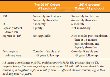
Bryant et al. [3], based on updated National Institute of Health and Care Excellence (NICE) guidelines [1], report on the effect of mpMRI incorporation into their institution’s AS protocols, specifically focusing on the time to treatment and number of biopsies required to trigger treatment. In 2014, they replaced protocol‐driven biannual prostate biopsies (PBs) with mpMRI ± cognitive targeted biopsy and systematic biopsy (TB). With a median follow‐up of 2.4 years, they found that more men who underwent TB progressed to treatment than men who underwent PB alone (44% vs 37%; P = 0.003). The median number of biopsies (beyond the original diagnostic biopsy) required to trigger intervention was 1.55. Based on these results, the authors conclude that mpMRI‐driven TB increases reclassification compared with protocol‐driven PB.

This is consistent with increasing evidence that mpMRI enhances, and sometimes, exceeds detection of clinically significant PCa over TRUS‐guided prostate biopsy alone. The PROMIS study [4], a multicentre paired validation study that compared mpMRI to TRUS‐guided biopsy in the diagnostic setting, found that mpMRI had better sensitivity (93% vs 43%; P < 0.001) and negative predictive value (89% vs 74%; P < 0.001) than TRUS‐guided biopsy in detecting clinically significant cancer (defined as Gleason grade ≥4 + 3). While the concerns about foregoing a systematic biopsy at the time of targeted biopsy in that study were warranted, there was consensus that prebiopsy mpMRI increased the yield for clinically significant PCa.

In the AS setting, unfortunately, randomized data are lacking; however, retrospective series and systematic reviews provide some guidance. In a systematic review, Schoots et al. [5] found that a positive mpMRI in the AS setting was associated with a higher risk of upgrading at the time of radical prostatectomy and a higher risk of reclassification at the time of confirmatory biopsy. Yet, a negative mpMRI did not preclude reclassification and upgrading, indicating the continued need for systematic biopsy. Recabal et al. [6] confirmed these conclusions in their retrospective assessment of an institutionally maintained prospective dataset. While MRI‐targeted biopsies detected higher grade cancer in 23% of men, they missed higher grade clinically significant cancers in 17%, 12% and 10% of patients with mpMRI scores of 3, 4 and 5, respectively. This suggests that both targeted and systematic biopsy should be used for the optimal detection of clinically significant PCa in men on AS.

The present study by Bryant et al. [3] reaffirms the value of mpMRI in the AS paradigm. Yet, some concerns about their study cohort and methodology should be noted. First, as the authors clearly note as a limitation, despite completing a targeted and systematic biopsy, all the samples were sent as a single specimen, precluding the ability to distinguish between targeted biopsy and systematic biopsy cores. As the absolute difference in the rate of progression to treatment between the PB and TB arms was only 7%, it is uncertain how much of that was attributable to the addition of targeted biopsy alone.

Additionally, in a closer analysis of their study population, it should be noted that 35% of the patients had Gleason Grade Group 2 disease or higher at the time of inclusion, representing a higher‐risk AS patient population than guideline recommendations. This may account for the higher rate of progression to treatment in this study cohort independent of grade progression – 24% of patients progressed to treatment based on PSA progression alone and an additional 10% were based on mpMRI findings alone.

Lastly, the median number of biopsies required to trigger intervention was 1.55 and, for the majority of patients, this was just one additional biopsy beyond the original diagnostic biopsy. Guideline recommendations indicate the importance of a confirmatory biopsy to exclude Gleason sampling error [2]; however, by definition, many of these patients were essentially upstaged or redirected to active treatment after a confirmatory biopsy. With 59% of the entire AS population never receiving a confirmatory biopsy beyond their original diagnostic biopsy and many progressing to treatment after a confirmatory biopsy, this study population may not reflect a well‐selected low‐risk PCa patient population for AS.

Despite these limitations, the work by Bryant et al. [3] adds to the growing body of evidence supporting the use of mpMRI‐targeted biopsies in addition to systematic biopsy to more accurately risk stratify men for AS, particularly at the time of diagnosis. It remains unknown how we can use mpMRI to individually tailor surveillance strategies or if mpMRI may ultimately replace surveillance biopsies over time.

References

  1. Graham J, Kirkbride P, Cann K, Hasler E, Prettyjohns M. Prostate cancer: summary of updated NICE guidance. BMJ (Clinical research ed.). 2014348: f7524
  2. Mottet N, Bellmunt J, Bolla M et al. EAU‐ESTRO‐SIOG Guidelines on Prostate Cancer. Part 1: screening, diagnosis, and local treatment with curative intentEur Urol 201771: 618–29


Article of the Month: Combined mpMRI Fusion and Systematic Biopsies Predict the Final Tumour Grading after RP

Every Month the Editor-in-Chief selects an Article of the Month from the current issue of BJUI. The abstract is reproduced below and you can click on the button to read the full article, which is freely available to all readers for at least 30 days from the time of this post.

In addition to the article itself, there is an accompanying editorial written by a prominent member of the urological community. This blog is intended to provoke comment and discussion and we invite you to use the comment tools at the bottom of each post to join the conversation.

Finally, the third post under the Article of the Week heading on the homepage will consist of additional material or media. This week we feature a video from Angelika Borkowetz, discussing her paper.

If you only have time to read one article this week, it should be this one.

Direct comparison of multiparametric magnetic resonance imaging (MRI) results with final histopathology in patients with proven prostate cancer in MRI/ultrasonography-fusion biopsy

Angelika Borkowetz*, Ivan Platzek, Marieta Toma, Theresa Renner*, Roman Herout*, Martin Baunacke*, Michael Laniado, Gustavo Baretton, Michael Froehner*, Stefan Zastrow* and Manfred Wirth*


*Department of Urology, Department of Radiology and Interventional Radiology, and
Department of Pathology, Technische Universitat Dresden, Dresden, Germany


Read the full article

Objective

To compare multiparametric magnetic resonance imaging (mpMRI) of the prostate and histological findings of both targeted MRI/ultrasonography-fusion prostate biopsy (PBx) and systematic PBx with final histology of the radical prostatectomy (RP) specimen.

Patients and Methods

A total of 105 patients with prostate cancer (PCa) histopathologically proven using a combination of fusion Pbx and systematic PBx, who underwent RP, were investigated. All patients had been examined using mpMRI, applying the European Society of Urogenital Radiology criteria. Histological findings from the RP specimen were compared with those from the PBx. Whole-mount RP specimen and mpMRI results were directly compared by a uro-pathologist and a uro-radiologist in step-section analysis.

AugAOTM1

Results

In the 105 patients with histopathologically proven PCa by combination of fusion PBx and systematic PBx, the detection rate of PCa was 90% (94/105) in fusion PBx alone and 68% (72/105) in systematic PBx alone (P = 0.001). The combination PBx detected 23 (22%) Gleason score (GS) 6, 69 (66%) GS 7 and 13 (12%) GS ≥8 tumours. Fusion PBx alone detected 25 (26%) GS 6, 57 (61%) GS 7 and 12 (13%) GS ≥8 tumours. Systematic PBx alone detected 17 (24%) GS 6, 49 (68%) GS 7 and 6 (8%) GS ≥8 tumours. Fusion PBx alone would have missed 11 tumours (4% [4/105] of GS 6, 6% [6/105] of GS 7 and 1% [1/105] of GS ≥8 tumours). Systematic PBx alone would have missed 33 tumours (10% [10/105] of GS 6, 20% [21/105] of GS 7 and 2% [2/105] of GS ≥8 tumours). The rates of concordance with regard to GS between the PBx and RP specimen were 63% (n = 65), 54% (n = 56) and 75% (n = 78) in fusion, systematic and combination PBx (fusion and systematic PBx combined), respectively. Upgrading of the GS between PBx and RP specimen occurred in 33% (n = 34), 44% (n = 46) and 18% (n = 19) in fusion, systematic and combination PBx, respectively. γ-correlation for detection of any cancer was 0.76 for combination PBx, 0.68 for fusion PBx alone and 0.23 for systematic PBx alone. In all, 84% (n = 88) of index tumours were identified by mpMRI; 86% (n = 91) of index lesions on the mpMRI were proven in the RP specimen.

Conclusions

Fusion PBx of tumour-suspicious lesions on mpMRI was associated with a higher detection rate of more aggressive PCa and a better tumour prediction in final histopathology than systematic PBx alone; however, combination PBx had the best concordance for the prediction of GS. Furthermore, the additional findings of systematic PBx reflect the multifocality of PCa, therefore, the combination of both biopsy methods would still represent the best approach for the prediction of the final tumour grading in PCa.

Read more articles of the week

Editorial: Role of systematic biopsy in the era of mpMRI and US fusion guidance

The success of multiparametric MRI (mpMRI) and MRI/ultrasound (US) fusion-guided biopsies in improving the detection of prostate cancer in patients with occult disease (elevated PSA level with prior negative biopsies) and optimising the detection of clinically significant cancer has been reported by centres that have served as early adopters of these techniques [1, 2]. Technological advances in MRI and associated imaging protocols, as well as increased clinical experience with MRI interpretation have led to increased prospective detection and characterisation of clinically significant prostate cancer. This, in conjunction with increasing experience with MRI/US fusion-guided prostate biopsy techniques, has led to the re-evaluation of the contributory role and utility of systematic template US-guided prostate biopsies in the diagnosis of prostate cancer. It is an attractive proposition to forego the systematic biopsy when performing MRI-directed fusion biopsy, as this would minimise the duration, morbidity, and overall cost of the biopsy procedure and post-biopsy pathology processing. However, before adopting this approach, it is important to first consider the potential possibility of missing clinically significant cancer diagnoses when relying on the targeted biopsy cores in isolation.

In this issue of BJUI, Borkowetz et al. [3] report their results of biopsy histological yields on systematic biopsies compared with MRI/US fusion biopsies in their series of patients who underwent radical prostatectomy (RP). These results corroborate previously reported comparisons of fusion biopsy of suspicious lesions on MRI performed concurrently with systematic biopsy, consistently showing an improved detection of both overall prostate cancer foci and, more importantly, an improved detection of clinically significant higher grade cancer foci [1, 2]. It is important to note that the overall detection rate and detection rate for clinically significant prostate cancer was highest when fusion and systematic biopsies were evaluated in conjunction with each other. Another important factor to consider when evaluating the utility and value of these biopsy techniques is the concordance of the pathology of the biopsy specimen with the final pathology of the RP specimen, the ‘gold standard’. The concordance of Gleason grade assigned on targeted fusion-biopsy cores and RP outperformed that of systematic biopsy cores and RP. This, in essence, suggests that targeted biopsy can perform as well, and likely better, than the systematic biopsy approach of sampling the prostate with a systematic-sextant approach, which has been the long standing standard of care for the diagnosis of prostate cancer. Again, it is important to note that the greatest concordance in this study was achieved when the results of the fusion and systematic biopsy cores were combined.

The question now arises regarding the ‘cost’ for the incremental improvement in cancer detection provided by the combination of both MRI-directed fusion biopsy and the systematic biopsy approach. The improved negative predictive value parallels the increased sensitivity for cancer detection by having a larger sampling of the prostate by augmenting the number of biopsy cores sampled and submitted for histopathological evaluation. The area under the curve for detection of clinically significant cancer reported by Borkowetz et al. [3] was not improved by adding systematic biopsies to the targeted biopsies. However, this experience described a mixed population of patients, most of whom had undergone prior prostate biopsy with benign pathology. This creates an enriched population who likely harbours prostate cancers that are more occult to the systematic biopsy approach, thus improving the diagnostic yield of MRI-directed biopsies even further. This is concordant with the work presented by Mendhiratta et al. [4], where systematic biopsies added little to the diagnosis of clinically significant prostate cancer in a population of men undergoing MRI/US fusion-guided biopsy after prior cancer-negative biopsy sessions.

Alternatively, current datasets for biopsy naïve patients have not shown the same degree of convincingly improved detection with targeted biopsies over systematic biopsies. In fact, Delongchamps et al. [5] recently reported a slightly lower rate of overall cancer detection with fusion-guided targeted biopsies vs systematic biopsy cores; however, the difference in detection of clinically significant prostate cancer was not statistically significant. Further study of the role of targeted biopsy in the biopsy naïve patient population is warranted, as there is suggestion that cancer detection efficiency per needle core is significantly improved with MRI-directed biopsies over systematic biopsies [6]. Alternatively, in patients with prior negative systematic biopsies and continued clinical suspicion for prostate cancer, a repeat biopsy session with targeted cores alone may be appropriate, particularly as these patients have previously undergone standard-of-care, extended sextant biopsy.

Read the full article
Jason A. Pietryga* and Soroush Rais-Bahrami*,
*Department of Radiology, and Department of Urology, University of Alabama at Birmingham, Birmingham, AL, USA


References


Video: Combined mpMRI Fusion and Systematic Biopsies Predict the Final Tumour Grading after RP

Direct comparison of multiparametric magnetic resonance imaging (MRI) results with final histopathology in patients with proven prostate cancer in MRI/ultrasonography-fusion biopsy

Angelika Borkowetz*, Ivan Platzek, Marieta Toma, Theresa Renner*, Roman Herout*, Martin Baunacke*, Michael Laniado, Gustavo Baretton, Michael Froehner*, Stefan Zastrow* and Manfred Wirth*


*Department of Urology, Department of Radiology and Interventional Radiology, and
Department of Pathology, Technische Universitat Dresden, Dresden, Germany


Read the full article

Objective

To compare multiparametric magnetic resonance imaging (mpMRI) of the prostate and histological findings of both targeted MRI/ultrasonography-fusion prostate biopsy (PBx) and systematic PBx with final histology of the radical prostatectomy (RP) specimen.

Patients and Methods

A total of 105 patients with prostate cancer (PCa) histopathologically proven using a combination of fusion Pbx and systematic PBx, who underwent RP, were investigated. All patients had been examined using mpMRI, applying the European Society of Urogenital Radiology criteria. Histological findings from the RP specimen were compared with those from the PBx. Whole-mount RP specimen and mpMRI results were directly compared by a uro-pathologist and a uro-radiologist in step-section analysis.

AugAOTM1

Results

In the 105 patients with histopathologically proven PCa by combination of fusion PBx and systematic PBx, the detection rate of PCa was 90% (94/105) in fusion PBx alone and 68% (72/105) in systematic PBx alone (P = 0.001). The combination PBx detected 23 (22%) Gleason score (GS) 6, 69 (66%) GS 7 and 13 (12%) GS ≥8 tumours. Fusion PBx alone detected 25 (26%) GS 6, 57 (61%) GS 7 and 12 (13%) GS ≥8 tumours. Systematic PBx alone detected 17 (24%) GS 6, 49 (68%) GS 7 and 6 (8%) GS ≥8 tumours. Fusion PBx alone would have missed 11 tumours (4% [4/105] of GS 6, 6% [6/105] of GS 7 and 1% [1/105] of GS ≥8 tumours). Systematic PBx alone would have missed 33 tumours (10% [10/105] of GS 6, 20% [21/105] of GS 7 and 2% [2/105] of GS ≥8 tumours). The rates of concordance with regard to GS between the PBx and RP specimen were 63% (n = 65), 54% (n = 56) and 75% (n = 78) in fusion, systematic and combination PBx (fusion and systematic PBx combined), respectively. Upgrading of the GS between PBx and RP specimen occurred in 33% (n = 34), 44% (n = 46) and 18% (n = 19) in fusion, systematic and combination PBx, respectively. γ-correlation for detection of any cancer was 0.76 for combination PBx, 0.68 for fusion PBx alone and 0.23 for systematic PBx alone. In all, 84% (n = 88) of index tumours were identified by mpMRI; 86% (n = 91) of index lesions on the mpMRI were proven in the RP specimen.

Conclusions

Fusion PBx of tumour-suspicious lesions on mpMRI was associated with a higher detection rate of more aggressive PCa and a better tumour prediction in final histopathology than systematic PBx alone; however, combination PBx had the best concordance for the prediction of GS. Furthermore, the additional findings of systematic PBx reflect the multifocality of PCa, therefore, the combination of both biopsy methods would still represent the best approach for the prediction of the final tumour grading in PCa.

Read more articles of the week

Article of the Week: TRUS-Guided RB PCa Detection – Reasons for Targeted Biopsy Failure

Every Week the Editor-in-Chief selects an Article of the Week from the current issue of BJUI. The abstract is reproduced below and you can click on the button to read the full article, which is freely available to all readers for at least 30 days from the time of this post.

In addition to the article itself, there is an accompanying editorial written by a prominent member of the urological community. This blog is intended to provoke comment and discussion and we invite you to use the comment tools at the bottom of each post to join the conversation.

Finally, the third post under the Article of the Week heading on the homepage will consist of additional material or media. This week we feature a video from Hannes Cash and Patrick Asbach, discussing their paper.

If you only have time to read one article this week, it should be this one.

Prostate cancer detection on transrectal ultrasonography-guided random biopsy despite negative real-time magnetic resonance imaging/ultrasonography fusion-guided targeted biopsy: reasons for targeted biopsy failure

Hannes Cash*, Karsten Gunzel*, Andreas Maxeiner*, Carsten Stephan*, Thomas Fischer, Tahir Durmus, Kurt Miller*, Patrick Asbach, Matthias Haas† and Carsten Kempkensteffen*


*Department of Urology, and Department of Radiology, ChariteUniversity of Medicine Berlin, Berlin, Germany M. H. and C.K. contributed equally to the study.


Read the full article

Objective

To examine the value of additional transrectal ultrasonography (TRUS)-guided random biopsy (RB) in patients with negative magnetic resonance imaging (MRI)/ultrasonography (US) fusion-guided targeted biopsy (TB) and to identify possible reasons for TB failure.

Patients and Methods

We conducted a subgroup analysis of 61 men with prostate cancer (PCa) detected by 10-core RB but with a negative TB, from a cohort of 408 men with suspicious multiparametric magnetic resonance imaging (mpMRI) between January 2012 and January 2015. A consensus re-reading of mpMRI results (using Prostate Imaging Reporting and Data System [PI-RADS] versions 1 and 2) for each suspicious lesion was performed, with the image reader blinded to the biopsy results, followed by an unblinded anatomical correlation of the lesion on mpMRI to the biopsy result. The potential reasons for TB failure were estimated for each lesion. We defined clinically significant PCa according to the Epstein criteria and stratified patients into risk groups according to the European Association of Urology guidelines.

JulAOTW3Results

Results

Our analysis showed that RB detected significant PCa in 64% of patients (39/61) and intermediate-/high-risk PCa in 57% of patients (35/61). The initial mpMRI reading identified 90 suspicious lesions in the cohort. Blinded consensus re-reading of the mpMRI led to PI-RADS score downgrading of 45 lesions (50%) and upgrading of 13 lesions (14%); thus, negative TB could be explained by falsely high initial PI-RADS scores for 32 lesions (34%) and sampling of the target lesion by RB in the corresponding anatomical site for 36 out of 90 lesions (40%) in 35 of 61 patients (57%). Sampling of the target lesion by RB was most likely for lesions with PI-RADS scores of 4/5 and Gleason scores (GS) of ≥7. A total of 70 PCa lesions (67% with GS 6) in 44 patients (72%) were sampled from prostatic sites with no abnormalities on mpMRI.

Conclusion

In cases of TB failure, RB still detected a high rate of significant PCa. The main reason for a negative TB was a TB error, compensated for by positive sampling of the target lesion by the additional RB, and the second reason for TB failure was a falsely high initial PI-RADS score. The challenges that arise for both MRI diagnostics and prostate lesion sampling are evident in our data and support the integration of RB into the TB workflow.

Read more articles of the week

Editorial: MRI-Fusion Biopsy – Behind the Scenes

MRI information of the prostate is increasingly used for improving the diagnostic yield of prostate biopsies [1]. However, increasing complexity of a procedure makes it prone to errors at multiple technical and human levels. Incorporating MRI information and ultrasonography (US) images for MRI-fusion biopsies is a technically challenging task. It involves various steps such as the acquisition and fusion of MRI and US images, the needle guidance during biopsy, and the diligence of the pathological evaluation of biopsy specimens. These different steps and interfaces between different medical professions influence the diagnostic performance of MRI-fusion biopsies.

For example, in daily clinical practice, MRIs from different institutions still harbour a great variance of sequences and reporting, despite the European Society of Urogenital Urology (ESUR) recently introducing acquisition and imaging protocols and a new and advanced version of the Prostate Imaging Reporting and Data System (PIRADS) version 2.0 [2]. The usefulness of such reporting schemes is evidenced by a moderate-to-good interobserver agreement between uro-radiologists for tumour lesion interpretation and corresponding κ values ranging from 0.55 to 0.80 [3]. Important pitfalls of image interpretation are benign lesions such as prostatitis, BPH and fibrosis, which might score similarly to prostate cancer lesions. This problem is further aggravated by a high proportion of patients that receive their first multiparametric MRI (mpMRI) of the prostate in the repeat-biopsy setting with a high burden of post-biopsy artefacts (haemorrhage, capsular irregularity) and lower overall cancer detection rate. Also, during MRI-fusion biopsy patient movement, prostate deformation by the US probe, and mismatch of image planes can lead to a biopsy error exceeding 4 mm. Moreover, targeting error might be aggravated by MRI underestimation of the tumour volume compared with final pathology [4]. After various authors reported the advantages and accuracy of MRI/US-fusion biopsy approaches, Cash et al. [5] address potential reasons for targeted biopsy failure to detect prostate cancer compared with random biopsy. Within their analyses the authors address potential limitations and technical considerations. Based on different technical biopsy strategies (with the patient placed within the MRI scanner (‘in-bore’) vs outside) and different technical approaches, these considerations are very important.

In contrast to cognitive fusion, most MRI/US platforms allow needle tracking by archiving the needle orientation, either by an electromagnetic, image-based or stepper-based mechanism [1]. However, lesion targeting by needle guidance is highly dependent on the dimensions of the primary lesion, numbers of relevant lesions, localisation, and overall prostate volume, making MRI-US fusion and cognitive fusion more error prone (i.e. aiming off the mark with the needle) than in-bore biopsies. Moreover, different technical fusion approaches provide different degrees of manual/automated adjustment tools, with for example either rigid or elastic image transformation to facilitate MRI/US image alignment.

In their analyses, Cash et al. [5] found that 34% of negative targeted biopsies could be explained by initially too high estimated PIRADS scores that were downgraded at re-reading. Interestingly, the remaining lesions were without an mpMRI correlate but within this group 92.9% showed a primary Gleason 3 pattern in biopsy pathology, suggesting a high degree of invisibility on mpMRI. Subanalyses did not show an association of targeted biopsy failures in the ventral location. Therefore, the study by Cash et al. [5] is an important precursor for further analyses to address other underlying reasons for targeted biopsy failure. Moreover, it reveals the need for a tight collaboration of radiologists, urologists, and pathologists as interdisciplinary partners involved in MRI-fusion biopsy. Consequently, the optimal diagnostic performance of MRI-fusion biopsies can only be achieved through standardised MRI performance, reading and reporting of MRI findings, as well as final correlation of MRI findings with histopathological work up.

Read the full article
Lars Budaus and Sami-Ramzi Leyh-Bannurah
Martini-Clinic University Hospital Hamburg-Eppendorf, Hamburg, Germany


References


Video: TRUS-Guided RB Prostate Cancer Detection – Reasons for Targeted Biopsy Failure

Prostate cancer detection on transrectal ultrasonography-guided random biopsy despite negative real-time magnetic resonance imaging/ultrasonography fusion-guided targeted biopsy: reasons for targeted biopsy failure

Hannes Cash*, Karsten Gunzel*, Andreas Maxeiner*, Carsten Stephan*, Thomas Fischer, Tahir Durmus, Kurt Miller*, Patrick Asbach, Matthias Haas† and Carsten Kempkensteffen*


*Department of Urology, and Department of Radiology, ChariteUniversity of Medicine Berlin, Berlin, Germany M. H. and C.K. contributed equally to the study.


Read the full article

Objective

To examine the value of additional transrectal ultrasonography (TRUS)-guided random biopsy (RB) in patients with negative magnetic resonance imaging (MRI)/ultrasonography (US) fusion-guided targeted biopsy (TB) and to identify possible reasons for TB failure.

Patients and Methods

We conducted a subgroup analysis of 61 men with prostate cancer (PCa) detected by 10-core RB but with a negative TB, from a cohort of 408 men with suspicious multiparametric magnetic resonance imaging (mpMRI) between January 2012 and January 2015. A consensus re-reading of mpMRI results (using Prostate Imaging Reporting and Data System [PI-RADS] versions 1 and 2) for each suspicious lesion was performed, with the image reader blinded to the biopsy results, followed by an unblinded anatomical correlation of the lesion on mpMRI to the biopsy result. The potential reasons for TB failure were estimated for each lesion. We defined clinically significant PCa according to the Epstein criteria and stratified patients into risk groups according to the European Association of Urology guidelines.

JulAOTW3Results

Results

Our analysis showed that RB detected significant PCa in 64% of patients (39/61) and intermediate-/high-risk PCa in 57% of patients (35/61). The initial mpMRI reading identified 90 suspicious lesions in the cohort. Blinded consensus re-reading of the mpMRI led to PI-RADS score downgrading of 45 lesions (50%) and upgrading of 13 lesions (14%); thus, negative TB could be explained by falsely high initial PI-RADS scores for 32 lesions (34%) and sampling of the target lesion by RB in the corresponding anatomical site for 36 out of 90 lesions (40%) in 35 of 61 patients (57%). Sampling of the target lesion by RB was most likely for lesions with PI-RADS scores of 4/5 and Gleason scores (GS) of ≥7. A total of 70 PCa lesions (67% with GS 6) in 44 patients (72%) were sampled from prostatic sites with no abnormalities on mpMRI.

Conclusion

In cases of TB failure, RB still detected a high rate of significant PCa. The main reason for a negative TB was a TB error, compensated for by positive sampling of the target lesion by the additional RB, and the second reason for TB failure was a falsely high initial PI-RADS score. The challenges that arise for both MRI diagnostics and prostate lesion sampling are evident in our data and support the integration of RB into the TB workflow.

Read more articles of the week
© 2024 BJU International. All Rights Reserved.