Tag Archive for: nomogram

Posts

Article of the week: A clinical prediction tool to determine the need for concurrent systematic sampling at the time of MRI‐guided biopsy

Every week, the Editor-in-Chief selects an Article of the Week from the current issue of BJUI. The abstract is reproduced below and you can click on the button to read the full article, which is freely available to all readers for at least 30 days from the time of this post.

In addition to the article itself, there is an editorial written by a prominent member of the urological community. These are intended to provoke comment and discussion and we invite you to use the comment tools at the bottom of each post to join the conversation. 

If you only have time to read one article this week, it should be this one.

A clinical prediction tool to determine the need for concurrent systematic sampling at the time of magnetic resonance imaging‐guided biopsy

Niranjan J. Sathianathen*, Christopher A. Warlick*, Christopher J. Weight*, Maria A. Ordonez*, Benjamin Spilseth, Gregory J. Metzger, Paari Muruganand Badrinath R. Konety*


Departments of *Urology, Radiology, and Pathology, University of Minnesota, Minneapolis, MN, USA


Abstract

Objective

To develop a clinical prediction tool that characterises the risk of missing significant prostate cancer by omitting systematic biopsy in men undergoing transrectal ultrasonography/magnetic resonance imaging (TRUS/MRI)‐fusion‐guided biopsy.

Patients and methods

A consecutive sample of men undergoing TRUS/MRI‐fusion‐guided biopsy with the UroNav® system (Invivo International, Best, The Netherlands) who also underwent concurrent systematic biopsy was included. By comparing the grade of cancer diagnosed on targeted and systematic biopsy cores, we identified cases where clinically significant disease (Gleason score ≥3+4) was only found on systematic and not targeted cores. Multivariable logistic regression analyses were used to identify predictive factors for finding significant cancer on systematic cores only. We then used these data to develop a nomogram and evaluated its utility using decision curve analysis.

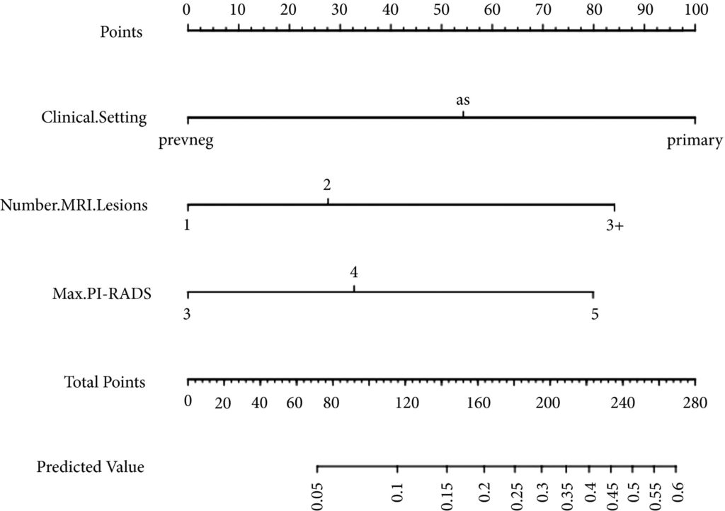
Fig 1. Nomogram for predicting the diagnosis of clinically significant on systematic biopsy only and missed on targeted biopsy.

Results

Of the 398 men undergoing TRUS/MRI‐fusion‐guided biopsy in our study, there were 46 (11.6%) cases in which clinically significant cancer was missed on targeted biopsy and detected on systematic biopsy. The clinical setting, number of MRI lesions identified, and the highest Prostate Imaging‐Reporting and Data System (PI‐RADS) score of the lesions, were all found to be predictors of this. Our model had a good discriminative ability (concordance index = 0.70). The results from our decision curve analysis show that this model provides a higher net clinical benefit than either biopsying all men or omitting biopsy in all patients when the threshold probability is <30%.

Conclusion

We found that omitting concurrent systematic biopsy in men undergoing TRUS/MRI‐fusion‐guided biopsy would miss significant disease in more than one in 10 patients. We propose a prediction model with good discriminative ability that can be used to improve patient selection for performing concurrent systematic biopsy in order to minimise the number of missed significant cancers. It is important that our model is validated in external cohorts before being employed in routine clinical practice.

Editorial: Can systematic biopsy be safely avoided at the time of MRI/ultrasonography fusion biopsy?

In clinical practice, the need for maximising prostate cancer detection is often balanced against the theoretical risks of infection, bleeding, and pain associated with taking additional cores. In this novel study, Sathianathen et al. [1] provide a tool for measuring the oncological benefit of including concurrent systematic biopsy (SB) at the time of MRI‐guided targeted biopsy (TB). There were several key findings: (i) Amongst patients undergoing MRI‐guided biopsy (all biopsy settings), 11.6% were found to have significant cancers detected by SB alone; (ii) Amongst patients who had clinically significant cancers detected by SB alone, 52.2% were sampled within sextants outside the targeted regions of interest; (iii) According to the proposed nomogram, patients with prior negative biopsies, fewer MRI lesions, and lower Prostate Imaging‐Reporting and Data System (PI‐RADS) scores were at the lowest risk of missing significant cancer when SB was omitted.

Based on the present study, biopsy setting appears to be a key factor for deciding whether to omit SB. In the subset of patients undergoing primary biopsy, the authors found that 18.5% of cancers were detected by SB alone. These results are consistent with those of the MRI‐FIRST trial, which showed 14% of cancers were detected by SB only, 20% by TB only, and 66% by combining both techniques [2]. MRI‐FIRST concluded that in the primary biopsy setting, there was no difference between SB and TB in detection of clinically significant prostate cancer, although combining both techniques provided the highest detection rate.

Prior negative biopsy cohorts are generally at lower risk of harbouring significant cancer, as many cancers have already been ‘selected out’ by initial biopsies. In this setting, TB plays an important role in sampling tumour foci in difficult‐to‐reach regions of the prostate (e.g., anterior and apical) [3]. According to the authors’ nomogram, prior negative biopsy patients were least likely to benefit from concurrent SB. While the authors suggest a paradigm of selectively omitting SB, some authors have proposed omitting both TB and SB altogether in select patients. A previously reported multi‐institutional nomogram can be used to predict benign pathology after MRI‐guided biopsy, which can help reduce the number of unnecessary biopsies after MRI in the prior negative biopsy setting [4]. This clinical tool was further externally validated and optimised by Bjurlin et al. [5].

The ‘active surveillance (AS)’ setting typically refers to a confirmatory MRI‐guided biopsy in men with Grade Group 1 prostate cancer prior to enrollment in AS. Recently, the presence of cribriform morphology in Grade Group 2 patients was confirmed to be a key poor prognostic feature that would exclude patients from AS [6]. The present study, however, did not account for different Gleason pattern 4 morphologies in their analysis, as ‘significant cancer’ was defined by Grade Group alone. Studies by independent groups have found that TB combined with SB was more accurate than either modality alone for detecting cribriform at the time of MRI‐guided biopsy [78]. Therefore, concurrent SB is required to properly sample cribriform cancers in patients who are considering AS.

In this study, Sathianathen et al. [1] provide clinicians with a clinical tool for quantifying the added oncological value of concurrent SB. However, concurrent SB is probably prudent for most patients, particularly for those considering AS or focal therapy for which accurate determination of whole gland grade, cancer volume, and cribriform status are essential. As reducing the number of cores has not yet been shown to reduce biopsy‐related complications, are we willing to suboptimise cancer sampling without proven compensation?

by Matthew Truong

References

  1. Sathianathen, NJWarlick, CAWeight, CJ et al. A clinical prediction tool to determine the need for concurrent systematic sampling at the time of magnetic resonance imaging‐guided biopsy. BJU 2019123612– 7
  2. Salami, SSBen‐Levi, EYaskiv, O et al. In patients with a previous negative prostate biopsy and a suspicious lesion on magnetic resonance imaging, is a 12‐core biopsy still necessary in addition to a targeted biopsy? BJU Int 2015115562– 70
  3. Truong, MWang, BGordetsky, JB et al. Multi‐institutional nomogram predicting benign prostate pathology on magnetic resonance/ultrasound fusion biopsy in men with a prior negative 12‐core systematic biopsy. Cancer 2018124278– 85
  4. Bjurlin, MARenson, ARais‐Bahrami, S et al. Predicting benign prostate pathology on magnetic resonance imaging/ultrasound fusion biopsy in men with a prior negative 12‐core systematic biopsy: external validation of a prognostic nomogram. Eur Urol Focus 2018. [Epub ahead of print] https://doi.org/10.1016/j.euf.2018.05.005
  5. Kweldam, CFKümmerlin, IPNieboer, D et al. Presence of invasive cribriform or intraductal growth at biopsy outperforms percentage grade 4 in predicting outcome of Gleason score 3+4=7 prostate cancer. Mod Pathol 2017301126– 32
  6. Truong, MFeng, CHollenberg, G et al. A comprehensive analysis of cribriform morphology on magnetic resonance imaging/ultrasound fusion biopsy correlated with radical prostatectomy specimens. J Urol 2018199106– 13
  7. Prendeville, SGertner, MMaganti, M et al. Role of magnetic resonance imaging targeted biopsy in detection of prostate cancer harboring adverse pathological features of intraductal carcinoma and invasive cribriform carcinoma. J Urol 2018200104– 13


Article of the week: Development of a side‐specific, mpMRI‐based nomogram for the prediction of extracapsular extension of PCa

Every week, the Editor-in-Chief selects an Article of the Week from the current issue of BJUI. The abstract is reproduced below and you can click on the button to read the full article, which is freely available to all readers for at least 30 days from the time of this post.

In addition to the article itself, there are two accompanying editorials written by prominent members of the urological community. These are intended to provoke comment and discussion and we invite you to use the comment tools at the bottom of each post to join the conversation. There is also a video produced by the authors. 

If you only have time to read one article this week, it should be this one.

Development and internal validation of a side‐specific, multiparametric magnetic resonance imaging‐based nomogram for the prediction of extracapsular extension of prostate cancer

Alberto Martini*, Akriti Gupta*, Sara C. Lewis, Shivaram Cumarasamy*, Kenneth G. Haines III§, Alberto Briganti, Francesco Montorsiand Ashutosh K. Tewari*


Departments of *Urology, Radiology, §Pathology, Icahn School of Medicine at Mount Sinai, New York, NY, USA and Department of Urology, Vita-Salute San Raffaele University, Milan, Italy
Read the full article

Abstract

Objectives

To develop a nomogram for predicting side‐specific extracapsular extension (ECE) for planning nerve‐sparing radical prostatectomy.

Materials and Methods

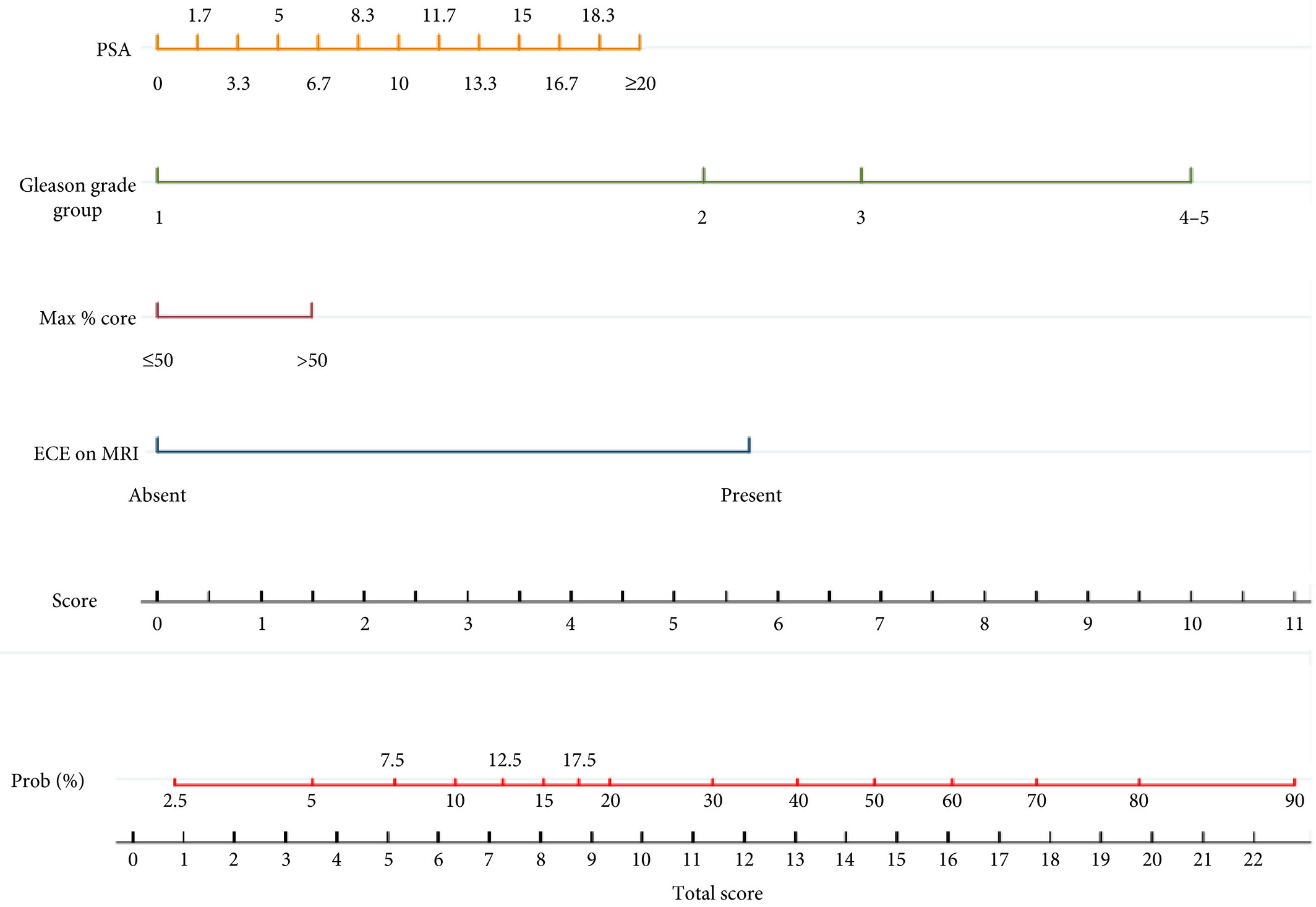
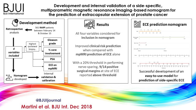
We retrospectively analysed data from 561 patients who underwent robot‐assisted radical prostatectomy between February 2014 and October 2015. To develop a side‐specific predictive model, we considered the prostatic lobes separately. Four variables were included: prostate‐specific antigen; highest ipsilateral biopsy Gleason grade; highest ipsilateral percentage core involvement; and ECE on multiparametric magnetic resonance imaging (mpMRI). A multivariable logistic regression analysis was fitted to predict side‐specific ECE. A nomogram was built based on the coefficients of the logit function. Internal validation was performed using ‘leave‐one‐out’ cross‐validation. Calibration was graphically investigated. The decision curve analysis was used to evaluate the net clinical benefit.

Results

The study population consisted of 829 side‐specific cases, after excluding negative biopsy observations (n = 293). ECE was reported on mpMRI and final pathology in 115 (14%) and 142 (17.1%) cases, respectively. Among these, mpMRI was able to predict ECE correctly in 57 (40.1%) cases. All variables in the model except highest percentage core involvement were predictors of ECE (all P ≤ 0.006). All variables were considered for inclusion in the nomogram. After internal validation, the area under the curve was 82.11%. The model demonstrated excellent calibration and improved clinical risk prediction, especially when compared with relying on mpMRI prediction of ECE alone. When retrospectively applying the nomogram‐derived probability, using a 20% threshold for performing nerve‐sparing, nine out of 14 positive surgical margins (PSMs) at the site of ECE resulted above the threshold.

Conclusion

We developed an easy‐to‐use model for the prediction of side‐specific ECE, and hope it serves as a tool for planning nerve‐sparing radical prostatectomy and in the reduction of PSM in future series.

Read more Articles of the week


Editorial: A novel nomogram for predicting ECE of prostate cancer

We read with great interest the publication on the side‐specific multiparametric magnetic resonance imaging (mpMRI)‐based nomogram from Martini et al. [1].

The prediction of extracapsular extension (ECE) of prostate cancer is of utmost importance to inform accurate surgical planning before radical prostatectomy (RP).

Today, surgical strategy is tailored to the patient’s characteristics, and the need for a correct prediction of ECE is of paramount importance to guarantee oncological safety, as well as optimal functional outcome. The most up‐to‐date guidelines suggest referring to nomograms to decide whether or not to perform nerve‐sparing (NS) surgery. Since the first version of the Partin Tables in 1993, several models have been developed based on PSA, Gleason score at prostate biopsy, and clinical staging, as the most used covariates.

Furthermore, mpMRI is increasingly used in the diagnostic pathway of prostate cancer to aid prostate biopsy targeting and to attain a more accurate diagnosis of clinically significant prostate cancer. Despite its recognised role in the detection of cancer, the accuracy for local staging is poor, providing a low and heterogeneous sensitivity for the detection of ECE [2].

Given this limitation, the addition of MRI to clinically derived nomograms might result in an improved assessment of preoperative local staging. In a retrospective analysis of 501 patients who underwent RP, MRI + clinical models outperformed clinical‐based models alone for all staging outcomes, with better discrimination in predicting ECE with MRI + Partin Tables and MRI + Cancer of the Prostate Risk Assessment (CAPRA) score than nomograms alone [3].

In the current article, Martini et al. [1] suggest a novel nomogram for predicting ECE that includes the presence of a ‘documented definite ECE at mpMRI’ as an additional variable beyond PSA, Gleason score, and maximum percentage of tumour in the biopsy core with the highest Gleason score. Readers should recognise that this is the first model integrating side‐specific MRI findings together with side‐specific biopsy data to provide a ‘MRI‐based side‐specific prediction of ECE’, in an effort to support the surgical decision for a uni‐ or bilateral NS approach.

However, given the frail generalisability of nomograms in different datasets even after external validation [4], a predictive tool has to be built on a rigorous methodology with clear reproducibility of all steps the covariates derive from.

In this respect, the current model raises some concerns.

The schedule of preoperative MRI assessment is arbitrary, with imaging being performed either before (23.9%) or after systematic biopsy (76.1%), and amongst patients with a MRI prior to biopsy, only 94 of 134 patients underwent additional targeted sampling. As a result, MRI is applied by chance in three different ways: before prostate biopsy without targeted sampling, before prostate biopsy with targeted sampling, and after prostate biopsy.

Based upon this heterogeneous MRI timing, the performance of such a model in a novel population may be biased depending on the diagnostic pathway applied at each institution.

The choice of the variables included represents another point of concern. The output of two out of four covariates, ECE depiction at mpMRI and the percentage of tumour in the biopsy core, have been deliberately dichotomised, without taking into account the continuous trend intrinsic to both variables.

Actually, local staging in the European Society of Urogenital Radiology (ESUR) guidelines has been scored on a 1–5 point scale to grade the likelihood of an ECE event. The authors deliberately dichotomised mpMRI findings, considering ‘the loss of prostate capsule and its irregularity’ as suggestive of ECE and ‘broad capsular contact, abutment or bulge without gross ECE’ evocative of organ‐confined disease. As a result, the included MRI covariate may account for a gross prediction of ECE, maintaining the inaccurate and inter‐reader subjective interpretation of local staging intrinsic to MRI.

Beyond those methodological concerns and the moderate sample size that may limit the reproducibility of the model, we wonder if such a prediction really assists the surgeon’s capability to perform a tailored surgery.

The ‘all or none’ era of NS surgery is over, and we are currently able to grade NS according to different approaches reported in the literature. Particularly, Tewari et al. [5] proposed a NS approach based on four grades of dissection, with the veins on the lateral aspect as vascular landmarks to gain the correct dissection planes. Patel et al. [6] described a five‐grade scale of dissection, using the arterial periprostatic vasculature as a landmark to the same purpose.

If we are able to grade a NS surgery, the prediction of ECE should be graded as well and should answer the prerequisite of knowing the amount of prostate cancer extent outside the capsule. How does a surgeon make the decision to follow a more or less conservative dissection otherwise?

We tried to address this issue by using a tool aimed at predicting the amount of ECE [the Predicting ExtraCapsular Extension in Prostate cancer tool] [6] and supporting the choice of the correct plane of dissection with a suggested decision rule. In our study, developed on a large sample of nearly 12 000 prostatic lobes and several combined clinicopathological variables, the absence of imaging characterization was the major point of weakness.

To date, the ideal predictive tool has yet to be described. However, in the modern era of precision surgery, we think that a model should encompass the surgical knowledge and techniques currently available.

Future developments will probably include three‐dimensional surgical navigation models displayed on the TilePro™ function of the robotic console (Intuitive Surgical Inc., Sunnyvale, CA, USA), based on the integration of MRI (for the number, size and location of disease) and predictive tools (to define the amount of ECE).


References

  1. Martini A, Gupta A, Lewis SC et al. Development and internal validation of a side‐specific, multiparametric magnetic resonance imaging‐based nomogram for the prediction of extracapsular extension of prostate cancer. BJU Int 2018; 122: 1025–33
  2. de Rooij M, Hamoen EH, Witjes JA, Barentsz JO, Rovers MM. Accuracy of magnetic resonance imaging for local staging of prostate cancer: a diagnostic meta‐analysis. Eur Urol 2016; 70: 233–45
  3. Morlacco A, Sharma V, Viers BR et al. The incremental role of magnetic resonance imaging for prostate cancer staging before radical prostatectomy. Eur Urol 2017; 71: 701–4
  4. Bleeker SE, Moll HA, Steyerberg EW et al. External validation is necessary in prediction research: a clinical example. J Clin Epidemiol 2003; 56: 826–32
  5. Tewari AK, Srivastava A, Huang MW et al. Anatomical grades of nerve sparing: a risk‐stratified approach to neural‐hammock sparing during robot‐assisted radical prostatectomy (RARP). BJU Int 2011; 108: 984–92
  6. Patel VR, Sandri M, Grasso AA et al. A novel tool for predicting extracapsular extension during graded partial nerve sparing in radical prostatectomy. BJU Int 2018; 121: 373–82


Article of the Week: A mpMRI-based risk model to determine the risk of prostate cancer prior to biopsy

Every Week the Editor-in-Chief selects an Article of the Week from the current issue of BJUI. The abstract is reproduced below and you can click on the button to read the full article, which is freely available to all readers for at least 30 days from the time of this post.

In addition to the article itself, there is an accompanying editorial written by a prominent member of the urological community. This blog is intended to provoke comment and discussion and we invite you to use the comment tools at the bottom of each post to join the conversation.

If you only have time to read one article this week, it should be this one.

A multiparametric magnetic resonance imaging-based risk model to determine the risk of significant prostate cancer prior to biopsy

Pim J. van Leeuwen*, Andrew Hayen, James E. Thompson*†‡, Daniel Moses§Ron Shnier§, Maret Bohm, Magdaline Abuodha, Anne-Maree HaynesFrancis Ting*†‡, Jelle Barentsz, Monique Roobol**, Justin Vass††, Krishan Rasiah††Warick Delprado‡‡ and Phillip D. Stricker*†‡


*St. Vincents Prostate Cancer Centre, Garvan Institute of Medical Research/The Kinghorn Cancer Centre, Darlinghurst, School of Public Health and Community Medicine, §School of Medicine, University of New South Wales, Kensington, New South Wales, Australia, Department of Radiology and Nuclear Medicine, Radboud University Medical Centre, Nijmegen, **Department of Urology, Erasmus University Medical Center, Rotterdam, the Netherlands, ††Department of Urology, Royal North Shore Private Hospital, St Leonards, and ‡‡Douglass Hanly Moir Pathology and University of Notre Dame, Darlinghurst, New South Wales, Australia


Read the full article

Abstract

Objective

To develop and externally validate a predictive model for detection of significant prostate cancer.

Patients and Methods

Development of the model was based on a prosp   ctive cohort including 393 men who underwent multiparametric magnetic resonance imaging (mpMRI) before biopsy. External validity of the model was then examined retrospectively in 198 men from a separate institution whom underwent mpMRI followed by biopsy for abnormal prostate-specific antigen (PSA) level or digital rectal examination (DRE). A model was developed with age, PSA level, DRE, prostate volume, previous biopsy, and Prostate Imaging Reporting and Data System (PIRADS) score, as predictors for significant prostate cancer (Gleason 7 with >5% grade 4, ≥20% cores positive or ≥7 mm of cancer in any core). Probability was studied via logistic regression. Discriminatory performance was quantified by concordance statistics and internally validated with bootstrap resampling.

Results

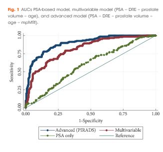
In all, 393 men had complete data and 149 (37.9%) had significant prostate cancer. While the variable model had good accuracy in predicting significant prostate cancer, area under the curve (AUC) of 0.80, the advanced model (incorporating mpMRI) had a significantly higher AUC of 0.88 (P < 0.001). The model was well calibrated in internal and external validation. Decision analysis showed that use of the advanced model in practice would improve biopsy outcome predictions. Clinical application of the model would reduce 28% of biopsies, whilst missing 2.6% significant prostate cancer.

Conclusions

Individualised risk assessment of significant prostate cancer using a predictive model that incorporates mpMRI PIRADS score and clinical data allows a considerable reduction in unnecessary biopsies and reduction of the risk of over-detection of insignificant prostate cancer at the cost of a very small increase in the number of significant cancers missed.

Read more articles of the week


Editorial: Novel risk stratification nomograms for counseling patients on the need for prostate biopsy

Contemporary recommendations for prostate screening incorporate the measurement of serum PSA levels into shared decision making [1]. PSA is limited by a low specificity for prostate cancer and exposes a certain number of men to unnecessary prostate biopsies. Moreover, it has been attributed to an over-diagnosis and over treatment of this disease, especially in indolent cancer that may never affect a man’s longevity [2].

Prostate cancer risk stratification and aggressiveness is necessary in both the pre-biopsy clinical counselling, as well as the decision-making process. It is clear that such an important approach cannot be accomplished based on PSA alone. In order to enhance prostate cancer screening and detection, other clinical variables such as PSA density, prostate volume, percentage free PSA, and DRE, are routinely considered in conjunction with PSA for determining the need for prostate biopsy.

Increasing evidence supports the use of MRI in prostate cancer detection when used as a localisation tool to guide MRI-targeted biopsy techniques such as MRI-ultrasonography fusion-targeted biopsy [3-5]. Pre-biopsy MRI not only allows accurate tumour localisation, but also provides an assessment of cancer suspicion using an MRI suspicion score, and thus provides accurate prediction of the likelihood of prostate cancer on prostate biopsy [6].

In this study, van Leeuwen et al. [7] developed and externally validated a set of nomograms predicting clinically significant prostate cancer by incorporating prostate MRI. The performance characteristics of the nomograms were maximised by inclusion of MRI results. The authors determined that clinical application of the model would reduce 28% of biopsies, while missing 2.6% of clinically significant prostate cancer. Ultimately incorporating these nomograms into the clinical decision-making process could result in a considerable reduction in unnecessary biopsies and reduction in the risk of over-detection of clinically insignificant disease at the cost of a small increase in the number of significant cancers missed.

The authors should be commended for their predictive nomograms, in that they may further aid in the decision to perform biopsy in men with clinical suspicion of prostate cancer. However, the findings of this study should be interpreted with caution. In formulating nomograms, the obvious clinical goal is the creation of a tool that improves the selection of men in need of biopsy. Unless nomograms are derived from general ‘at risk’ populations, including men with low- and high-risk of prostate cancer, the tool may be limited in its prediction. As an example, if all men in the training cohort have an elevated PSA level, the predictive value of PSA in the nomogram may be reduced. In this case, the training and validation cohort are not well described, but it seems to be a referral population, and the PSA range is relatively narrow. As such, it’s applicability to all men presenting for prostate cancer may be questionable.

We also have had difficulty modelling a nomogram from our MRI-targeted biopsy dataset because the power of MRI-suspicion score in predicting cancer tends to minimise the effect of other variables such as PSA, age, and gland size. The authors did not compare their multivariable predictive models to MRI alone, and this may have been the most relevant comparison. In this study, the gland size and PSA level contribute significantly to the nomogram score, but one might question the findings. For example, a man with a Prostate Imaging Reporting and Data System (PI-RADS) score of 4 or 5, a large gland and a low PSA level, has a similar or lower risk as a man with a PSA level of 15 ng/mL, a moderate gland, and a PI-RADS score of 3. This is not consistent with our clinical experience and draws concern in the reliability of the nomogram at extremes of PSA and age. Men with PI-RADS 5 have high rates of clinically significant prostate cancer, regardless of PSA or age. This also may reflect variability in the predictive accuracy of MRI depending upon MRI interpretation.

Another limitation of the study, as the authors cite, is that patients were biopsied using a transperineal mapping with a median of 30 cores. This biopsy strategy is not routinely used in most institutions, and consequently limits the generalisability of the nomograms.

Selective use of prostate biopsy among men with elevated PSA levels through further refinement of cancer risk is highly desirable. The novel risk stratification nomograms developed by van Leeuwen et al. [7] add to the tools we may use to counsel our patients on the need for prostate biopsy. Further evaluation of these nomograms on additional independent patient cohorts is warranted prior to implementation in clinical practice.

Marc A. Bjurlinand Samir S. Taneja
*Division of Urology, Department of Surgery, NYU Lutheran Medical Center, NYU Langone Health System, New York, NY, USA and Division of Urologic Oncology, Department of Urology, NYU Langone Medical Center, New York, NY, US


References


1 Carter HBAlbertsen PCBarry MJ et al. Early detection of prostate cancer: AUA Guideline. J Urol2013; 190: 41926


2 Loeb SBjurlin MANicholson J et al. Overdiagnosis and overtreatment of prostate cancer. Eur Urol
2014; 65: 104655


3 Mendhiratta NMeng XRosenkrantz AB et al. Prebiopsy MRI and MRI-ultrasound fusion-targeted prostate biopsy in men with previous negative biopsies: impact on repeat biopsy strategies. Urology 2015; 86: 11928


5 Meng XRosenkrantz ABMendhiratta N et al. Relationship between prebiopsy multiparametric magnetic resonance imaging (MRI), biopsy indication, and MRI-ultrasound fusion-targeted prostate biopsy outcomes. Eur Urol 2016; 69: 51217


7 van Leeuwen PJHayan AThompson JE et al. A multiparametric magnetic resonance imaging-based risk model to determine the risk of significant prostate cancer prior to biopsy. BJU Int 2017; 120: 7748


Article of the Week: Predicting pathological outcomes in patients undergoing RARP for high-risk prostate cancer

Every Week the Editor-in-Chief selects an Article of the Week from the current issue of BJUI. The abstract is reproduced below and you can click on the button to read the full article, which is freely available to all readers for at least 30 days from the time of this post.

In addition to the article itself, there is an accompanying editorial written by a prominent member of the urological community. This blog is intended to provoke comment and discussion and we invite you to use the comment tools at the bottom of each post to join the conversation.

Finally, the third post under the Article of the Week heading on the homepage will consist of additional material or media. This week we feature a video from Dr. Firas Abdollah, discussing his paper. 

If you only have time to read one article this week, it should be this one.

Predicting Pathologic Outcomes in Patients Undergoing Robot-Assisted Radical Prostatectomy for High Risk Prostate Cancer:  A Preoperative Nomogram

Firas Abdollah, Dane E. Klett, Akshay Sood, Jesse D. Sammon, Daniel PucherilDeepansh Dalela, Mireya Diaz, James O. Peabody, Quoc-Dien Trinh* and Mani Menon


Vattikuti Urology Institute, Center for Outcomes Research Analytics and Evaluation, Henry Ford Health System, Detroit, MI, and *Division of Urologic Surgery/Center for Surgery and Public Health, Brigham and Womens Hospital, Harvard Medical School, Boston, MA, USA


Read the full article
OBJECTIVE

To identify which high-risk patients with prostate cancer may harbour favourable pathological outcomes at radical prostatectomy (RP).

PATIENTS AND METHODS

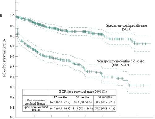
We evaluated 810 patients with high-risk prostate cancer, defined as having one or more of the following: PSA level of >20 ng/mL, Gleason score ≥8, clinical stage ≥T2c. Patients underwent robot-assisted RP (RARP) with pelvic lymph node dissection, between 2003 and 2012, in one centre. Only 1.6% (13/810) of patients received any adjuvant treatment. Favourable pathological outcome was defined as specimen-confined disease (SCD; pT2–T3a, node negative, and negative surgical margins) at RARP-specimen. Logistic regression models were used to test the relationship among all available predicators and harbouring SCD. A logistic regression coefficient-based nomogram was constructed and internally validated using 200 bootstrap resamples. Kaplan–Meier method estimated biochemical recurrence (BCR)-free and cancer-specific mortality (CSM)-free survival rates, after stratification according to pathological disease status.

RESULTS

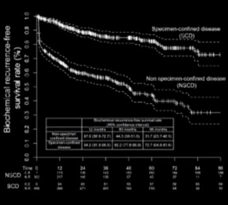
Overall, 55.2% patients harboured SCD at RARP. At multivariable analysis, PSA level, clinical stage, primary/secondary Gleason scores, and maximum percentage tumour quartiles were all independent predictors of SCD (all P < 0.04). A nomogram based on these variables showed 76% discrimination accuracy in predicting SCD, and very favourable calibration characteristics. Patients with SCD had significantly higher 8-year BCR- (72.7% vs 31.7%, P < 0.001) and CSM-free survival rates (100% vs 86.9%, P < 0.001) than patients with non-SCD.

CONCLUSIONS

We developed a novel nomogram predicting SCD at RARP. Patients with SCD achieved favourable long-term BCR- and CSM-free survival rates after RARP. The nomogram may be used to support clinical decision-making, and aid in selection of patients with high-risk prostate cancer most likely to benefit from RARP.

Read more articles of the week

Editorial: More Nomograms or Better Lymph node dissection – What do we need in Prostate Cancer?

The publication of nomograms to predict radical prostatectomy (RP) outcome using preoperative parameters were important steps in urological oncology. Abdollah et al. [1], in this issue of BJU International, present a new nomogram to predict specimen-confined disease (SCD; pT2–3a, pN0 R0) in men with high-risk prostate cancer undergoing pelvic lymph node dissection (PLND) and robot-assisted RP (RARP). They used statistical logistic regression to measure the impact of various preoperatively available clinicopathological parameters on the likelihood of pathological outcome and tumour recurrence. The final nomogram accurately identified SCD (pT2–3a, pN0 R0) in 76% of the patients. It is intuitive that these patients have good long-term oncological outcomes after surgery. Consequently, Abdollah et al. found excellent 8-year cancer-specific survival rates in these patients. Because nomograms provide individualised risk prediction for patients in an easily applicable manner, they have become very popular among clinicians. Nomograms are now being applied for almost every aspect of prostate cancer. These are freely available and both patients and physicians are encouraged to use them.

Although nomograms undoubtedly have improved our perspective of disease behaviour and individual patient prediction, several key questions remain. First, how good are the input data to a nomogram? Abdollah et al. [1] evaluated 810 patients with high-risk prostate cancer treated in a single large centre between 2003 and 2012. Impressively, more than half of the patients (55%) harboured SCD at RARP. Such a high chance of having SCD will probably encourage many physicians and patients to choose surgery, even without using a nomogram, because this approach may avoid the need for hormonal treatment, which is obligatory for radiation therapy in high-risk prostate cancer. Second, is the predictive accuracy safe within clinical practice? Most nomograms using clinicopathological data generate predictive accuracies within the range of 75–90% (including the nomogram presented by Abdollah et al. [1]). It is of special importance to consider that 64/447 (14%) of the patients with SCD in the series reported by Abdollah et al. [1] received salvage treatment, which was initiated after a median (interquartile range, IQR) of 4.8 (1.4–9.3) months, and the indication to initiate this salvage therapy was PSA recurrence. Obviously, these patients did not have specimen confined disease and were misclassified. In this case, one might postulate a persistence of nodal disease, given an inadequate extent of PLND. Abdollah et al. [1] reported on a median (IQR) of 5 (3.0–11.0) lymph nodes removed.

In their landmark paper on extended PLND (ePLND) in cadavers, Weingartner et al. [2] demonstrated that a mean lymph node yield of 20 serves as a guideline for sufficient ePLND. More than 10 years ago, Heidenreich et al. [3] reported on a 15% higher rate of lymph node metastasis detection when comparing ePLND with the standard LND (obturator). Bader et al. [4] provided further evidence that an ePLND is needed to provide adequate clinical staging and potential therapeutic benefit. Of 365 patients with clinically localised prostate cancer, 88 (24%) had positive lymph nodes. In this series, a pelvic LND that spared the internal iliac bed would have left 58% of patients with positive nodes with residual disease and 19% would have been incorrectly staged as lymph node-negative for cancer. These data were recently confirmed by several authors when analysing retrospective series. Furthermore, Seiler et al. [5] updated their series of 88 patients and recently reported on the long-term outcome after a median follow-up of 15.6 years. They showed that 18% of those patients with one positive node remained biochemical recurrence free, 28% showed biochemical recurrence only, and 54% had clinical progression. Of these 39 patients, 57% never required deferred androgen-deprivation therapy. In contrast, patients with multiple positive nodes are likely to experience rapid progression and, thus, may benefit from early adjuvant therapies. International clinical practice guidelines recommend the performance of an anatomically ePLND at RP in men with high-risk prostate cancer, for both staging and therapeutic purposes.

Nowadays, most urologists claim to perform an ePLND. However, a recent analysis among 50 671 men who were surgically treated with RP from 2010 to 2011 in the USA showed that, overall, only 69.3% of the high-risk patients underwent concomitant PLND [6]. Surgical approach and hospital characteristics were associated with treatment with PLND and detection of lymph node metastasis. More specifically, patients with prostate cancer undergoing open RP or surgically treated at high-volume centres were more likely to undergo PLND than those undergoing RARP or surgically treated at low-volume centres.

Despite the strong evidence that ePLND positively affects survival in men with limited lymph node involvement, this procedure is not commonly performed. The reasons for this are multiple and include expertise, stage migration and functional and oncological outcomes, as well as economics and the introduction of laparoscopic and laparoscopic RARP. However, this is no reason not to offer the patient, if possible, an operation which has the highest chance of cure

Read the full article
Martin Spahn
Department of Urology, University Hospital Bern , InselspitalBern, Switzerland


References


Video: Predicting pathological outcomes in patients undergoing RARP for high-risk prostate cancer: A Preoperative Nomogram

Predicting Pathologic Outcomes in Patients Undergoing Robot-Assisted Radical Prostatectomy for High Risk Prostate Cancer:  A Preoperative Nomogram

Firas Abdollah, Dane E. Klett, Akshay Sood, Jesse D. Sammon, Daniel PucherilDeepansh Dalela, Mireya Diaz, James O. Peabody, Quoc-Dien Trinh* and Mani Menon


Vattikuti Urology Institute, Center for Outcomes Research Analytics and Evaluation, Henry Ford Health System, Detroit, MI, and *Division of Urologic Surgery/Center for Surgery and Public Health, Brigham and Womens Hospital, Harvard Medical School, Boston, MA, USA


Read the full article
OBJECTIVE

To identify which high-risk patients with prostate cancer may harbour favourable pathological outcomes at radical prostatectomy (RP).

PATIENTS AND METHODS

We evaluated 810 patients with high-risk prostate cancer, defined as having one or more of the following: PSA level of >20 ng/mL, Gleason score ≥8, clinical stage ≥T2c. Patients underwent robot-assisted RP (RARP) with pelvic lymph node dissection, between 2003 and 2012, in one centre. Only 1.6% (13/810) of patients received any adjuvant treatment. Favourable pathological outcome was defined as specimen-confined disease (SCD; pT2–T3a, node negative, and negative surgical margins) at RARP-specimen. Logistic regression models were used to test the relationship among all available predicators and harbouring SCD. A logistic regression coefficient-based nomogram was constructed and internally validated using 200 bootstrap resamples. Kaplan–Meier method estimated biochemical recurrence (BCR)-free and cancer-specific mortality (CSM)-free survival rates, after stratification according to pathological disease status.

RESULTS

Overall, 55.2% patients harboured SCD at RARP. At multivariable analysis, PSA level, clinical stage, primary/secondary Gleason scores, and maximum percentage tumour quartiles were all independent predictors of SCD (all P < 0.04). A nomogram based on these variables showed 76% discrimination accuracy in predicting SCD, and very favourable calibration characteristics. Patients with SCD had significantly higher 8-year BCR- (72.7% vs 31.7%, P < 0.001) and CSM-free survival rates (100% vs 86.9%, P < 0.001) than patients with non-SCD.

CONCLUSIONS

We developed a novel nomogram predicting SCD at RARP. Patients with SCD achieved favourable long-term BCR- and CSM-free survival rates after RARP. The nomogram may be used to support clinical decision-making, and aid in selection of patients with high-risk prostate cancer most likely to benefit from RARP.

Read more articles of the week

Article of the Week: Am I normal? A systematic review for penis length and circumference

Every Week the Editor-in-Chief selects the Article of the Week from the current issue of BJUI. The abstract is reproduced below and you can click on the button to read the full article, which is freely available to all readers for at least 30 days from the time of this post.

In addition to the article itself, there is an accompanying editorial written by a prominent member of the urological community. This blog is intended to provoke comment and discussion and we invite you to use the comment tools at the bottom of each post to join the conversation.

If you only have time to read one article this week, it should be this one.

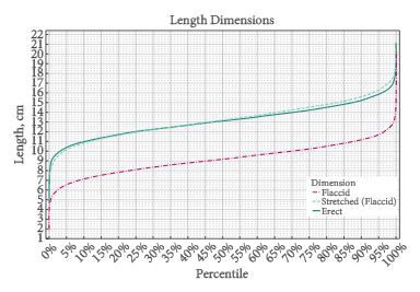
Am I normal? A systematic review and construction of nomograms for flaccid and erect penis length and circumference in up to 15,521 men

David Veale*, Sarah Miles*, Sally Bramley, Gordon Muir§ and John Hodsoll*


*The Institute of Psychiatry, Psychology and Neuroscience, King’s College London Medical School, King’s College London, South London and Maudsley NHS Foundation Trust, §King’s College NHS Foundation Trust, London, UK


Read the full article
OBJECTIVE

To systematically review and create nomograms of flaccid and erect penile size measurements.

METHODS

Study key eligibility criteria: measurement of penis size by a health professional using a standard procedure; a minimum of 50 participants per sample. Exclusion criteria: samples with a congenital or acquired penile abnormality, previous surgery, complaint of small penis size or erectile dysfunction. Synthesis methods: calculation of a weighted mean and pooled standard deviation (sd) and simulation of 20 000 observations from the normal distribution to generate nomograms of penis size.

RESULTS

Nomograms for flaccid pendulous [n = 10 704, mean (sd) 9.16 (1.57) cm] and stretched length [n = 14 160, mean (sd) 13.24 (1.89) cm], erect length [n = 692, mean (sd) 13.12 (1.66) cm], flaccid circumference [n = 9407, mean (sd) 9.31 (0.90) cm], and erect circumference [n= 381, mean (sd) 11.66 (1.10) cm] were constructed. Consistent and strongest significant correlation was between flaccid stretched or erect length and height, which ranged from r = 0.2 to 0.6. Limitations: relatively few erect measurements were conducted in a clinical setting and the greatest variability between studies was seen with flaccid stretched length.

CONCLUSIONS

Penis size nomograms may be useful in clinical and therapeutic settings to counsel men and for academic research.

Read more articles of the week
© 2024 BJU International. All Rights Reserved.