Tag Archive for: urolithiasis

Posts

Video abstract: Is low-dose tadalafil better than tamsulosin?

Is low-dose tadalafil better than tamsulosin? A randomized controlled trial in shockwave lithotripsy for solitary upper tract calculi

The aim of this work is to ascertain whether low-dose tadalafil (5 mg) is more efficient than tamsulosin (0.4 mg) in facilitating calculus expulsion in those receiving extracorporeal shockwave lithotripsy for solitary upper urinary tract calculi.

Madhuri Evangeline Sadanala, Anuj Deep Dangi, Geetha Rajendran, Antonisamy Balavendra, Subramanian Annadurai, Rajiv Paul Mukha, J. Chandra Singh, Antony Devasia and Santosh Kumar

Residents’ podcast: MIMIC Study

Part of the BURST/BJUI Podcast Series

Mr Chuanyu Gao is a Core Surgical Trainee in KSS Deanery. He graduated from UCL Medical School and obtained his iBSc in Surgical Sciences before completing his Academic Foundation Years in East of England Foundation School. Chuanyu first became involved with BURST on the MIMIC Study as an international site coordinator and has been part of the BURST committee ever since. 

Factors associated with spontaneous stone passage in a contemporary cohort of patients presenting with acute ureteric colic: results from the Multi‐centre cohort study evaluating the role of Inflammatory Markers In patients presenting with acute ureteric Colic (MIMIC) study

Taimur T. Shah*†‡§, Chuanyu Gao*, Max Peters, Todd Manning**, Sophia Cashman*, Arjun Nambiar*, Marcus Cumberbatch*††, Ben Lamb*, Anthony Peacock‡‡, Marieke J. Van Son, Peter S. N. van Rossum, Robert Pickard§§, Paul Erotocritou¶¶, Daron Smith***, Veeru Kasivisvanathan*‡ and British Urology Researchers in Surgical Training (BURST) Collaborative MIMIC Study Group


*British Urology Researchers in Surgical Training (BURST), London, UK, Division of Surgery and Cancer, Imperial College London, Division of Surgery and Interventional Science, University College London, §Charing Cross Hospital, Imperial Health NHS Trust, London, UK, Department of Radiation Oncology, Cancer Center, University Medical Center Utrecht, Utrecht, The Netherlands, **Australian Young Urology Researchers Organisation (YURO), Heidelberg, Victoria, Australia, ††Academic Urology Unit, University of Shefeld, Shefeld, ‡‡Information Services Division, University College London (UCL), London, §§Department of Urology, Newcastle University, Newcastle, UK, ¶¶Department of Urology, Whittington Hospital, and ***Department of Urology, UCL Hospital, London, UK


Read the full article

Article of the week: Ultrasound guidance can be used safely for renal tract dilatation during percutaneous nephrolithotomy

Every week, the Editor-in-Chief selects an Article of the Week from the current issue of BJUI. The abstract is reproduced below and you can click on the button to read the full article, which is freely available to all readers for at least 30 days from the time of this post.

In addition to the article itself, there is an editorial written by a prominent member of the urological community and a visual abstract prepared by a trainee urologist; we invite you to use the comment tools at the bottom of each post to join the conversation. 

If you only have time to read one article this week, we recommend this one. 

Ultrasound guidance can be used safely for renal tract dilatation during percutaneous nephrolithotomy

Manuel Armas-Phan*, David T. Tzou*, David B. Bayne*, Scott V. Wiener*, Marshall L. Stoller* and Thomas Chi*

*Department of Urology, University of California, San Francisco, CA and Division of Urology, University of Arizona, Tucson, AZ, USA

Read the full article

Abstract

Objectives

To compare clinical outcomes in patients who underwent percutaneous nephrolithotomy (PCNL) with renal tract dilatation performed under fluoroscopic guidance vs renal tract dilatation with ultrasound guidance.

Patients and Methods

We conducted a prospective observational cohort study, enrolling successive patients undergoing PCNL between July 2015 and March 2018. Included in this retrospective analysis were cases where the renal puncture was successfully obtained with ultrasound guidance. Cases were then grouped according to whether fluoroscopy was used to guide renal tract dilatation or not. All statistical analyses were performed using Stata version 15.1 including univariate (Fisher’s exact test, Welch’s t‐test) and multivariate analyses (binomial logistic regression, ordinal logistic regression, and linear regression).

Results

A total of 176 patients underwent PCNL with successful ultrasonography‐guided renal puncture, of whom 38 and 138 underwent renal tract dilatation with fluoroscopic vs ultrasound guidance, respectively. There were no statistically significant differences in patient age, gender, body mass index (BMI), preoperative hydronephrosis, stone burden, procedure laterality, number of dilated tracts, and calyceal puncture location between the two groups. Among ultrasound tract dilatations, a higher proportion of patients were placed in the modified dorsal lithotomy position as opposed to prone, and a significantly shorter operating time was observed. Only modified dorsal lithotomy position remained statistically significant after multivariate regression. There were no statistically significant differences in postoperative stone clearance, complication rate, or intra‐operative estimated blood loss. A 5‐unit increase in a patient’s BMI was associated with 30% greater odds of increasingly severe Clavien–Dindo complications. A 5‐mm decrease in the preoperative stone burden was associated with 20% greater odds of stone‐free status. No variables predicted estimated blood loss with statistical significance.

Conclusions

Renal tract dilatation can be safely performed in the absence of fluoroscopic guidance. Compared to using fluoroscopy, the present study demonstrated that ultrasonography‐guided dilatations can be safely performed without higher complication or bleeding rates. This can be done using a variety of surgical positions, and future studies centred on improving dilatation techniques could be of impactful clinical value.

Read more Articles of the week

Editorial: Zero‐radiation stone treatment

In this month’s BJUI, Armas‐Phan et al. [1] report on a prospective observational trial of fluoroscopic vs ultrasound (US)‐guided tract dilatation during percutaneous nephrolithotomy (PCNL). A total of 176 patients underwent successful initial US‐only guided puncture; of these patients, 138 had US‐only dilatation, while in 38 fluoroscopy was required. The authors found no difference in patient factors (e.g. age, gender, body mass index [BMI]) or stone factors (hydronephrosis, stone burden, number of tracts or puncture location). On multivariate analysis, US dilatation was more likely to be performed in the modified dorsal lithotomy position (compared to prone), but there was no significant difference in important outcomes such as stone clearance, complication rates or blood loss.

Whilst only reporting on access (and not necessarily dilatation), the Clinical Research Office of the Endourological Society PCNL Global Study shows us that worldwide fluoroscopic access is by far the most common (88.3% of cases) [2] and there are relatively few reports of US‐guided dilatation in the literature. The technique does produce technical challenges as the surgeon needs to confidently identify the depth of the dilators or balloon and be sure of its location relative to calyceal anatomy. Whilst dilating short is not usually a problem as simply re‐dilating can be done, dilating too far carries serious risk of perforation of the pelvicalyceal system and vascular injury. The authors’ described technique does rely on good kidney and guidewire visualisation, and if this is not possible then fluoroscopy is used instead. Thus, even in this series with experts at this technique, 38 (22%) underwent fluoroscopic dilatation after US‐guided puncture, and of the 138 with intended US dilatation, seven (5%) were converted to fluoroscopy. Furthermore, 115 patients never entered this series as they underwent initial fluoroscopic‐guided puncture. Thus, it is important to realise that this is a series of select patients being treated by expert enthusiasts of this technique and fluoroscopy should be available in the operating theatre, as it is not possible to do this technique for all patients. In particular, obesity limits the visualisation under US and the authors have previously shown that renal access drops from 76.9% of normal‐weight patients (BMI <25 kg/m2) to 45.6% for those classified as obese (BMI >30 kg/m2) [3]. An alternative strategy to avoid radiation is to use endoscopic combined intrarenal surgery (ECIRS), as the depth of dilatation can be monitored by direct visualisation via the flexible ureteroscope.

Patients and healthcare professionals are increasingly aware of the risks posed by ionising radiation. Ferrandino et al. [4] analysed radiation exposure of patients presenting with acute stone episodes in an American setting. The mean dose was a staggering 29.7 mSv and 20% of patients received >50 mSV. There is also awareness of risk to the operating staff from endourological procedures and although doses are relatively low [5], these can accumulate during a lifetime of operating, with risks of not only malignancy but also cataract formation [6]. Whilst I am sure we all wear protective lead gowns in the operating theatre, how many people wear lead glasses? A recent study showed that, at typical workload, the annual dose to the lens of the eye was 29 mSv in interventional endourology [7].

As urologists, we should all be aware of these risks and follow the ALARA (As Low As Reasonably Achievable) principals of keeping doses to a minimum. Thus, this paper [1] is particularly welcome and shows zero‐radiation procedures can be safely performed. The authors now attempt this technique for all PCNL procedures and achieve US‐only puncture and dilatation in over half of their patients. Hopefully, this paper will inspire us all to look at reducing or eliminating radiation usage in our stone procedures and this will be good for patients and surgeons alike.

by Matt Bultitude


References

  1. Armas‐Phan MTzou DTBayne DB et al. Ultrasound guidance can be used safely for renal tract dilatation during percutaneous nephrolithotomy. BJUI 2019; 125: 284-91
  2. De La Rosette JAssimos DDesai M et al. The Clinical Research Office of the Endourological Society Percutaneous Nephrolithotomy Global Study: indications, complications, and outcomes in 5803 patients. J Endourol 20112511– 7
  3. Usawachintachit MMasic SChang HAllen IChi TUltrasound guidance to assist percutaneous nephrolithotomy reduces radiation exposure in obese patients. Urology 20169832– 8
  4. Ferrandino MNBagrodia APierre SA et al. Radiation exposure in the acute and short‐term management of urolithiasis at 2 academic centers. J Urol 2009181668– 72
  5. Galonnier FTraxer ORosec M et al. Surgical staff radiation protection during fluoroscopy‐guided urologic interventions. J Endourol 201630638– 43
  6. Hartmann JDistler FBaumuller M et al. Risk of radiation‐induced cataracts: investigation of radiation exposure to the eye lens during endourologic procedures. J Endourol 201832897– 903
  7. Hristova‐Popova JZagorska ASaltirov I et al. Risk of radiation exposure to medical staff involved in interventional endourology. Radiat Prot Dosimetry 2015165268– 71


Visual abstract: Ultrasound guidance can be used safely for renal tract dilatation during percutaneous nephrolithotomy


See more infographics


Trustworthy ‘Rapid Recommendations’ for Urology

BJU International has a longstanding track record of promoting the principles of evidence-based clinical practice to an international audience of urologists. Recent initiatives include the “guidelines of guidelines” series which appraises and contrasts clinical practice guidelines from different professional organizations on the same topic, for example on microscopic hematuria and non-muscle-invasive bladder cancer. It also co-publishes high quality, urology-relevant guidance by the UK’s National Institute for Health and Care Excellence (NICE), for example on the preoperative testing for elective surgery (https://www.bjuinternational.com/learning-2/urology-guidelines/nice-guidance-routine-preoperative-tests-elective-surgery/).

In collaboration with the MAGIC research and innovation program (www.magicproject.org), BJU International has published its first Rapid Recommendation guidance document on the use of medical expulsive therapy (MET) with alpha-blockers that was triggered by the recent rigorous Cochrane review on the same topic. Its purpose is to provide trustworthy, timely and practical guidance on this topic based on the entire body of evidence, given several recently published trials with contradictory findings. To develop this trustworthy guidance, an international team that included patients with a personal history of ureteral stones, general practitioners (GPs), emergency clinicians, urologists familiar with treating renal colic, epidemiologists, and methodologists followed a rigorous and transparent GRADE-based process in accordance with The National Academy of Science, Engineering and Medicine (formerly: Institute of Medicine) (https://www.nationalacademies.org/hmd/Reports/2011/Clinical-Practice-Guidelines-We-Can-Trust/Standards.aspx) standards for trustworthy guidelines. Panel member had no financial conflicts of interest and intellectual and professional conflicts of interests were described and carefully minimized. All meetings were conducted by web conference and the process was completed within 90 days of publication of the Cochrane review, which is co-published in BJU International in this same issue.

Initially pioneered in collaboration with the BMJ for questions of broader interest (https://www.bmj.com/rapid-recommendations) such as the use of corticosteroids for the treatment of a sore throat, this Rapid Recommendation breaks new ground for evidence-based guidance in urology, complementing the efforts by professional organizations such as the European Association of Urology (EAU) and American Urological Association (AUA). Rapid Recommendations stand out for their focus on patient-important outcomes, the use of an explicit and transparent process for moving from evidence to recommendations and its timely development process. Rapid Recommendations provide actionable guidance as well as information on the underlying evidence and supporting judgments that are summarized in an infographic that is easily understood by patients. The Rapid Recommendation on MET is intended to be the first of many to help inform patients, providers and policy-makers but also to seeks to provide a strong impetus for more trustworthy and useful guidelines in urology in general.


By Philipp Dahm1 2 and Per Olav Vandvik3 4 5

1 Minneapolis VA Medical Center, Urology Section, Minneapolis, MN, USA

2 University of Minnesota, Department of Urology, Minneapolis, MN, USA

3 Norwegian Institute of Public Health, Oslo, Norway


Disclosures:

Philipp Dahm serves as Coordinating Editor of Cochrane Urology, is member of the GRADE Working Group and served as a panel member of this Rapid Recommendation project

Per Olav Vandvik is member of the GRADE Working Group, is the leader of the MAGIC Foundation and BMJ Rapid Recommendations project and served as a panel member of this Rapid Recommendation project.


Highlights from the 6th International Alliance of Urolithiasis annual meeting 2017

There is absolutely no doubt that #urolithiasis is a truly global disease. It is extremely rare in medicine to have a single disease entity with enough breadth and variety to generate such immense interest across the world that it merits a 3 days meeting on its own.

The International Alliance of Urolithiasis (IAU) was founded by Professor Zhangqun Ye, Professor Guohua Zeng (both from China), and Professor Kemal Sarica (Turkey, current chair of #EULIS) in 2010. The aim of the association is to provide a platform for urologists across the world to exchange knowledge on urinary tract stone disease, and to establish professional links for research.

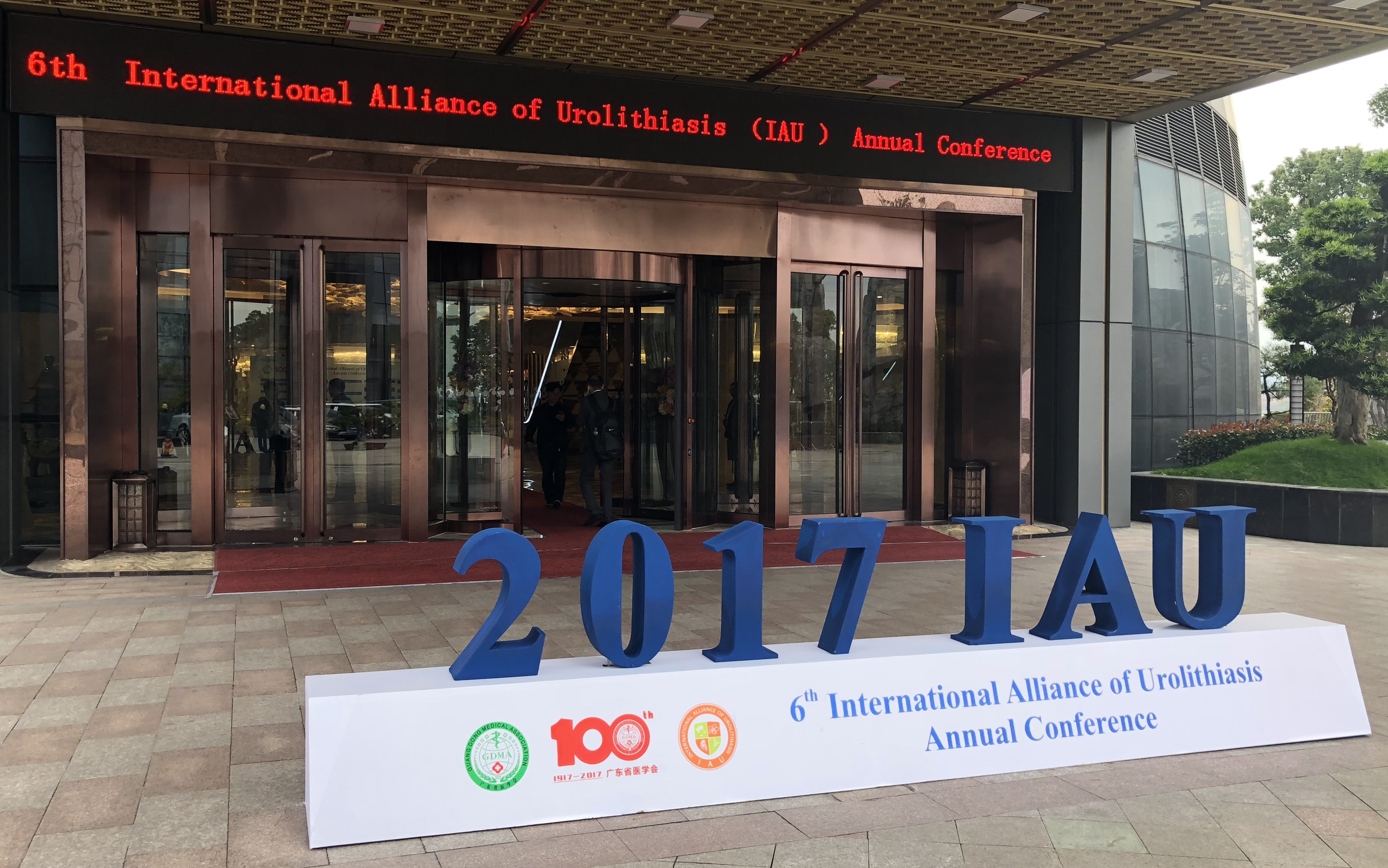
Famous for the fine yellow rice wine produced in the region, Shaoxing in Southeast China was chosen as the host city of this year’s 6th IAU annual conference. This was our first time attending the IAU, and we were both honoured to be invited to speak at the conference. We must congratulate the association and the organising committee in putting together a truly excellent program, which included many thought-stimulating and inspiring talks by eminent local and international stone experts, provocative debates, and many live surgeries demonstrating latest endoscopic techniques in the management of challenging stone diseases.

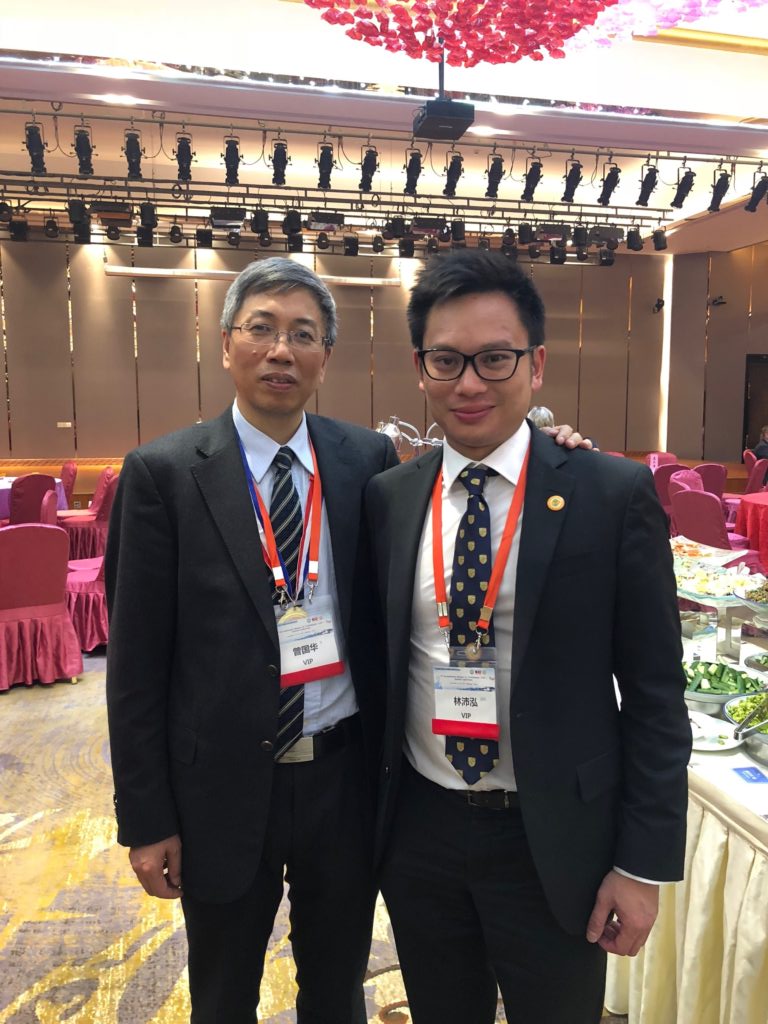
Professor Guohua Zeng and @WayneLam_Urol

#IAU17 Day 1

#IAU17 started off with the Young Urologists sessions. These sessions not only provided an opportunity for young urologists from all over the world to present their work, but also set a stage for debates on controversial stone topics and a platform to interact with experienced and established eminent stone surgeons.

One of the first sessions were talks on #PCNL. All speakers agreed choice and accurate access is key to a successful and effective #PCNL. It is interesting to know that in China, the vast majority of punctures are performed by urologists. And with experience due to high prevalence of stone disease in particular in the southern part of the country, the practice of pure ultrasound-guided puncture has gained popularity in the past decade. Dr Zhiyong Chen and Dr Xiao Yu, both from China, provided some tips and tricks on pure ultrasound-guided puncture for access for #PCNL, in particular when treating patients with complex or staghorn stones. Both sagittal and coronal planes should be used to assess all major and minor calyces, and they also interestingly showed that the benefit of aiming stones with multiple branches as the most effective first puncture point in patients with complex or staghorn stones.

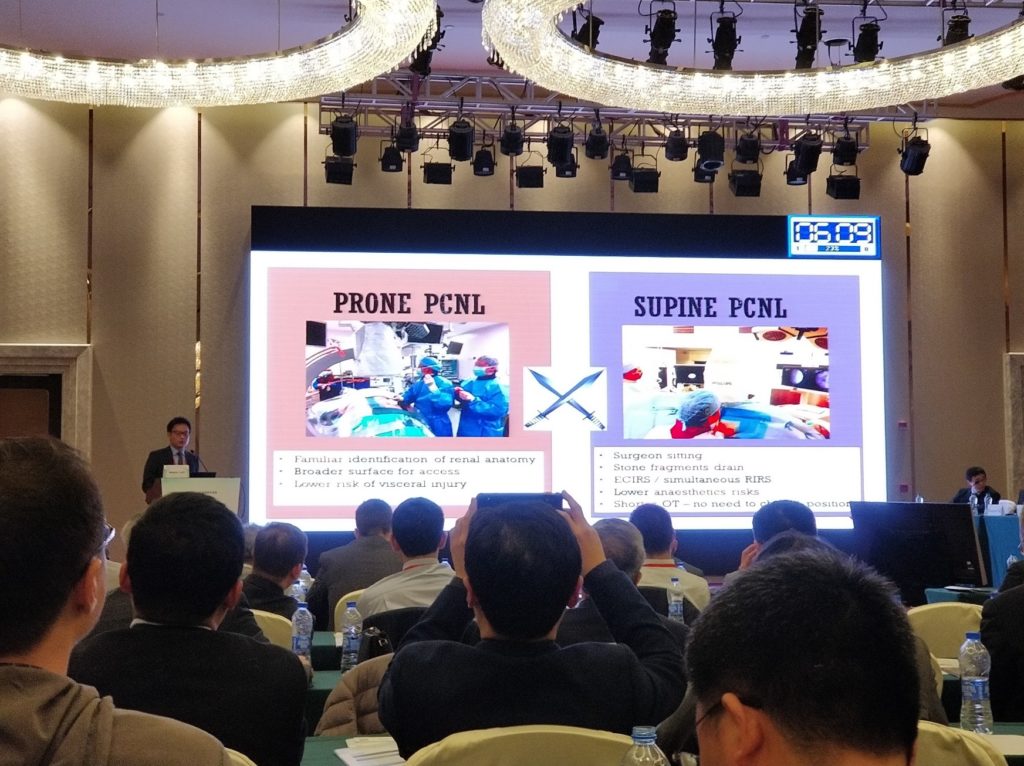
Position of #PCNL has been a regular debate in stone conferences, and few speakers in the young fellow sessions presented their findings and reviews on the topic. Both #supine and #prone positions have their pros and cons but all presenters agreed that surgeon’s preference to offer best chance of achieving best outcome is the most important determining factor.

Another eye-catching presentation of the day was a randomised study conducted in China comparing standard #PCNL against mini-PCNL in the management of 2-4cm renal stone. The study randomised 800 patients and demonstrated that mini-PCNL was superior in terms of reduced bleeding, post-operative pain, shorter hospital length of stay, and more patients were ‘tubeless’ after the procedure. It is a well-designed study that will add much-needed high-quality data to the argument on #PCNL sizing.

#Sepsis remains one of the most worrying complications during endoscopic surgery for urolithiasis. Optimal method of culture technique was discussed, with Dr Kremena Petkova of Bulgaria arguing that renal, pelvis, urine and stone cultures are more specific and sensitive in predicting post-op complications. Both are superior to #MSU, with higher concordance between pathogens and antibiotic sensitivities. However, their results are not often available pre-operatively, and it’s best to use them as guidance on choice of anti-bacterial treatment if sepsis develops post-operatively. However, in patients with high risk of post-operative sepsis despite peri-operative prophylaxis; renal, pelvis, urine and stone culture should be considered.

Another very interesting study was a randomised trial presented by Dr Wei Zhu from Guangzhou, China, on investigating dosage required for prophylactic antibiotics for patients undergoing retrograde intrarenal surgery for stones. Their study suggested that stone size is a determining factor of whether patients require prophylactic antibiotics, with risk of post-operative sepsis being low if <200 mm2.

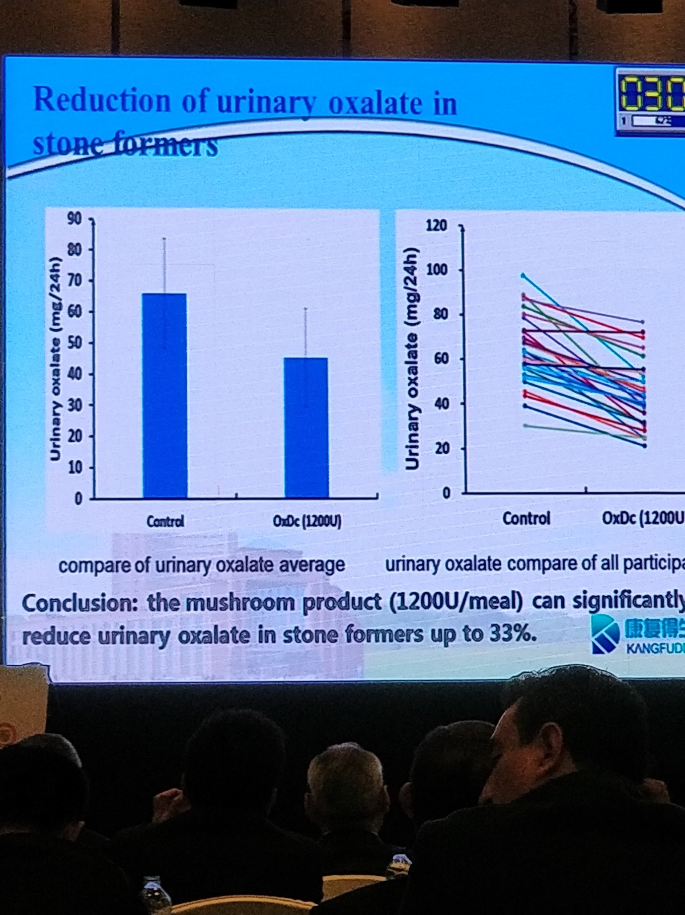
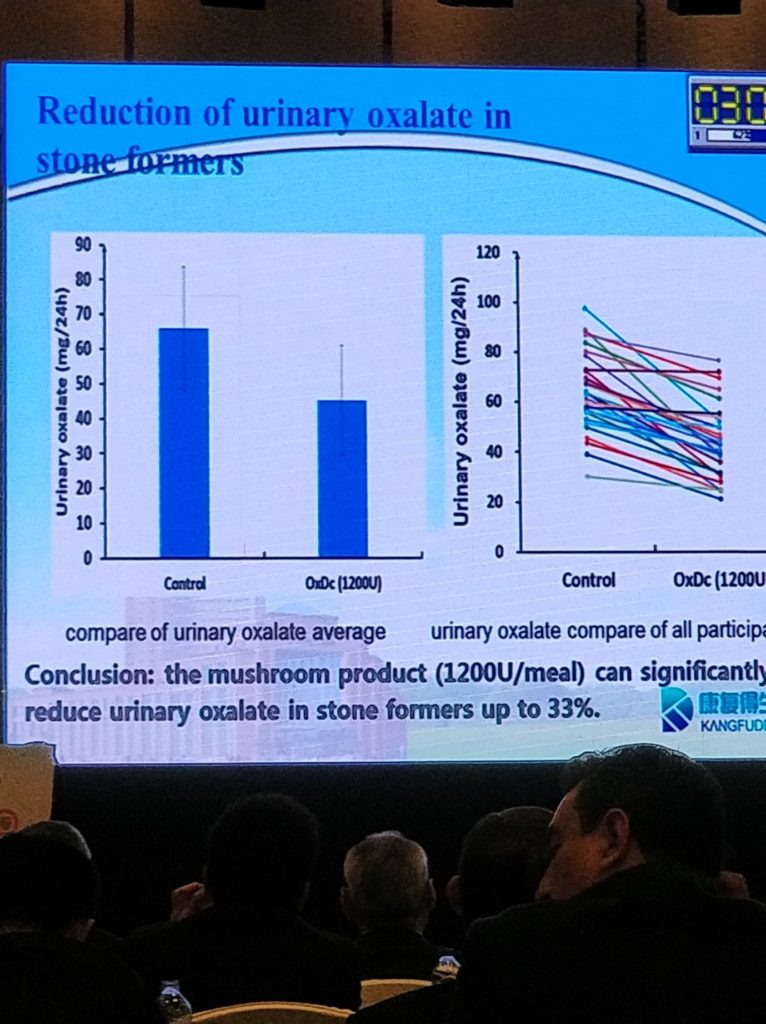
In the stone prevention session chaired by Professor Hans-Goran Tiselius from Sweden, Dr Guohao Li from China presented a study on reduction of urinary oxalate. Diet in general contributes to urinary oxalate concentration, and they discovered the use of a mushroom powder is able to reduce oxalate contents by degradation in traditionally oxalate-rich things such as spinach and tea. Their study found that the mushroom product is able to reduce urinary oxalate in stone-formers by up to 33%!

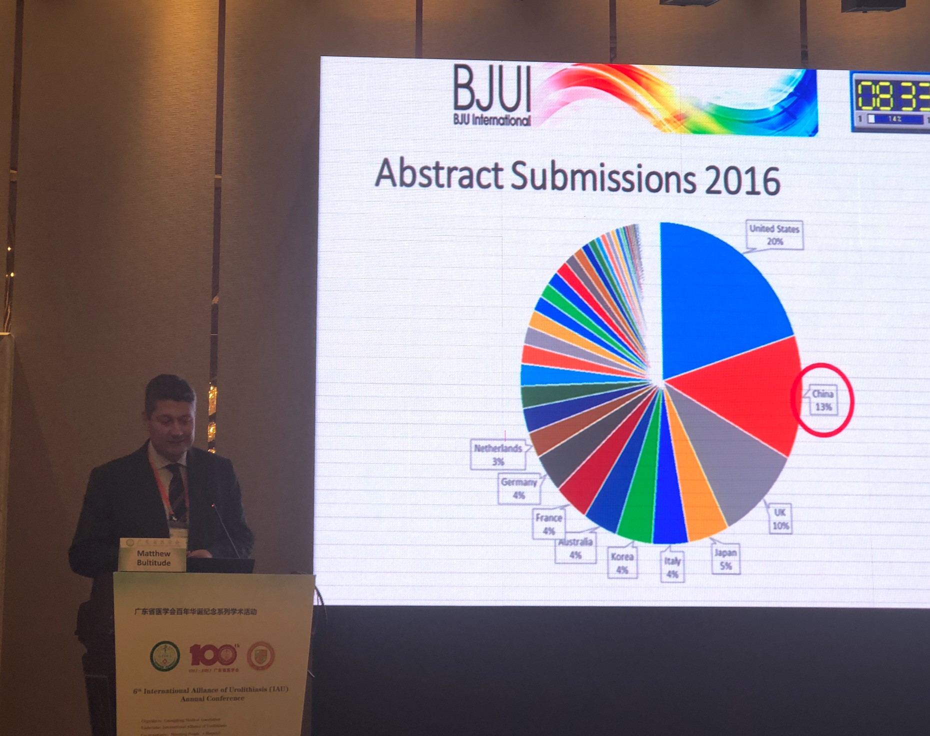
@Mattbultitude, representing @BJUinternational, was invited to give a talk on tips for submitting manuscripts to the journal to maximise the chances of publication. An interesting fact was that China submitted more papers to @BJUinternational in 2016 than the UK, and came second only to the USA. There are now services from the Journal’s publisher,Wiley, to help improve fluency of manuscripts for papers written by authors whose first language isn’t English, and perhaps this may further increase acceptance rates from countries such as China in the future (https://wileyeditingservices.com/en/translation-service/).

Heavy weights in #urolithiasis closed the last session of the first day of #IAU17. Professor Pal from India, with over 30 years of experience in PCNL, offered tips to young urologists on a procedure not often talked about in textbooks. Short, straight puncture track through a papilla into the most peripheral calyx harbouring or leading to the stone is what we should be aiming for when performing the puncture, and he suggested that meticulous alignment of the C-arm is crucial to provide the spatial information to guide us to do that. This was followed by Professor Jean de la Rosette from the Netherlands, who gave a provocative but strong arguments on why he felt #MET should not be recommended. This discussion came as a heated debate has been going on with regards to the recent publication of a very large, multi-centre randomised controlled trial from China suggesting the use of MET is beneficial in patients with larger distal ureteric stones, and of course much debate was initiated after his talk amongst the audience and on social media (see The Drugs Don’t Work … Or Do They? https://www.bjuinternational.com/bjui-blog/drugs-dont-work/).

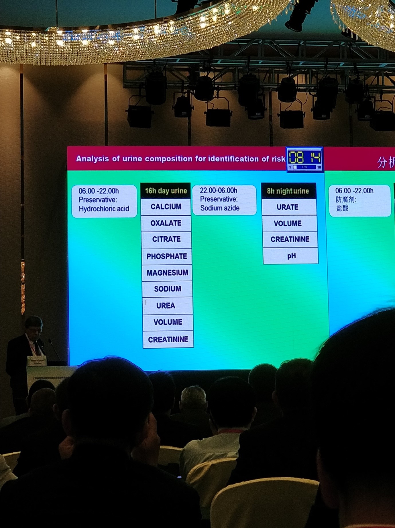
As we all know, 24-hour urine work-up is dreaded by most recurrent stone-formers.  It takes up a lot of the patient’s time and thus may result in incomplete collection or just simply be forgotten.  Professor Hans-Göran Tiselius described an abbreviated form of the 24-hour urine work-up that he uses in order to reduce patient inconvenience when collecting the urine samples. The first 16 hours are used to collect most of the common urine biochemistry of calcium, oxalate, citrate, etc while the last 8 hours are used to measure for urate and pH. Each portion is then extrapolated to achieve the final data. Through three examples, Professor Tiselius shows how it is easy to apply the results from the 24-hour urine to give specific dietary instructions and treatment to the patients in the prevention of stones.

One of the final talks of the day was presented by Professor Thomas Knoll from Germany on the use of miniaturised PCNL. Various high-quality comparative studies have demonstrated the benefit of miniaturised equipment for PCNL (in particular reduced morbidities), and interestingly the availability of miniaturised systems appeared to have increased the use of PCNL instead of using RIRS and ESWL for patients with renal stones.

This final session, with master stone surgeons sharing their experience and knowledge in the Young Urology Section, allowed the up-and-coming urologists a chance to pick the mind of the masters.  It set a great tone for the first day and created anticipation for the coming two days of the conference!


#IAU17 Day 2

Day 2 of the conference was full of exciting talks and live surgery, spanning over 12 hours from 07:50 in the morning to 20:00 in the evening!

This year @BJUinternational became an Affiliated journal of the IAU. To celebrate this, a virtual issue comprising the 10 best stone papers published in the journal over the past 2 years was published online (Best of Urolithiasis VI), and @mattbultitude, Consultant Urologist and head of stone unit at @guysurology, was invited to present these selected papers in the meeting.

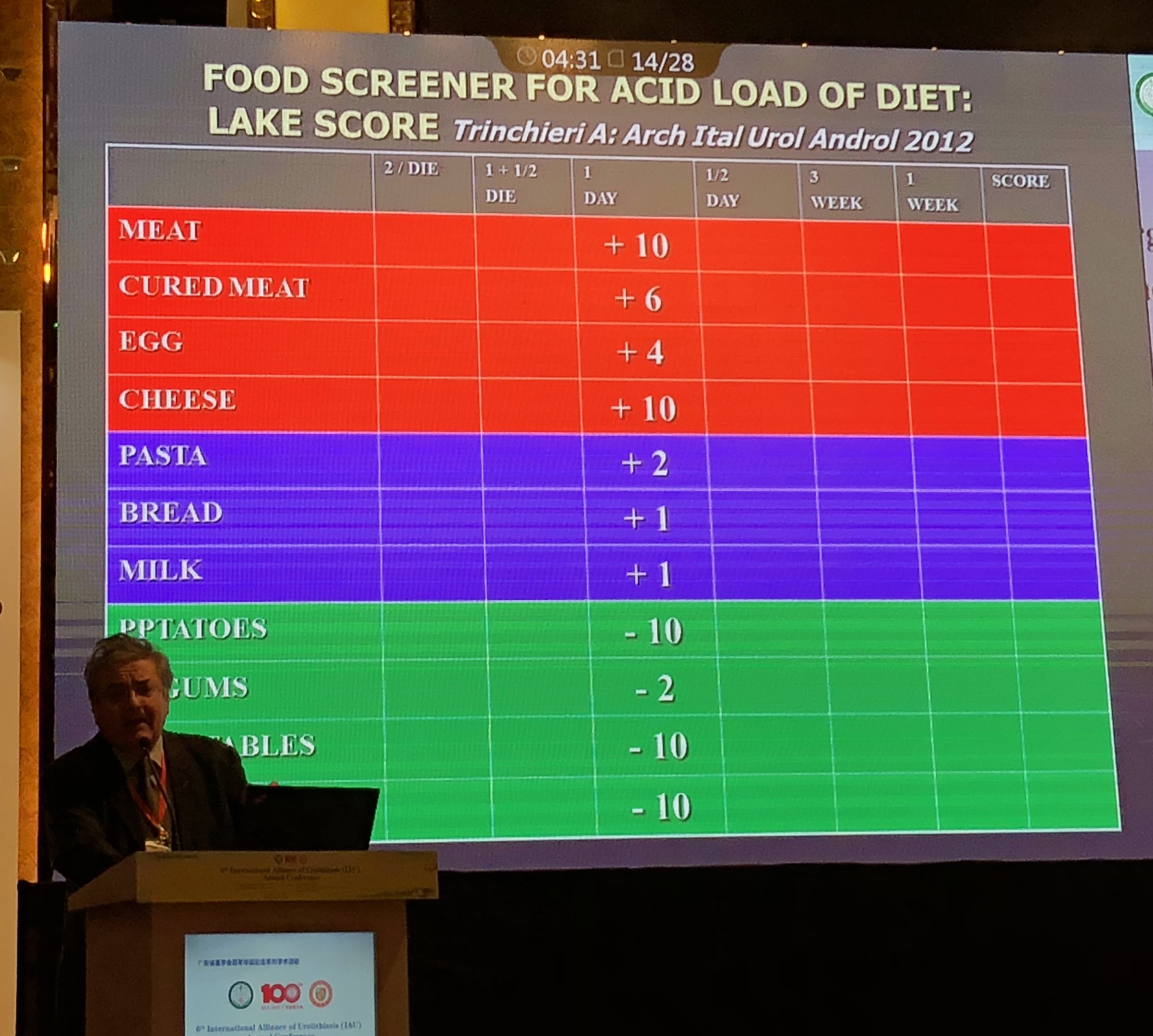
Professor Alberto Trinchieri from Italy then provided an informative talk on the role of acid load in citrate excretion. Hypocitraturia is a common feature in up to 68% of calcium stone-formers. He argued that the acid load of the diet could decrease renal citrate excretion, and the LAKE score, could be a useful tool to be used as a food screener for acid load of diet.

The LAKE score by Professor Trinchieri.

He also argued that the use of oral alkaline citrate can potentially treat uric acid stones by dissolution and prevent calcium oxalate renal stones formation.

Dr Ravi Kulkani from the UK gave an interesting talk on the management of stones in the elderly population. He presented a study conducted at his institution of 60 patients with a median age of 84.6 years with low morbidity, post-op ITU stay and a median length of stay of only 1 day for the cohort. Complete stone clearance rate was still high in the elderly population studied. It is important to assess co-morbidity pre-operatively and optimise them before any surgical treatment, together with extensive anaesthetic input and assessment. Patient selection is crucial and a good outcome can still be achieved in the ever-growing geriatric population.

Following live surgery session demonstrating various tips and tricks of RIRS, the afternoon session on day 2 of #IAU continued to be comprehensive and informative. Professor Peter Alken from Germany gave a provocative talk on the underuse of chemolysis in treatment of patients with uric acid stones, of which most of the audience were in agreement. He argued that evidence suggests it is effective and should be recommended in the guidelines!

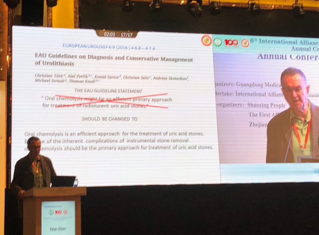
Professor Peter Alken from Germany on chemolysis for treatment of uric acid stone.

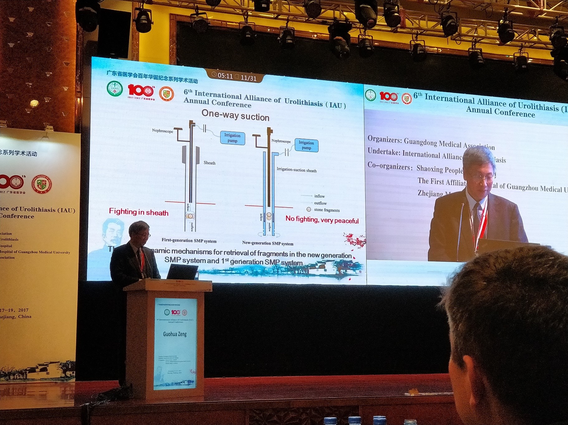
Professor Guohua Zeng, inventor of super-mini PCNL (SMP), gave a lecture on his experience using the second generation SMP. With the modified sheath enabling efficient irrigation-suction system, he found that the intrarenal pressure intraoperatively remained stably low, with shorter operative time and good stone clearance rate. Undoubtedly, SMP is useful in particular in the management of stones up to 3cm in size, and can be used as an adjunct to standard PCNL when multi-tracts are required. He has also presented his experience in using the technique in the paediatric population with good stone clearance rate and safe. (See The new generation super-mini percutaneous nephrolithotomy (SMP) system: a step-by-step guide)


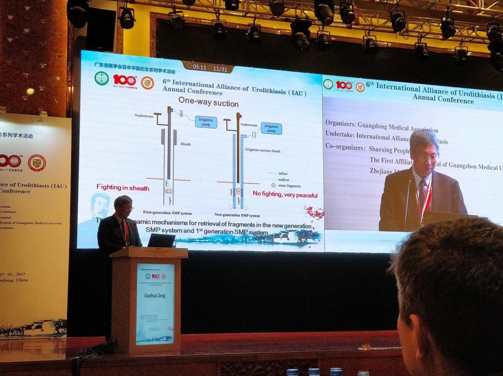
Professor Guohua Zeng on the use of second generation SMP, with an improved
irrigation/suction system.

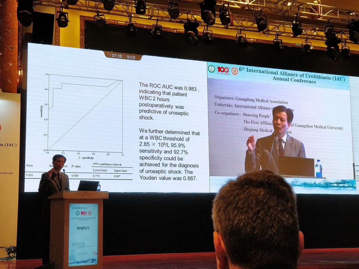
Another interesting study from Professor Gonghui Li revealed that post-endoscopic lithotripsy septic shock was heralded by a White Cell Count of <2.85 x 10⁹/L at 2 hours post-operation, with sensitivity & specificity over 90%.  Stepping up the antibiotics and aggressive fluid resuscitation at this point could stave off significant hypotension or even mortality in his study.

Professor Gonghui Li of China on early detection of risk of septic shock post-surgery with White Cell Count at 2 hours post-op.

In the evening, the conference became heated with various debates. Management of calyceal diverticulum stones has always been challenging. Mr Simon Choong from UCLH in London, UK, presented good arguments in the use of PCNL with high success rates, but Dr Yi Zhang also showed good clearance rate in experienced surgeons’ hands. And in selective cases, both minimally invasive and open surgery may have a role, presented by Dr Gang Zhu from China and Dr Zinelabidine Abouelfadel from Africa.

Debate on optimal management of 1-2cm lower pole renal stone has been a hot topic for years. Brian Eisner (@BEendourology) from the USA argued that with experience, RIRS stone clearance rate is approaching that of PCNL but with lesser morbidity. However, seeing a live surgery of clearing a >3 cm stone with a miniaturised PCNL technique on day 3 of the meeting, with the patient left completely tubeless post-operatively, may have changed his mind!


#IAU17 Day 3

The final day of the conference included 13 live #PCNL surgeries, demonstrating various puncture techniques, tips on how to improve accuracy for access, and advanced surgical techniques including various miniaturised #PCNL by local and international experts.

Professor Guohua Zeng from Guangzhou Medical University First Affiliated Hospital in China demonstrated the treatment of a 3.5cm lower pole renal stone using the second generation super-mini PCNL (SMP), which he invented. With its innovative sheath, which provides effective irrigation and controlled suction, he completed the surgery within 20 minutes. It was bloodless and tubeless, with visually complete stone clearance. Stones were completely extracted via its suction system for stone analysis.

Professor Qu Chen of China demonstrated the use of ultra-mini-PCNL, effectively clearing a medium sized renal stone in a matter of minutes. Interestingly, many surgeons in China prefers to use the ureteric catheter as inflow for irrigation, which generates a flow pressure to help flush stone fragments out – great tip!

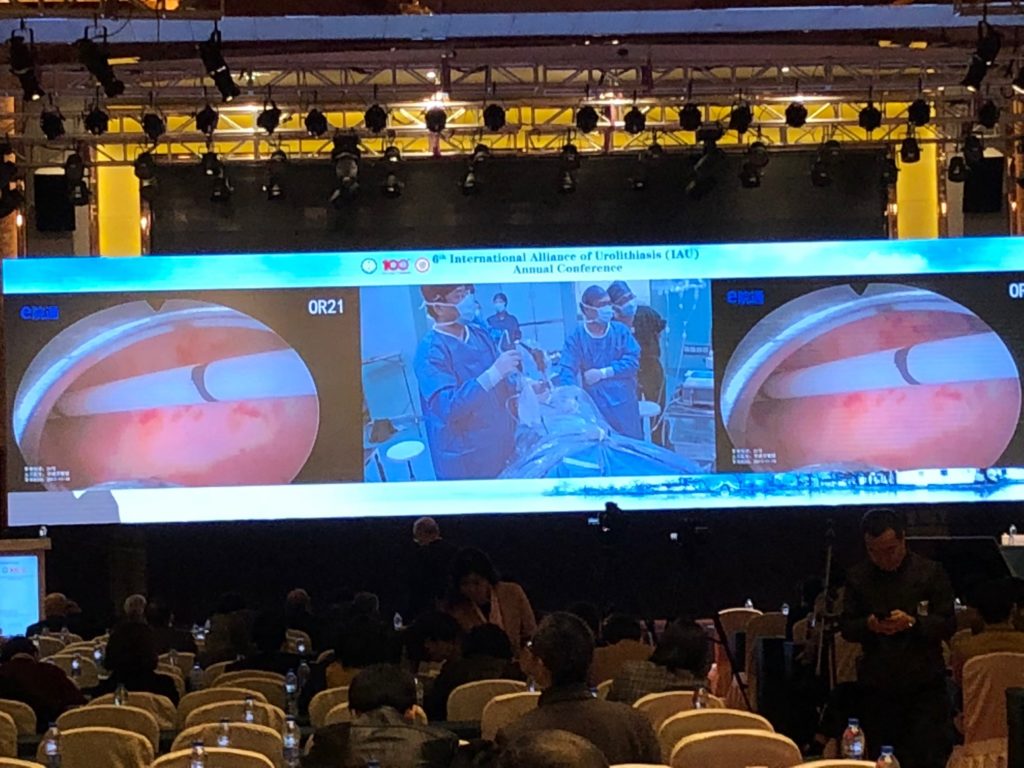
13 live stone surgeries were broadcasted to the audience on day 2 of IAU2017

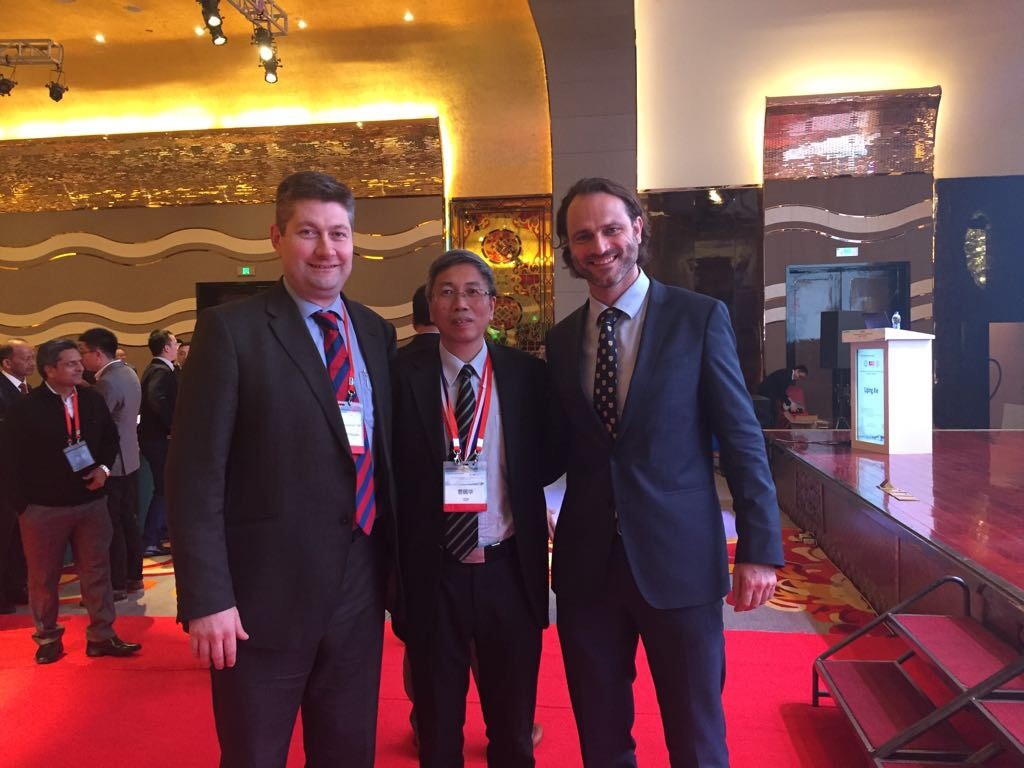
Mr Matt Bultitidue @mattbultitide (left) with Professor Guohua Zeng (middle) and
Dr Christian Seitz @SEITZ_C_C (right)

We must congratulate the #IAU17 organisers’ incredible effort in making the conference an inspiring and valuable learning experience to all who attended. The short duration (8-15mins) of presentations ensured that all the meaty details were packed in with very little fluff!  It was also a great opportunity to build bridges to network and collaborate research in #urolithiasis. We thoroughly enjoyed it and would definitely recommend any urologist with an interest in #urolithiasis to attend its future meetings, and we very much look forward to #IAU18 in Istanbul!


Dr Wayne Lam

Assistant Professor in Urology, Queen Mary Hospital, University of Hong Kong

Twitter: @WayneLam_Urol


Dr Brian Ho

Associate Consultant in Urology, Queen Mary Hospital, University of Hong Kong


Urolithiasis around the world

Stone disease is a highly prevalent condition that unites all countries around the world, although surgical management will depend on many factors including availability of different technologies. However, percutaneous nephrolithotomy (PCNL) remains the cornerstone for the management of larger renal stones in all parts of the world, and Rizvi et al. [1] report on a huge cohort of PCNL procedures – 3 402 to be precise from Karachi. This is a single-centre series, over an 18-year period, reporting real-life data and showing a stone clearance rate of ~80%, as assessed by plain abdominal radiograph of the kidneys, ureters and bladder, and ultrasonography (US). Whilst the definition of stone-free and imaging modality used to judge it remains a contentious issue, this paper reflects the excellence of high-volume surgery in specialist centres.

Recently, the BJUI became the affiliated journal for the International Alliance of Urolithiasis (IAU), whose annual meeting takes place in Shaoxing this month. To celebrate this, we are proud to publish a ‘Best of Urolithiasis’ issue, which features some of the top stone papers published in the BJUI over the last few years [2]. Choosing articles for this was quite a task given the quality and whilst we have attempted to recognise submissions that potentially change practice, the geographical diversity of the work shows not only the global nature of stone disease but also the excellent research that is being done worldwide and in different healthcare systems to improve care and outcomes. Of particular importance are randomised trials that are often lacking in surgical areas. One such paper from China addressed the question of US vs fluoroscopy for PCNL access during mini-PCNL [3]. Whilst the truth is that surgeons should use whatever gives the best outcomes, the authors in a very high-volume centre were able to demonstrate the effectiveness of US-only punctures, although a combination may be better in complex stone burdens. Another randomised controlled trial (RCT) of clinical importance was from the USA, where the authors conducted a good quality double-blind RCT of NSAID use before ureteric stent removal under local anaesthesia [4]. Whilst a small study, the incidence of severe pain in the 24 h after stent removal was 55% in the placebo group vs 0% in the NSAID group – as such this simple study should have changed practice for all who perform this procedure.

Other papers worthy of inclusion include a single-centre experience of the conservative management of staghorn calculi, which challenges the dogma that all staghorn stones should be treated [5]. This single-centre series showed a conservative policy could be adopted in highly selected patients. Is this practice changing? Maybe … but it certainly gives an evidence base for stone surgeons in making decisions in very high-risk patients. Manoj Monga and his group recently reported on the accuracy of US for the detection of renal stones [6]. This again is a very important topic and a question that commonly arises. In a series of >500 patients with US-detected stones who subsequently underwent CT scanning, 22% of patients would have been inappropriately counselled about their stone based on US alone. Again, the message is clear … US is a good screening tool but do not rely on it for treatment decisions.

I hope you take the time to check out the virtual issue on urolithiasis and read the other papers I could not mention here. Please continue to send your high-quality stone papers to the BJUI and maybe your submission will feature in our next ‘Best of Urolithiasis’ issue.

Matthew Bultitude, BJUI Associate Editor


Guys and St Thomas NHS Foundation Trust, London, UK


References


1 Rizvi SA, Hussain M, Askari SH, Hashmi A, Lal M, Zafar MN. Surgical outcomes of percutaneous nephrolithotomy in 3402 patients and results of stone analysis in 1559 patients. BJU Int 2017; 120: 7029


2 BJU International. Virtual Issues Page. Available at: https://bit.ly/BJUI-VIs. Accessed September 2017


4 Tadros NN, Bland L, Legg E, Olyaei A, Conlin MJ. A single dose of non-steroidal anti-inammatory drug (NSAID) prevents severe pain after ureteric stent removal: a prospective, randomised, double-blind, placebo-  controlled trial. BJU Int 2013; 111: 1015


5 Deutsch PG, Subramonian K. Conservative management of staghorn calculi: a single-centre experience. BJU Int 2016; 118: 44450


6 Ganesan V, De S, Greene D, Torricelli FC, Monga M. Accuracy of ultrasonography for renal stone detection and size determination: is it good enough for management decisions? BJU Int 2017; 119: 4649


Article of the Week: Comparing FG, USG and CG for renal access in mini-PCNL

Every week the Editor-in-Chief selects an Article of the Week from the current issue of BJUI. The abstract is reproduced below and you can click on the button to read the full article, which is freely available to all readers for at least 30 days from the time of this post.

In addition to the article itself, there is an accompanying editorial written by a prominent member of the urological community. This blog is intended to provoke comment and discussion and we invite you to use the comment tools at the bottom of each post to join the conversation.

Finally, the third post under the Article of the Week heading on the homepage will consist of additional material or media. This week we feature a video discussing the paper.

If you only have time to read one article this week, it should be this one.

A prospective and randomised trial comparing fluoroscopic, total ultrasonographic, and combined guidance for renal access in mini-percutaneous nephrolithotomy

Wei Zhu*, Jiasheng Li*, Jian Yuan*, Yongda Liu*, Shaw P.Wan*, Guanzhao Liu*† Wenzhong Chen*, Wenqi Wu*, Jintai Luo*, Dongliang Zhong*, Defeng Qi*, Ming
Lei*, Wen Zhong*, Ze Zhang*, Zhaohui He*, Zhijian Zhao*, Suilin Lu*, Yuji Wu*
and Guohua Zeng*


*Department of Urology, Minimally Invasive Surgery Center, The First Afliated Hospital of Guangzhou Medical University, and Guangdong Key Laboratory of Urology, Guangzhou, Guangdong, China


Read the full article

Abstract

Objective

To compare the safety and efficacy of fluoroscopic guidance (FG), total ultrasonographic guidance (USG), and combined ultrasonographic and fluoroscopic guidance (CG) for percutaneous renal access in mini-percutaneous nephrolithotomy (mini-PCNL).

Patients and methods

The present study was conducted between July 2014 and May 2015 as a prospective randomised trial at the First Affiliated Hospital of Guangzhou Medical University. In all, 450 consecutive patients with renal stones of >2 cm were randomised to undergo FG, USG, or CG mini-PCNL (150 patients for each group). The primary endpoints were the stone-free rate (SFR) and blood loss (haemoglobin decrease during the operation and transfusion rate). Secondary endpoints included access failure rate, operating time, and complications. S.T.O.N.E. score was used to document the complexity of the renal stones. The study was registered at https://clinicaltrials.gov/ (NCT02266381).

apr-saotw-4

Results

The three groups had similar baseline characteristics. With S.T.O.N.E. scores of 5–6 or 9–13, the SFRs were comparable between the three groups. For S.T.O.N.E. scores of 7–8, FG and CG achieved significantly better SFRs than USG (one-session SFR 85.1% vs 88.5% vs 66.7%, P = 0.006; overall SFR at 3 months postoperatively 89.4% vs 90.2% vs 69.8%, P = 0.002). Multiple-tracts mini-PCNL was used more frequently in the FG and CG groups than in the USG group (20.7% vs 17.1% vs 9.5%, P = 0.028). The mean total radiation exposure time was significantly greater for FG than for CG (47.5 vs 17.9 s, P < 0.001). The USG had zero radiation exposure. There was no significant difference in the haemoglobin decrease, transfusion rate, access failure rate, operating time, nephrostomy drainage time, and hospital stay among the groups. The overall operative complication rates using the Clavien–Dindo grading system were similar between the groups.

Conclusions

Mini-PCNL under USG is as safe and effective as FG or CG in the treatment of simple kidney stones (S.T.O.N.E. scores 5–6) but with no radiation exposure. FG or CG is more effective for patients with S.T.O.N.E. scores of 7–8, where multiple percutaneous tracts may be necessary.

Read more articles of the week

Editorial: Renal access during PCNL: increasing value of USG for a safer and successful procedure

Renal access to the pelvicalyceal system is the initial but highly important and crucial step of percutaneous nephrolithotomy (PCNL), which can significantly affect the final outcome of the procedure. Although the puncture of the kidney and subsequently dilatation of the tract has been commonly performed under fluoroscopic guidance [1]; renal access can also be established under ultrasonographic guidance (USG) with or without fluoroscopy.

To give a further insight into the role of both methods; in a prospective and randomised study published in this issue of the BJUI, Zhu et al. [2] have compared the safety and efficacy of fluoroscopic (FG), total ultrasonographic (USG), and combined ultrasonographic and fluoroscopic guidance (CG) for percutaneous renal access during mini-PCNL (mini-PCNL). In all, 450 consecutive patients with renal stones of >2 cm were randomised to undergo three different approaches during mini-PCNL. In addition to the stone-free rate (SFR) and blood loss as primary endpoints; access failure rate, operative time and complications were also evaluated. The S.T.O.N.E. [stone size (S), tract length (T), obstruction (O), number of involved calices (N), and essence or stone density (E)] scoring system was used for stone assessment [3] and the scores were further categorised into three grades (5–6, 7–8 and 9–13) for comparison.

While the overall operative complication rates, using the Clavien–Dindo grading system, were similar between the three groups; colonic injury treated with a temporary colostomy occurred in one case in the CG group. Although the SFRs were similar between the groups with S.T.O.N.E. scores of 5–6 and 9–13; the FG and CG approaches achieved significantly better SFRs than USG in patients with scores of 7–8, (P = 0.006). Multiple-tracts PCNL were used more frequently in the FG and CG group than USG group (P = 0.028). While the access failure rate was similar in the groups, the mean access time was longer in the CG group than in the FG and USG groups (P = 0.003). However, the mean total radiation exposure time was significantly greater for FG than for CG (47.5 vs 17.9 s, P < 0.001). The USG had zero radiation exposure. The operative time, hospital stay, nephrostomy drainage time, and the changes in the haemoglobin and creatinine levels were all similar in the three groups. The authors [1] concluded that mini-PCNL under total USG is as safe and effective as FG or CG in the treatment of simple kidney stones (S.T.O.N.E. scores 5–6) with no risk of radiation exposure. FG or CG is more effective for patients with S.T.O.N.E. scores of 7–8 where multiple percutaneous tracts may be necessary.

Percutaneous nephrolithotomy is now the preferred treatment method for larger stones (>2 cm) with successful outcomes. However, despite the high SFR obtained in a single session this approach can be associated with some severe complications such as bleeding, organ perforation, and sepsis. Such complications could be encountered during all steps of PCNL among which renal access seems to be the most critical one [4]. An appropriate puncture aiming a direct path from the skin through the papilla of the desired calyx of the kidney is of paramount importance to limit the above mentioned complications. Such an access to the renal collecting system can be established by either FG and/or USG. Although FG has been used commonly in the past; increasing experience in US applications has enabled endourologists to use this approach more often with some certain advantages in preventing renal puncture-related complications. When compared with FG, use of USG in establishing an access under vision allows the surgeon to identify the kidney pelvicalyceal system as well as the surrounding organs in a precise manner [5], with the benefit of minimising the risk of injury to such organs. Moreover, in addition to being free of ionising radiation; USG results in fewer punctures, has shorter operating times, and avoids contrast-related complications [1, 2]. Apart from helping to identify non-opaque residual stones at the end of the procedure; colour Doppler US can be used as a tool to localise the intrarenal arteries and avoid their puncture. However, the use of USG is an operator-dependent procedure requiring sufficient experience before routine performance and it may not be as efficient in the extremely obese patient and patients without hydronephrosis.

For the use of USG access in clinical practice, Agarwal et al. [5] reported a shorter mean time for successful puncture and significantly lower radiation exposure, yielding complete stone clearance with no substantial morbidity when compared with the FG technique. USG access was found also to increase puncture accuracy to a certain extent with a 96.5% SFR in another trial [6].

In conclusion, each of these techniques mentioned above have their own advantages and disadvantages. Despite its high success rate, radiation exposure and risk of multiple punctures are the main risks of the FG approach. USG renal access in experienced hands can produce high success rates following an appropriate puncture, lower risk of radiation exposure, and the ability to monitor all organs in the path of the puncture [7]. Depending on the surgeon’s experience, patient and stone-related factors, as well as the technical infrastructure, each approach may be used either alone or in combination for a complication-free and successful procedure. However, taking the above mentioned advantages of USG access into account, it is clear that all young urologist need to increase their experience in USG puncture to use it in appropriate cases (children, pregnant cases, dilated kidneys etc.) to lower the radiation risk and shorten the procedural duration.

Read the full article
Kemal Sarica, Professor of Urology, Chief


Department of Urology, Health Sciences University, Dr Lut Kirdar Kartal Research and Training Hospital, Istanbul, Turkey


References


1 Michel MS, Trojan L, Rassweiler JJ. Complications in percutaneous nephrolithotomy. Eur Urol 2007; 51: 899906


3 Okhunov Z, Friedlander JI, George AK et al. S.T.O.N.E. nephrolithometry: novel surgical classication system for kidney calculi. Urology 2013; 81: 115460


4 Aslam MZ, Thwaini A, Duggan B et al. Urologists versus radiologists made PCNL tracts: the UK experience. Urol Res 2011; 39: 21721


5 Agarwal M, Agrawal MS, Jaiswal A, Kumar D, Yadav H, Lavania PSafety and efcacy of ultrasonography as an adjunct to uoroscopy for renal access in percutaneous nephrolithotomy. BJU Int 2011; 108: 13469


6 BasiriA, Ziaee AM, Kianian HR, Mehrabi S, Ka rami H, Moghaddam SM. Ultrasonographic versus u oroscopic access for percutaneounephrolithotomy, a randomized clinical trial. J Enodourol 2008; 22: 28 14


7 Osman M, Wendt-Nordahl G, Heger K, Michel MS, Alken P, Knoll TPercutaneous nephrolithotomy with ultrasonography-guided renal access: experience from over 300 cases. BJU Int 2005; 96: 8758


© 2024 BJU International. All Rights Reserved.