Archive for category: BJUI Blog

Residents’ podcast: Artificial intelligence applications in urology

Maria Uloko is a Urology Resident at the University of Minnesota Hospital. In this podcast she is joined by Dr Christopher Weight, an Associate Professor in the Department of Urology at the University of Minnesota. They are discussing a recent BJUI Article of the month:

Current status of artificial intelligence applications in urology and their potential to influence clinical practice

Abstract

Objective

To investigate the applications of artificial intelligence (AI) in diagnosis, treatment and outcome prediction in urologic diseases and evaluate its advantages over traditional models and methods.

Materials and methods

A literature search was performed after PROSPERO registration (CRD42018103701) and in compliance with Preferred Reported Items for Systematic Reviews and Meta‐Analyses (PRISMA) methods. Articles between 1994 and 2018 using the search terms “urology”, “artificial intelligence”, “machine learning” were included and categorized by the application of AI in urology. Review articles, editorial comments, articles with no full‐text access, and nonurologic studies were excluded.

Results

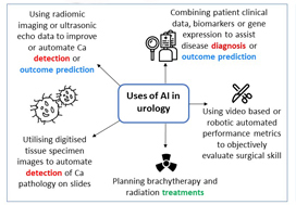
Initial search yielded 231 articles, but after excluding duplicates and following full‐text review and examination of article references, only 111 articles were included in the final analysis. AI applications in urology include: utilizing radiomic imaging or ultrasonic echo data to improve or automate cancer detection or outcome prediction, utilizing digitized tissue specimen images to automate detection of cancer on pathology slides, and combining patient clinical data, biomarkers, or gene expression to assist disease diagnosis or outcome prediction. Some studies employed AI to plan brachytherapy and radiation treatments while others used video-based or robotic automated performance metrics to objectively evaluate surgical skill. Compared to conventional statistical analysis, 71.8% of studies concluded that AI is superior in diagnosis and outcome prediction.

Conclusion

AI has been widely adopted in urology. Compared to conventional statistics AI approaches are more accurate in prediction and more explorative for analyzing large data cohorts. With an increasing library of patient data accessible to clinicians, AI may help facilitate evidence‐based and individualized patient care.

BJUI Podcasts now available on iTunes, subscribe here https://itunes.apple.com/gb/podcast/bju-international/id1309570262


Dr Weight specializes in the surgical treatment of urologic cancers including prostate, bladder, kidney, adrenal, testis and penile cancer. He performs open, endoscopic, laparoscopic, robotic (da Vinci) and retroperineoscopic surgery.

Dr Weight completed his residency training at Cleveland Clinic where he received several awards including the George and Grace Crile Traveling Fellowship Award, the Society of Laparoendoscopic Surgeons Resident Achievement Award and the ASCO Genitourinary Cancer Symposium Merit Award. Dr. Weight then completed a fellowship in Urologic Oncology at Mayo Clinic, where he also completed a Masters degree in Clinical and Translational Research from Mayo Graduate School and was awarded the Mayo Fellows Association Humanitarian Award.

Dr Weight believes that medical research is a key component to offering excellent patient care. His research is focused on improving patient outcomes and the use of artificial intelligence in different urologic applications. He is an author of more than 45 peer-reviewed publications and book chapters and has been invited to speak at regional, national and international conferences. 

November 2019 – About the cover

November’s Article of the Month was written by researchers primarily from New York City, USA: Guideline of Guidelines: Testosterone Replacement Therapy for Testosterone Deficiency

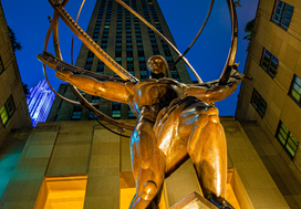
The cover image shows the statue of Atlas located within the Rockefeller Center. This “city within a city” was conceived by John D. Rockefeller Jr. and was built during the 1930s, providing valuable jobs during the Great Depression. The first buildings were opened in 1933 providing a center of art, style and entertainment.

The statue of Atlas – a half man/half god giant from Greek mythology – was built in 1937 by Lee Laurie and Rene Paul Chambellan. It is 45 feet (14 metres) tall and weighs 7 tonnes.


BJUI at the Indonesian Urological Association Annual Scientific Meeting

The Indonesian Urological Association Annual Scientific meeting was held at The Golden Tulip Hotel, Banjarmasin – 3-5 October 2019.


The main conference was preceded by pre-congress workshops at the University of Indonesia Medical Education and Research Institute (IMERI) in Jakarta.

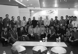
Masterclass with Consultant Urologist Mr Brian Chaplin

Furthermore, the BJUI held a plenary lecture entitled: High Risk Non-Muscle-Invasive Bladder Cancer : The Promise of New Therapies by Consultant Urologist Miss Jo Cresswell, also from the South Tees Hospitals NHS Foundation trust in the UK.

[caption id=”attachment_40134″ align=”aligncenter” width=”243′ label=’ Promoting knowledge: Miss Jo Cresswell at the Masterclass

The conference also featured the increasingly popular 10 and 5 Km Uroruns and a Urowalk starting at 6 and 7am on the Saturday morning.


Residents’ podcast: NICE guidelines – renal and ureteric stones

Nikita Bhatt is a Specialist Trainee in Urology in the East of England Deanery and a BURST Committee member @BURSTUrology

NICE Guideline – Renal and ureteric stones: assessment and management

Read the full article

Context

Renal and ureteric stones usually present as an acute episode with severe pain, although some stones are picked up incidentally during imaging or may present as a history of infection. The initial diagnosis is made by taking a clinical history and examination and carrying out imaging; initial management is with painkillers and treatment of any infection.

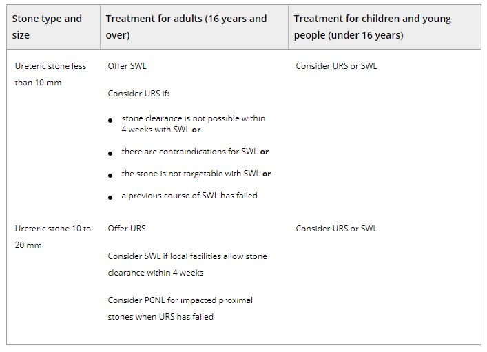
Ongoing treatment of renal and ureteric stones depends on the site of the stone and size of the stone (less than 10 mm, 10 to 20 mm, greater than 20 mm; staghorn stones). Options for treatment range from observation with pain relief to surgical intervention. Open surgery is performed very infrequently; most surgical stone management is minimally invasive and the interventions include shockwave lithotripsy (SWL), ureteroscopy (URS) and percutaneous stone removal (surgery). As well as the site and size of the stone, treatment also depends on local facilities and expertise. Most centres have access to SWL, but many use a mobile machine on a sessional basis rather than a fixed‐site machine, which has easier access during the working week. The use of a mobile machine may affect options for emergency treatment, but may also add to waiting times for non‐emergency treatment.

Although URS for renal and ureteric stones is increasing (there has been a 49% increase from 12,062 treatments in 2009/10, to 18,066 in 2014/15 [Hospital Episode Statistics data]), there is a trend towards day‐case/ambulatory care, with this increasing by 10% to 31,000 cases a year between 2010 and 2015. The total number of bed‐days used for renal stone disease has fallen by 15% since 2009/10. However, waiting times for treatment are increasing and this means that patient satisfaction is likely to be lower.

Because the incidence of renal and ureteric stones and the rate of intervention are increasing, there is a need to reduce recurrences through patient education and lifestyle changes. Assessing dietary factors and changing lifestyle have been shown to reduce the number of episodes in people with renal stone disease.

Adults, children and young people using services, their families and carers, and the public will be able to use the guideline to find out more about what NICE recommends, and help them make decisions. These recommendations apply to all settings in which NHS‐commissioned care is provided.


Table 2.Surgical treatment (including SWL) of ureteric stones in adults, children and young people Abbreviations: PCNL, percutaneous nephrolithotomy; SWL, shockwave lithotripsy; URS, ureteroscopy.


BJUI Podcasts now available on iTunes, subscribe here https://itunes.apple.com/gb/podcast/bju-international/id1309570262


A taster week in urology and renal transplant in the UK

A taster week is a training opportunity offered to UK doctors in their first two years of clinical practice to try a new specialty. They are an important learning experience for doctors at this stage, who will have experienced working in six different specialties at most. While taster weeks are only five days long, they offer a unique insight into a new specialty, as well as the chance to network with registrars and consultants.

During medical school I was interested in transplantation, as I found the combination between surgery and immunology interesting. This led me to complete a Master of Research in Transplantation while at medical school. During this degree, I looked at urinary tract infection in transplant patients and this started my interest in urology.

In the UK, the majority of trainees enter the field of transplantation following training in General Surgery. In clinical practice, it is good to have urologists with an active interest in Renal Transplantation for the betterment of these patients but there are few centres where this can be learnt. However, the Freeman Hospital in Newcastle, UK offers a one to two-year fellowship in renal transplantation which can be completed at the end of urology training. I contacted one of the urology and renal transplant surgeons and organised a week’s visit to the Freeman Hospital.


During my taster week I had the opportunity to shadow a urology and renal transplant surgeon. I joined urology and transplant ward rounds, including a renal transplant grand round, and I also attended a transplant nephrology clinic where I saw the long-term management of patients who had received kidney transplants.  I observed theatre lists in both urology and transplant and saw the wide variety of operations that urology and transplant surgeons are involved in, such as renal access surgery for dialysis and robotic partial nephrectomy for renal cancer.

I also had a chance to attend multi-disciplinary team meetings about new transplant recipients as well as an x-ray imaging meeting concerning live kidney donors.

Speaking to urology and renal transplant surgeons was an invaluable experience and helped me plan the next steps in my career as well as solidify it as a preferred career choice.

The highlight of my taster week was attending regional surgical teaching. I spent a day in one of the few world-class cadaveric training laboratories in the UK and learnt how to perform an orchidopexy for testicular torsion and vascular anastomosis; two operations that are no doubt necessary for a urology and renal transplant surgeon.

I am very glad I completed a taster week in urology and renal transplantation. It allowed me to experience the variety of work involved in this niche specialty. It was an experience that would have only been available much later in my career otherwise, which would be at a point too late for a career change.

by Matthew Byrne


Matthew Byrne recently completed two years as an Academic Foundation Doctor in Cambridge, UK. He graduated MBBS from Newcastle where he also completed a Master of Research in Transplantation. He is now a Urology Clinical Fellow in Cambridge, UK.


October 2019 – About the cover

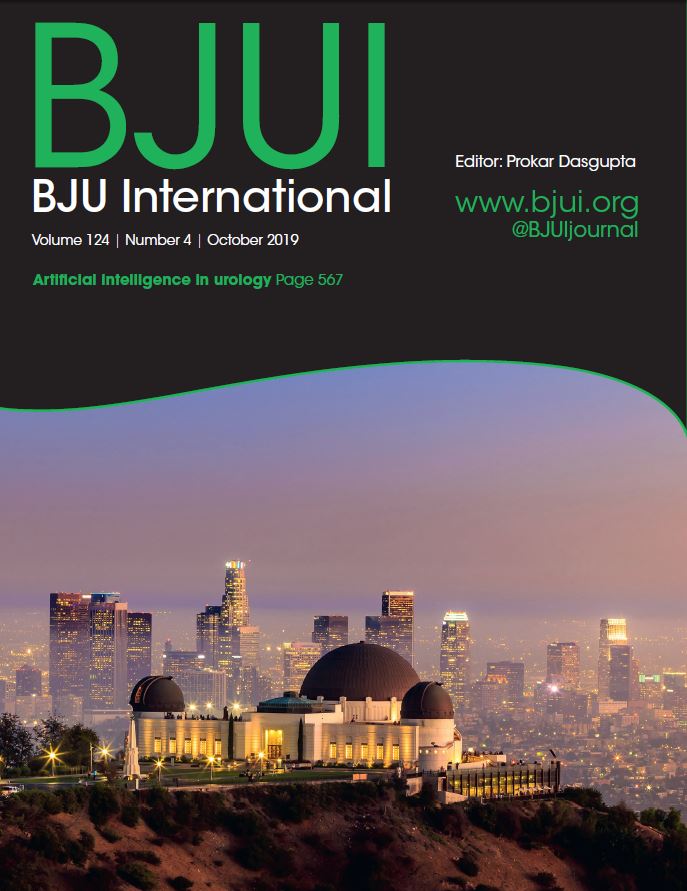
The Article of the Month for October was written by researchers primarily from Los Angeles, California, USA: Current status of artificial intelligence applications in urology and their potential to influence clinical practice

The cover image shows the Griffith Observatory, with the surrounding view of LA. The observatory is “Southern California’s gateway to the cosmos”. It is named after its creator and funder Griffith J. Griffith who wanted a free public observatory – it is now a world-leader in public astronomy and has received over 80 million visitors in its nearly 90-year history. It is also one of the best vantage points from which to view the Hollywood sign.


Video: Current status of artificial intelligence applications in urology

Current status of artificial intelligence applications in urology and their potential to influence clinical practice

Abstract

Objective

To investigate the applications of artificial intelligence (AI) in diagnosis, treatment and outcome prediction in urologic diseases and evaluate its advantages over traditional models and methods.

Materials and methods

A literature search was performed after PROSPERO registration (CRD42018103701) and in compliance with Preferred Reported Items for Systematic Reviews and Meta‐Analyses (PRISMA) methods. Articles between 1994 and 2018 using the search terms “urology”, “artificial intelligence”, “machine learning” were included and categorized by the application of AI in urology. Review articles, editorial comments, articles with no full‐text access, and non-urologic studies were excluded.

Results

Initial search yielded 231 articles, but after excluding duplicates and following full‐text review and examination of article references, only 111 articles were included in the final analysis. AI applications in urology include: utilizing radiomic imaging or ultrasonic echo data to improve or automate cancer detection or outcome prediction, utilizing digitized tissue specimen images to automate detection of cancer on pathology slides, and combining patient clinical data, biomarkers, or gene expression to assist disease diagnosis or outcome prediction. Some studies employed AI to plan brachytherapy and radiation treatments while others used video based or robotic automated performance metrics to objectively evaluate surgical skill. Compared to conventional statistical analysis, 71.8% of studies concluded that AI is superior in diagnosis and outcome prediction.

Conclusion

AI has been widely adopted in urology. Compared to conventional statistics AI approaches are more accurate in prediction and more explorative for analyzing large data cohorts. With an increasing library of patient data accessible to clinicians, AI may help facilitate evidence‐based and individualized patient care.

View more videos

Visual abstract: Current status of artificial intelligence applications in urology and their potential to influence clinical practice

See more infographics

Making real change where it is needed! The HSIB investigation into a case of testicular torsion

This week saw the first Health Service Investigation Branch (HSIB) investigation into a urological condition. The HSIB is the health services version of the Air Investigation Branch, which investigate air crashes, and the case that it was investigating was one of testicular loss from torsion.

The investigation followed the best principles of human factors theory and causal analysis. It was not looking to assign blame but instead to constructively implement better process and systems that do not relay solely on one individual, as humans are notoriously fallible. The outcome of any investigation is to make it easier for medical teams and administrators to perform well and to mitigate the risk of errors, in an inherently complex area such as medicine.

Only a small number of HSIB investigations have taken place so far so we are fortunate that a Urology case was chosen. The report concentrated on the community aspects of the testicular pain pathway, and the investigating team had fruitful meetings with NHS 111 that led to changes in the questions and prompts that were asked of callers with testicular pain who dialled in. The Royal College of GPs, as a result of the investigation, has convened a group to review the communication standards between practices running telephone services and emergency departments; and NICE has agreed to improve the on-line guidance on testicular torsion and scrotal pain to make it more accessible to clinicians, patients and their carers.

The fact that this came about after an investigation of a single case shows the power of this investigative process and the rigour with which it was carried out.

I would encourage others who may want to be involved with this type of work. I was lucky enough to be approached by the HSIB to be the subject matter expert (SME) on this case as I have a known interest in both Quality Improvement (QI) and Torsion. Anyone approached to help with investigations of this type should be reassured of the professionalism under which a case is undertaken: no individuals or organisations are named; no fingers are pointed but instead the HSIB are able to open a lot of doors and instigate change by negotiating agreements from departments and institutions that most clinicians involved in QI could only dream of getting.

Maybe we need a few more investigations of this type in Urology; retained stents spring immediately to mind as a strong candidate as the HSIB is also experienced in talking to industry. Wouldn’t retained stents be so much easier to avoid if each stent had an individualised barcode that could be scanned and tracked? The companies making stents could perhaps be encouraged to be more involved in making sure that they were easier to track across the whole of the UK (or the world) so patients wouldn’t have so many problems with stents in the future.  Every component of a jet is tracked in a similar way so why shouldn’t we look for the same standard in Urology Healthcare!

by Tony Tien & James Green

Twitter: @greenxmedical


James S. A. Green is a Urological Surgeon, Network Lead for Urology at Barts Health NHS Trust, Quality Improvement Director at Whipps Cross University Hospital and visiting Professor in Health Services Research at Kings College, London. His interest in medical education and improvement started when developing medical support for the British Army and he has published extensively on team-working and improving clinical care. He was SME for the HSIB investigation into a case of delayed testicular torsion.

Mr Tony Tien MRCS is a clinical fellow in Urology at Whipps Cross Hospital and a champion for Quality Improvement.


© 2023 BJU International. All Rights Reserved.