Tag Archive for: biochemical recurrence

Posts

BJUI journal prizes

Every year the BJUI awards three prizes to trainee urologists who have played a significant role in contributing to the work published in the journal. The prizes go towards travel costs enabling the trainees to visit international conferences. In 2020, due to the coronavirus pandemic leading to the cancellation of many of these conferences, the usual prize-giving ceremonies have not taken place so here we are introducing you to the prize winners and their work. We hope they will be able to spend their prize money in 2021.

Global prize

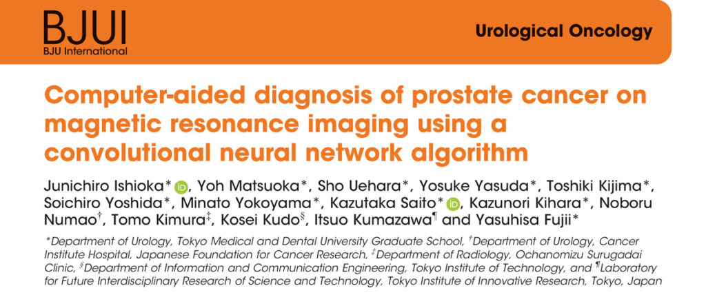
This is awarded to authors who are trainees based anywhere in the world other than the Americas and Europe. Usually presented at the USANZ annual meeting. In 2020 the prize was awarded to Sho Uehara for his work on artificial intelligence in prostate cancer diagnosis.

Sho Uehara MD Ph.D Tokyo, Japan
Assistant professor, Department of Urology
Tokyo Medical and Dental University

Email: [email protected]


Sho Uehara received a Ph.D. from the graduate school of Tokyo Medical and Dental University, Tokyo, Japan, in 2018.  He is now working as a urologist and an assistant professor at the university hospital. His research interests include prostate cancer diagnostics, and utilization of machine learning for them.

Membership of academic societies:

JUA (The Japanese Urological Association), EAU (European Association of Urology) and AUA (American Urological Association)


Coffey-Krane prize

The Coffey-Krane prize is awarded to an author who is a trainee based in The Americas. Normally presented at the AUA annual conference. Dr Nathan Wong received this year’s award for his work on using machine learning to predict biochemical cancer recurrence following prostatectomy.

Dr Nathan Wong
Associate Professor
Westchester Medical Center and New York Medical College

Dr Nathan Wong is an assistant professor and associate program director in the Department of Urology at Westchester Medical Center and New York Medical College. He specializes in urologic oncology and robotics surgery. His main interests are in technology, clinical trials and surgical education. He completed a Society of Urologic Oncology fellowship at Memorial Sloan Kettering Cancer Center in New York City and urology residency at McMaster University in Hamilton, Ontario in Canada. 


John Blandy prize

This prize is for authors who are trainees based in Europe. Presented at the BAUS annual conference; the winner gives a presentation. This year the prize went to Nicholas Raison for his work on a RCT on cognitive training in robotic surgery.

Nicholas Raison is Vattikuti fellow at the MRC Centre for Transplantation and Mucosal Cell Biology, King’s College London and a Urology Specialist Registrar in the London Deanery.

Article of the month: Long‐term oncological and functional follow‐up in low‐dose‐rate brachytherapy for PCa: results from the prospective nationwide Swiss registry

Every month, the Editor-in-Chief selects an Article of the Month from the current issue of BJUI. The abstract is reproduced below and you can click on the button to read the full article, which is freely available to all readers for at least 30 days from the time of this post.

In addition to this post there is also an Editorial written by a prominent member of the urological community and a visual abstract created by Cora Griffin at King’s College London. We invite you to use the comment tools at the bottom of each post to join the conversation. 

If you only have time to read one article this month, we recommend this one. 

Long‐term oncological and functional follow‐up in low‐dose‐rate brachytherapy for prostate cancer: results from the prospective nationwide Swiss registry

Pascal Viktorin-Baier*, Paul M. Putora‡§, Hans-Peter Schmid*, Ludwig Plasswilm‡§, Christoph Schwab*, Armin Thoeni, Werner Hochreiter**, Ladislav Prikler††, Stefan Suter‡‡, Patrick Stucki, Michael Müntener§§, Nadja Blick§§, Hans Schiefer, Sabine Güsewell¶¶, Karin Zürn* and Daniel Engeler*

*Department of Urology, St. Gallen Cantonal Hospital, St. Gallen, Urology Clinic, Cantonal Hospital Lucerne, Lucerne, Department of Radiation Oncology, St. Gallen Cantonal Hospital, St. Gallen, §Department of Radiation Oncology, University of Berne, Clinic for Radiation-Oncology, Lindenhof Hospital Berne, Berne, **Urology Clinic, Hirslanden Clinic Aarau, Aarau, ††Urology Clinic, Uroviva Clinic Buelach, Buelach, ‡‡Urology Clinic Zug, Zug, §§Urology Clinic, Triemli Hospital, Zurich, and ¶¶Clinical Trial Unit, St. Gallen Cantonal Hospital, St. Gallen, Switzerland

Read the full article

Abstract

Objective

To evaluate the long‐term oncological, functional and toxicity outcomes of low‐dose‐rate brachytherapy (LDR‐BT) in relation to risk factors and radiation dose in a prospective multicentre cohort.

Patients and Methods

Data of patients from 12 Swiss centres undergoing LDR‐BT from September 2004 to March 2018 were prospectively collected. Patients with a follow‐up of ≥3 months were analysed. Functional and oncological outcomes were assessed at ~6 weeks, 6 and 12 months after implantation and annually thereafter. LDR‐BT was performed with 125I seeds. Dosimetry was done 6 weeks after implantation based on the European Society for Radiotherapy and Oncology recommendations. The Kaplan–Meier method was used for biochemical recurrence‐free survival (BRFS). A prostate‐specific antigen (PSA) rise above the PSA nadir + 2 was defined as biochemical failure. Functional outcomes were assessed by urodynamic measurement parameters and questionnaires.

Results

Of 1580 patients in the database, 1291 (81.7%) were evaluable for therapy outcome. The median (range) follow‐up was 37.1 (3.0–141.6) months. Better BRFS was found for Gleason score ≤3+4 ( = 0.03, log‐rank test) and initial PSA level of <10 ng/mL ( < 0.001). D’Amico Risk groups were significantly associated with BRFS ( < 0.001), with a hazard ratio of 2.38 for intermediate‐ and high‐risk patients vs low‐risk patients. The radiation dose covering 90% of the prostate volume (D90) after 6 weeks was significantly lower in patients with recurrence. Functional outcomes returned close to baseline levels after 2–3 years. A major limitation of these findings is a substantial loss to follow‐up.

Conclusion

Our results are in line with other studies showing that LDR‐BT is associated with good oncological outcomes together with good functional results.

Read more Articles of the week

Editorial: Low-dose-rate brachytherapy for prostate cancer stands the test of time – the Swiss experience

The clinical results from 12 Swiss centres reaffirm the benefits of Low Dose Rate Brachytherapy (LDR-BT) for the treatment of localised prostate cancer [1]. The authors are to be commended for collating and analysing prospective, countrywide, long-term data. This is an excellent example of Good Clinical Practice for the urology community, patients, commissioning groups and for governance purposes. Prostate brachytherapy offers suitable men with prostate cancer a high chance of long-term cure but with a low risk of urinary incontinence and most retaining erectile dysfunction [2].

Two thirds of the patients reported in the Swiss series had low-risk cancer who would now more commonly be offered active surveillance as an initial treatment option. However our own and other large mature series have shown similar treatment efficacy of LDR-BT, either as monotherapy as in the Swiss study, or as a boost to external-beam radiotherapy, for the treatment of patients with intermediate and high risk of disease relapse [3, 4]. Indeed the ASCENDE-RT trial recently showed that men with unfavourable intermediate or high-risk prostate cancer randomised to an LDR-BT boost arm, relative to a dose-escalated external-beam radiotherapy boost, were twice as likely to be free of biochemical failure at a median follow-up of 6.5 years. A slight increase in urinary toxicity was observed which may have been an issue related to implant technique [5].

The authors show LDR-BT affords excellent disease control that associates with post-implant dosimetry in keeping with current treatment guidelines. They also report an association between biochemical control and seed loss. It therefore becomes unclear the extent to which implant quality or implant technique, i.e. the use of loose or stranded seeds, influenced the oncological outcome, as it would appear that more than one brachytherapy technique has been used.

In this series no prostate cancer-related deaths were reported. However the median follow-up length of 37 months is relatively short. Examples from more mature series show longer follow-up is needed to begin to document the low rates of prostate cancer-related deaths following LDR-BT. Lazarev et al [6] in a similar risk group distribution to the Swiss population, reported 97% prostate cancer-specific survival at 17-years with all deaths occurring more than 10 years after treatment. Morris et al [4] reported 99.1% cause-specific survival at 10 years with death events 9 years after treatment in low and intermediate-risk disease. Our own series showed 98% prostate-cancer-specific survival at 7 and 9 years post-implantation in high-risk (as defined by NICE) patients treated with monotherapy [3].

Treatment-related toxicity assessments in the Swiss series showed that baseline values are crucial to understand the impact of treatment on patient-reported outcomes. Higher post-implant scores were consistently observed in those patients with higher baseline scores. The patient-reported outcomes were similar to those from our series where sexual potency was preserved in 70-80% of men who were ≤60 years old at time of implant [7].

Salvage therapies are seldom given after LDR-BT as the local failure rate is low and the surgery complex. It was undertaken in only two patients in the Swiss series. In the era of mp-MRI and PSMA PET/CT scans and targeted biopsies, tumour recurrence can be better assessed.  Salvage surgery has been offered to approximately 0.5% (27/4200) of our patients, by either robotic-assisted radical prostatectomy or seminal vesiculectomy if the recurrence is localised to the seminal vesicle alone.

This nation-wide report from the 12 Swiss centres is a welcome addition to the extensive body of evidence that attests to the excellent results and generalisability of prostate LDR-BT. The treatment is efficacious and convenient for patients with a low toxicity profile. It is a cost effective option that should be offered to all suitable patients with localised prostate cancer.

by Stephen Langley

References

1. Viktorin-Baier P, Putora PM, Schmid HP, et al. Long-term oncological and functional follow-up in low-dose-rate brachytherapy for prostate cancer: results from the prospective nationwide Swiss registry. BJU Int 2020: 125(6).

2. Punnen S, Cowan JE, Chan JM, Carroll PR, Cooperberg MR. Long-term health-related quality of life after primary treatment for localized prostate cancer: results from the CaPSURE registry. European urology 2015; 68: 600-608.

3. Laing R, Uribe J, Uribe-Lewis S, et al. Low-dose-rate brachytherapy for the treatment of localised prostate cancer in men with a high risk of disease relapse. BJU Int 2018; 122: 610-617.

4. Morris WJ, Keyes M, Spadinger I, et al. Population-based 10-year oncologic outcomes after low-dose-rate brachytherapy for low-risk and intermediate-risk prostate cancer. Cancer 2013; 119: 1537-1546.

5. Morris WJ, Tyldesley S, Rodda S, et al. Androgen Suppression Combined with Elective Nodal and Dose Escalated Radiation Therapy (the ASCENDE-RT Trial): An Analysis of Survival Endpoints for a Randomized Trial Comparing a Low-Dose-Rate Brachytherapy Boost to a Dose-Escalated External Beam Boost for High- and Intermediate-risk Prostate Cancer. Int J Rad Onc Bio Phys 2017; 98: 275-285.

6. Lazarev S, Thompson MR, Stone NN, Stock RG. Low-dose-rate brachytherapy for prostate cancer: outcomes at >10 years of follow-up. BJU Int 2018; 121: 781-790.

7. Langley SEM, Soares R, Uribe J, et al. Long-term oncological outcomes and toxicity in 597 men aged ≤60 years at time of low-dose-rate brachytherapy for localised prostate cancer. BJU Int 2018; 121: 38-45.

Visual abstract: Long-term oncological and functional follow-up in low dose rate brachytherapy for PCa: results from the prospective nation-wide Swiss Registry

Read the full article
See more infographics

Article of the week: 68Ga‐PSMA PET/CT predicts complete biochemical response from RP and lymph node dissection in intermediate‐ and high‐risk PCa

Every week, the Editor-in-Chief selects an Article of the Week from the current issue of BJUI. The abstract is reproduced below and you can click on the button to read the full article, which is freely available to all readers for at least 30 days from the time of this post.

In addition to the article itself, there is an editorial written by a prominent member of the urological community. These are intended to provoke comment and discussion and we invite you to use the comment tools at the bottom of each post to join the conversation. 

If you only have time to read one article this month, it should be this one.

Gallium‐68‐prostate‐specific membrane antigen (68Ga‐PSMA) positron emission tomography (PET)/computed tomography (CT) predicts complete biochemical response from radical prostatectomy and lymph node dissection in intermediate‐ and high‐risk prostate cancer


Pim J. van Leeuwen*, Maarten Donswijk, Rohan Nandurkar, Phillip Stricker§¶Bao Ho**, Stijn Heijmink††, Esther M.K. Wit*, Corinne Tillier*, Erik van Muilenkom*, Quoc Nguyen§, Henk G. van der Poel* and Louise Emmett§**


*Department of Urology, Department of Nuclear Medicine, The Netherlands Cancer Institute, Amsterdam, The Netherlands, Faculty of Medicine, University of New South Wales Sydney, §The Australian Prostate Cancer Research Centre-NSW, The Garvan Institute of Medical Research, St Vincents Clinic, **Department of Theranostics and Nuclear Medicine, St Vincents Hospital Sydney, Sydney, New South Wales, Australia and ††Department of Radiology, The Netherlands Cancer Institute, Amsterdam, The Netherlands

Read the full article

Abstract

Objective

To determine the value of gallium‐68‐prostate‐specific membrane antigen (68Ga‐PSMA)‐11 positron emission tomography (PET) /computed tomography (CT) in men with newly diagnosed prostate cancer.

Patients and methods

We analysed results of 140 men with intermediate‐ and high‐risk prostate cancer. All men underwent 68Ga‐PSMA‐11 PET/CT and multiparametric magnetic resonance imaging (mpMRI) before radical prostatectomy (RP) with extended pelvic lymph node (LN) dissection. For each patient, the clinical and pathological features were recorded. Prostate‐specific antigen (PSA) was documented at staging scan, and after RP, at a median (interquartile range) of 110 (49–132) days. A PSA level of ≥0.03 ng/mL was classified as biochemical persistence (BCP). Logistic regression was performed for association of clinical variables and BCP.

Results

In these 140 patients with intermediate‐ and high‐risk prostate cancer, 27.1% had PSMA PET/CT‐positive findings in the pelvic LNs. Sensitivity and specificity for detection of LN metastases were 53% and 88% (PSMA PET/CT) and 14% and 99% (mpMRI), respectively. The overall BCP rate was 25.7%. The BCP rate was 16.7% in men who were PSMA PET/CT LN‐negative compared to 50% in men who were PSMA PET/CT LN‐positive (P < 0.05). The presence of PSMA‐positive pelvic LNs was more predictive of BCP after RP than cT‐stage, PSA level, and the Gleason score, adjusted for surgical margins status.

Conclusions

68Ga‐PSMA‐11 PET/CT is highly predictive of BCP after RP, and should play an important role informing men with intermediate‐ or high‐risk prostate cancer.


Read more Articles of the week

Editorial: Preoperative PSMA‐targeted PET imaging: more than just a tool for prostate cancer staging?

The presence of lymph node metastases at the time of prostate cancer diagnosis has significant implications for treatment. According to current guidelines from the National Comprehensive Cancer Network, men with positive lymph nodes on initial staging imaging should be offered treatment with androgen deprivation (± abiraterone) along with consideration for external beam radiation therapy [1]. In contrast, men with clinically localised high‐ or very‐high‐risk prostate cancer have the option of undergoing radical prostatectomy. Unfortunately, currently available diagnostic imaging modalities (i.e. contrast‐enhanced CT and MRI) fall short in their ability to accurately identify lymph node metastases, which are often small and difficult to discern from other structures within the pelvis. Thus, there exists a conundrum: if we cannot accurately detect lymph node involvement, how can we appropriately manage it?

In this edition of the BJUI, Leeuwen et al. [2] report on the utility of molecular imaging with 68Ga‐PSMA‐11 positron emission tomography (PET)/CT in the preoperative staging of men with prostate cancer. To date, the greatest clinical utility of PSMA‐targeted PET has been in the management of men with biochemically recurrent prostate cancer [3]. In the present study by Leeuwen et al. [2], 140 patients with newly diagnosed intermediate‐ or high‐ risk prostate cancer underwent 68Ga‐PSMA‐11 PET/CT before radical prostatectomy with extended pelvic lymph node dissection. Surgical pathology served as the reference standard to which findings on 68Ga‐PSMA‐11 PET/CT were compared. In total, 27.1% of men were found to have radiotracer uptake in their pelvic lymph nodes, resulting in a sensitivity of 53% and a specificity of 88%. In contrast, multiparametric MRI had a sensitivity of only 14%, albeit with a higher specificity of 99%. These findings are in line with prior studies evaluating the diagnostic performance of PSMA‐targeted PET imaging for preoperative prostate cancer staging [4]. Of greater interest, however, is the authors’ observation that positivity on 68Ga‐PSMA‐11 PET/CT was strongly associated with postoperative PSA persistence (i.e. failure to cure). More specifically, after controlling for Gleason score, surgical margin status, and preoperative PSA level, positivity on PET/CT had an odds ratio of 5.87 (95% CI 1.30–26.59) for biochemical persistence. Furthermore, men with pN1 disease and a positive preoperative PET/CT (i.e. true positives) were over three times more likely to experience biochemical persistence than patients with pN1 disease and negative imaging (71.4% vs 21.4%). Thus, PSMA‐targeted PET not only stands to inform clinical staging, but also has the potential to offer independent prognostic information.

A future line of investigation is to explore the biological basis of the authors’ observation regarding PSMA as a prognostic marker. One explanation is that PET/CT identified men with higher volume lymph node metastases (a known prognostic factor), whilst patients with smaller more curable nodes were negative on imaging. After all, the authors state that the imaging test did not detect any pathologically positive lymph nodes <2 mm. Furthermore, only 27% of positive lymph nodes between 2 and 4 mm showed radiotracer uptake. Unfortunately, the authors did not account for differences in the volume of nodal metastases in their analysis. A second possible explanation for the authors’ observation is that PSMA is upregulated through the same signaling pathways that drive an aggressive prostate cancer phenotype, allowing for PSMA expression to provide prognostic information independent of tumour volume. Indeed, others have previously shown that PSMA expression, as measured by immunohistochemistry, corresponds with increasing tumour grade, stage and risk of biochemical failure [5]. Of course, these concepts are not mutually exclusive and further investigation is needed in order for PSMA‐targeted imaging to be rationally applied as a prognostic test.

References

  1. NCCN Clinical Practice Guidelines in Oncology: Prostate Cancer (Version 4.2018)2018. Accessed November 2018. Available at: https://www.nccn.org/professionals/physician_gls/pdf/prostate.pdf.
  2. Leeuwen, PJDonswijk, MNandurkar, R et al. Gallium‐68‐prostate‐specific membrane antigen (68Ga‐PSMA) positron emission tomography (PET)/computed tomography (CT) predicts complete biochemical response from radical prostatectomy and lymph node dissection in intermediate‐ and high‐risk prostate cancer. BJU Int 201912462– 8
  3. Han, SWoo, SKim, YJSuh, CHImpact of 68Ga‐PSMA PET on the management of patients with prostate cancer: a systematic review and meta‐analysis. Eur Urol 201874179– 90
  4. Gorin, MARowe, SPPatel, HD et al. Prostate specific membrane antigen targeted 18F‐DCFPyL positron emission tomography/computerized tomography for the preoperative staging of high risk prostate cancer: results of a prospective, phase II, single center study. J Urol 2018199126– 32
  5. Minner, SWittmer, CGraefen, M et al. High level PSMA expression is associated with early PSA recurrence in surgically treated prostate cancer. Prostate 201171281– 8


Why attend Advanced Prostate Cancer Consensus Conference APCCC 2019?

From the 29th to the 31st of August 2019 the next Advanced Prostate Cancer Consensus Conference #APCCC19 will take place in Basel, Switzerland. The consensus conference was inspired by the very successful and pioneering early breast cancer consensus conference that was started in 1978 in St Gallen.

APCCC was initiated in 2015 because of the rapid developments in the field of advanced prostate cancer with the aim to discuss the clinical management of men with advanced prostate cancer, with a special focus on situations with a lack of or only weak evidence from the literature or conflicting evidence. Prostate cancer is such a common disease that the majority of men across the globe are not treated in expert centers but rather in smaller hospitals or community-based practices. APCCC wants to help the process of knowledge translation by assembling a large group of international prostate cancer experts that hold highly educational lectures summarizing the available literature and evidence, and discussing controversial questions.

The recommendations from the previous two conferences in 2015 and 2017 have been published open access in renowned scientific journals and have been widely read:

APCCC 2015 report

APCCC 2017 report

The expert panel in action at APCCC 2017

The following topics have been chosen for discussion at the 2019 conference:

  1. Locally advanced prostate cancer
  2. Biochemical recurrence of prostate cancer after local therapy
  3. Management of primary tumour in the metastatic situation
  4. Newly diagnosed metastatic prostate cancer, including oligometastatic prostate cancer
  5. Management of nmCRPC
  6. Management of mCRPC
  7. Bone and bone metastases
  8. Molecular characterization: tissue and blood
  9. Heterogeneity of men with prostate cancer (ethnicity, elderly)
  10. Side effects of hormonal treatments and their management

Difficult questions at APCCC17

Importantly all participants of APCCC can attend the consensus discussion and voting on Saturday morning. The questions and voting results will again form the basis for a report that will be published soon after the conference.

Why attend APCCC 2019?

The management of men with advanced prostate cancer keeps changing rapidly. Practice changing results have been recently presented and controversially discussed at ESMO (e.g. radiation therapy of the primary in the metastatic situation) and important new data will be presented at ASCO 2019 (e.g. results of standard-of-care therapy with or without enzalutamide or apalutamide for metastatic hormone-sensitive prostate cancer), making the choice of treatment in this situation even more challenging.

There are many reasons to attend APCCC 2019:

  • A unique opportunity to focus on the important topic of advanced prostate cancer management for two and a half days and be updated on current standards and state of the art of the care of men with advanced prostate cancer
  • Be involved in a global discussion on questions relevant to daily clinical practice but not investigated well

Expert interactions at APCCC17

  • Help to translate the outcomes from clinical trials into the management of men with prostate cancer in daily practice especially for men treated outside of large prostate cancer centers.
  • Be inspired and organize an APCCC satellite meeting for your country or region as has been done after the previous consensus conferences, with manuscripts published here in the BJUI.
    • Chiong et al. The Asia-Pacific Satellite of the APCCC BJUI 2019
    • Ma WK et al. Consensus statements on the management of clinically localized prostate cancer from the Hong Kong Urological Association and the Hong Kong Society of Uro-Oncology BJU Int. 2019
    • Omlin A, Gillessen S. The Advanced Prostate Cancer Consensus on a regional level – what can we learn? BJU Int. 2019
  • And of course, enjoy a few wonderful summer days in the beautiful city of Basel!!!

Further information and links

APCCC 2019

Preview of APCCC 2019: Silke Gillessen, in conversation with Alicia Morgans, discusses many of the changes we have experienced in clinical practice since the last APCCC in 2017


APCCC on the THE “NEW” PROSTATE CANCER INFOLINK


by Dr Aurelius Omlin

Twitter: @apccc19
 

Article of the week: The impact on oncological outcomes after RP for PCa of converting soft tissue margins at the apex and bladder neck from tumour‐positive to ‐negative

Every week, the Editor-in-Chief selects an Article of the Week from the current issue of BJUI. The abstract is reproduced below and you can click on the button to read the full article, which is freely available to all readers for at least 30 days from the time of this post.

In addition to the article itself, there is an editorial written by a prominent member of the urological community and the authors have also kindly produced a video describing their work. These are intended to provoke comment and discussion and we invite you to use the comment tools at the bottom of each post to join the conversation. 

If you only have time to read one article this week, it should be this one.

The impact on oncological outcomes after radical prostatectomy for prostate cancer of converting soft tissue margins at the apex and bladder neck from tumour‐positive to ‐negative

Sahyun Pak*, Sejun Park, Myong Kim*, Heounjeong Go, Yong Mee Choand Hanjong Ahn*


*Department of Urology, Asan Medical Center, University of Ulsan College of Medicine, Seoul, Department of Urology, Ulsan University Hospital, University of Ulsan College of Medicine, Ulsan and Department of Pathology, Asan Medical Center, University of Ulsan College of Medicine, Seoul, Korea


Read the full article

Abstract

Objectives

To assess the impact of conversion from histologically positive to negative soft tissue margins at the apex and bladder neck on biochemical recurrence‐free survival (BCRFS) and distant metastasis‐free survival (DMFS) after radical prostatectomy (RP) for prostate cancer.

Materials and Methods

The records of 2 013 patients who underwent RP and intra‐operative frozen section (IFS) analysis between July 2007 and June 2016 were reviewed. IFS analysis of the urethra and bladder neck was performed, and if malignant or atypical cells remained, further resection with the aim of achieving histological negativity was carried out. Patients were divided into three groups according to the findings: those with a negative surgical margin (NSM), a positive surgical margin converted to negative (NCSM) and a persistent positive surgical margin (PSM).

Table 4. Impact of converting margins from tumour‐positive to ‐negative on biochemical recurrence

Results

Among the 2 013 patients, rates of NSMs, NCSMs and PSMs were 75.1%, 4.9%, and 20.0%, respectively. The 5‐year BCRFS rates of patients with NSMs, NCSMs and PSMs were 89.6%, 85.1% and 57.1%, respectively (P < 0.001). In both pathological (p)T2 and pT3 cancers, the 5‐year BCRFS rate for patients with NCSMs was similar to that for patients with NSMs, and higher than for patients with PSMs. The 7‐year DMFS rates of patients with NSMs, NCSMs and PSMs were 97.8%, 99.1% and 89.4%, respectively (P < 0.001). Among patients with pT3 cancers, the 7‐year DMFS rate was significantly higher in the NCSM group than in the PSM group (98.0% vs 86.7%; P = 0.023), but not among those with pT2 cancers (100% vs 96.9%; P = 0.616). The 5‐year BCRFS rate for the NCSM group was not significantly different from that of the NSM group among the patients with low‐ (96.3% vs 95.8%) and intermediate‐risk disease (91.1% vs 82.8%), but was lower than that of the NSM group among patients in the high‐risk group (73.2% vs 54.7%).

Conclusions

Conversion of the soft tissue margin at the prostate apex and bladder neck from histologically positive to negative improved the BCRFS and DMFS after RP for prostate cancer; however, the benefit of conversion was not apparent in patients in the high‐risk group.

Read more Articles of the week


Editorial: Conversion to negative surgical margin after intraoperative frozen section – (un)necessary effort and relevance in 2019?

The assessment and impact of positive surgical margins (PSMs) at the time of radical prostatectomy (RP) have been discussed for many decades. The determination and reporting should be performed in a standardised fashion according to the International Society of Urological Pathology [1]. The SM is considered positive if tumour cells touch the inked surface of the RP specimen. However, reasons for difficulty in truly differentiating between negative SMs (NSMs) and PSMs include iatrogenic disruption of the prostatic capsule, penetration of ink into small cracks on the outside, or cases in which prostate cancer cells are very close to, but not definitely touching, the inked margins.

A systematic review by Yossepowitch et al. [2] found a contemporary PSM rate of 15% (range 6.5–32%), which increases with extracapsular extension. In addition, the likelihood of PSM is strongly influenced by surgeon experience, independent of the surgical technique. Although PSM is considered an adverse pathological outcome and associated with an increased risk of biochemical recurrence (BCR), the impact on long‐term survival and actual prognostic value remains debatable. The association with other endpoints, such as prostate‐cancer specific mortality and overall survival, is controversial and may be primarily influenced by other risk factors, such as preoperative PSA level, Gleason score, and pathological T‐stage [2].

The role of intraoperative frozen section analysis in order to reduce the PSM rate continues to evolve. In a study by von Bodman et al. [3], 92.3% of patients with a PSM on frozen‐section analysis could ultimately be converted to a NSM. Similar findings were reported by Schlomm et al. [4] in 5392 patients using the intraoperative neurovascular structure‐adjacent frozen section examination (NeuroSAFE) technique, PSMs were detected in 25%, leading to re‐resection and conversion to definitive NSMs in 86% of these patients. In the setting of increasing experience with intraoperative frozen section analysis, a false‐positive SM status was found in only 48 patients (3.3%).

The study by Pak et al. [5], published in this issue of the BJUI, reported that specimens with initial PSMs were converted to NSMs upon permanent specimen evaluation (NCSM) in 4.9% of 2013 men undergoing RP. In this subgroup, the 5‐year BCR‐free survival (BCRFS) rates did not differ from those observed in National Comprehensive Cancer Network (NCCN) low‐ and intermediate‐risk patients with an initially NSM. However, the benefit of conversion from an initial PSM to final NSM was not apparent in high‐risk patients, as the authors found a significantly lower rate of BCRFS amongst this NCSM group. In multivariate analysis, NCSM status was independently associated (hazard ratio 0.624, P = 0.033) with BCR but not distant metastasis. These findings corroborate the findings of the Schlomm et al. [4] study, in which the BCRFS rates of propensity score‐based matched patients with conversion to NSMs did not differ significantly from patients with primarily NSMs.

What is the current role of intraoperative frozen section analysis during RP? How important is it to achieve NSMs in contemporary practice? In whom and how should the assessment be performed? Although it is clearly desirable to completely remove the entire tumour at the time of surgery, and NSMs are a surrogate marker of adequate local excision, the devil is in the details. First, in this study [5], the authors only assessed SMs at the bladder neck and apex. Although the apex is one of the most frequent locations for PSMs, other and/or multiple sites of PSMs are possible and could have been missed. Alternatively, the NeuroSAFE method is able to assess the entire laterorectal circumference albeit with the trade‐off of more extensive pathological involvement and assessment. Second, intraoperative frozen section analysis, and manoeuvers for NCSM, may ultimately be necessary and beneficial in only a small number of patients currently undergoing RP. An increasing proportion of men harbour more aggressive, higher‐risk disease in whom PSMs may have no impact on oncological outcomes or treatment decisions. In these men, long‐term cancer outcomes are probably more related to risks of unsuspected metastatic disease rather than residual, microscopic cancer within the prostatic fossa. As suggested in this study [5], an initial PSM in high‐risk men, independent of ultimate NCSM, may be a surrogate for non‐localised disease and poorer outcomes; PSMs were found in 53% of men with pT3b. In low‐risk men, the issues are whether active surveillance is a more appropriate initial management strategy and that routine intraoperative frozen section analysis may not be worthwhile with a PSM rate of only 10%. How does this alter the decision for adjuvant therapy? Adjuvant radiotherapy is probably under‐utilised in men with PSMs after RP (~11%), and NCSM may spare men from unnecessary treatment, particularly with lower‐risk disease [6]. However, men with PSMs and additional adverse pathological features, such as extraprostatic extension or seminal vesicle invasion, should probably receive adjuvant therapy, primarily driven by T stage.

The incremental value and potential clinical benefit of intraoperative frozen section analysis to achieve NSMs remain to be determined. Although one would suspect that PSM leading to excision of additional tissue could lead to worse functional outcomes, the study from Mirmilstein et al. [7] is reassuring. Despite higher Gleason score and pT stage in those undergoing the NeuroSAFE approach, the PSM rate was lower in this group (9.2%) compared with those undergoing standard intraoperative nerve‐sparing while leading to greater bilateral nerve preservation, higher potency rates at 12 months, and pad‐free continence.

In the future, other methods may guide surgical decision‐making and may eventually alter PSM rate including preoperative MRI of the prostate to evaluate extracapsular extension, genomic risk scores, or real‐time, near‐infrared fluorescent surgical guidance with prostate‐specific membrane antigen ligands [8]. However, one should not forget that outcomes are not solely based on the SM status. Various pathological and clinical factors and patients’ comorbidities and preference should be taken into consideration in the surgical management and that evaluation of validated oncological and functional outcomes is critical.

by Annika Herlemann and Maxwell Meng

References

  1. Tan, PHCheng, LSrigley, JR et al. International Society of Urological Pathology (ISUP) Consensus Conference on Handling and Staging of Radical Prostatectomy Specimens. Working group 5: surgical margins. Mod Pathol 20112448– 57
  2. Yossepowitch, OBriganti, AEastham, JA et al. Positive surgical margins after radical prostatectomy: a systematic review and contemporary update. Eur Urol 201465303– 13
  3. Bodman, CBrock, MRoghmann, F et al. Intraoperative frozen section of the prostate decreases positive margin rate while ensuring nerve sparing procedure during radical prostatectomy. J Urol 2013190515– 20
  4. Schlomm, TTennstedt, PHuxhold, C et al. Neurovascular structure‐adjacent frozen‐section examination (NeuroSAFE) increases nerve‐sparing frequency and reduces positive surgical margins in open and robot‐assisted laparoscopic radical prostatectomy: experience after 11,069 consecutive patients. Eur Urol 201262333– 40
  5. Pak, SPark, SKim, MGo, HCho, YMAhn, HThe impact on oncological outcomes after radical prostatectomy for prostate cancer of converting soft tissue margins at the apex and bladder neck from tumour‐positive to ‐negative. BJU Int 2019123811– 7
  6. Ghabili, KNguyen, KHsiang, W et al. National trends in the management of patients with positive surgical margins at the time of radical prostatectomy. J Clin Oncol 201836 (Suppl.)111
  7. Mirmilstein, GRai, BPGbolahan, O et al. The neurovascular structure‐adjacent frozen‐section examination (NeuroSAFE) approach to nerve sparing in robot‐assisted laparoscopic radical prostatectomy in a British setting – a prospective observational comparative study. BJU Int 2018;121854– 62
  8. Neuman, BPEifler, JBCastanares, M et al. Real‐time, near‐infrared fluorescence imaging with an optimized dye/light source/camera combination for surgical guidance of prostate cancer. Clin Cancer Res 201521771– 80


Video: Prostatic capsular incision during RP has important oncological implications. A systematic review and meta‐analysis

Prostatic capsular incision during radical prostatectomy has important oncological implications. A systematic review and meta‐analysis

Abstract

Introduction

Capsular Incision (CapI) is an iatrogenic breach of the prostatic capsule during radical prostatectomy that can cause positive surgical margins (PSM) in organ‐confined (pT2) prostate cancer (PCa), or the retention of benign prostatic tissue. We systematically interrogated the literature in order to clarify the definition of CapI, and the implications of this event for rates of PSM and biochemical recurrence (BCR).

Methods

A literature search was conducted according to PRISMA criteria using the search terms ‘CapI’ AND ‘prostatectomy’ and variations of each. 18 studies were eligible for inclusion.

Results

A total of 51,057 radical prostatectomy specimens were included. The incidence of CapI ranged from 1.3‐54.3%. CapI definitions varied, and included a breach of the prostatic capsule “exposing both benign or malignant PCa cells”, “malignant tissue only”, or “benign tissue only”. The incidence of PSM due to CapI ranged from 2.8 – 71.7%. Our meta‐analysis results found that when CapI was defined as “exposing malignant tissue only in organ‐confined prostate cancer” there was an increased risk of BCR compared to patients with pT2 disease and no CapI (RR 3.53, 95%CI 2.82‐4.41; p < 0.00001).

Conclusions

The absolute impact of CapI on oncological outcomes is currently unclear due to inconsistent definitions. However, the data implies an association between CapI and PSM and BCR. Reporting of possible areas of CapI on the operation note, or marking areas of concern on the specimen, are critical to assist CapI recognition by the pathologist.


View more videos

© 2024 BJU International. All Rights Reserved.