Tag Archive for: haematuria

Posts

Podcast: The IDENTIFY Study

Part of the BURST/BJUI podcast series

Podcast:  The IDENTIFY Study: The investigation and detection of urological neoplasia in patients referred with suspected urinary tract cancer; a multicentre observational study 

Mr Sinan Khadhouri is a Specialty Registrar in Urology in the East of Scotland and currently doing his PhD at the University of Aberdeen. He is also the co-vice chair of BURST and the lead trainee on IDENTIFY.

Guideline of guidelines: Asymptomatic Microscopic Haematuria

Abstract

The aim of the present study was to review major organizational guidelines on the evaluation and management of asymptomatic microscopic haematuria (AMH). We reviewed the haematuria guidelines from: the American Urological Association; the consensus statement by the Canadian Urological Association, Canadian Urologic Oncology Group and Bladder Cancer Canada; the American College of Physicians; the Joint Consensus Statement of the Renal Association and British Association of Urological Surgeons; and the National Institute for Health and Care Excellence. All guidelines reviewed recommend evaluation for AMH in the absence of potential benign aetiologies, with the evaluation including cystoscopy and upper urinary tract imaging. Existing guidelines vary in their definition of AMH (role of urine dipstick vs urine microscopy), the age threshold for recommending evaluation, and the optimal imaging method (computed tomography vs ultrasonography). Of the reviewed guidelines, none recommended the use of urine cytology or urine markers during the initial AMH evaluation. Patients should have ongoing follow-up after a negative initial AMH evaluation. Significant variation exists among current guidelines for AMH with respect to who should be evaluated and in what manner. Given the patient and health system implications of balancing appropriately focused and effective diagnostic evaluation, AMH represents a valuable future research opportunity.

Access the full article

What’s the Diagnosis?

This 61-year-old man presented with pelvic discomfort and haematuria. His PSA was 0.96ug/L. He had an abnormal prostate on DRE.

No such quiz/survey/poll

Article of the Week: Be Clear on Cancer – Blood in Pee

Every Week the Editor-in-Chief selects an Article of the Week from the current issue of BJUI. The abstract is reproduced below and you can click on the button to read the full article, which is freely available to all readers for at least 30 days from the time of this post.

In addition to the article itself, there is an accompanying editorial written by a prominent member of the urological community. This blog is intended to provoke comment and discussion and we invite you to use the comment tools at the bottom of each post to join the conversation.

Finally, the third post under the Article of the Week heading on the homepage will consist of additional material or media. This week we feature a video from Mr. Archie Hughes-Hallett,, discussing his paper.

If you only have time to read one article this week, it should be this one.

Assessing the impact of mass media public health campaigns. ‘Be Clear on Cancer: Blood in Pee’ a case in point

Archie Hughes-Hallett*, Daisy Browne, Elsie Mensah*, Justin Vale*† and Erik Mayer*†‡


*Department of Surgery and Cancer, Imperial College London, Department of Urology, Imperial College Healthcare Trust, and Institute of Global Health Innovation, Imperial College London, London, UK


Read the full article

Objectives

To assess the impact on suspected cancer referral burden and new cancer diagnosis of Public Health England’s recent Be Clear on Cancer ‘blood in pee’ mass media campaign.

Methods

A retrospective cohort study design was used. For two distinct time periods, August 2012 to May 2013 and August 2013 to May 2014, all referrals of patients deemed to be at risk of urological cancer by the referring primary healthcare physician to Imperial College NHS Healthcare Trust were screened. Data were collected on age and sex and whether the referral was for visible haematuria, non-visible haematuria or other suspected urological cancer. In addition to referral data, hospital episode data for all new renal cell (RCC) and upper and lower tract transitional cell carcinoma (TCC), as well as testicular and prostate cancer diagnoses for the same time periods were obtained.

Results

Over the campaign period and the subsequent 3 months, the number of haematuria referrals increased by 92% (P = 0.013) when compared with the same period a year earlier. This increase in referrals was not associated with a significant corresponding rise in cancer diagnosis; instead changes of 26.8% (P = 0.56) and −3.3% (P = 0.84) were seen in RCC and TCC, respectively.

AOTW2Apr


Conclusions

This study has shown that the Be Clear on Cancer ‘blood in pee’ mass media campaign significantly increased the number of new suspected cancer referrals, but there was no significant change in the diagnosis of target cancers across a large catchment. Mass media campaigns are expensive, require significant planning and appropriate implementation and, while the findings of this study do not challenge their fundamental objective, more work needs to be done to understand why no significant change in target cancers was observed. Further consideration should also be given to the increased referral burden that results from these campaigns, such that pre-emptive strategies, including educational and process mapping, across primary and secondary care can be implemented.

Read more articles of the week

Editorial: Be better with public health campaigns (and taxpayers’ money)

In this month’s issue of the BJUI, Hughes-Hallette et al. [1] report on the impact of a mass media public health campaign for gross haematuria. The authors performed a retrospective analysis evaluating the effectiveness of the ‘Be Clear on Cancer: “Blood in the pee”’ campaign. Similar campaigns for colorectal cancer have shown increased referrals and cost, without increasing the number of cancer diagnoses [2, 3]. In the current study [1], cancer diagnosis similarly did not rise. The two questions that therefore needs to be asked are:

  1. Is gross i.e. visible haematuria a predictor for urological malignancy?
  2. Does a mass media public health campaign constitue an effective means of improving early diagnosis of cancer?

Recent data from large integrative datasets have shown that visible haematuria is a significant predictor for bladder cancer [4, 5]. If gross haematuria is a predictor for urological malignancy, however, why did the authors [1] fail to find an increase in diagnosis of urological malignancy in their study? While the authors indicate that the study may have been underpowered, and that the use of an unlinked dataset may have interfered with proper accounting of cancer incidence, one must also consider that mass media outreach may not be an effective method for cancer outreach.

The ‘Be Clear on Cancer’ campaign involved the use of television adverts, print media and ‘out of home’ advertisements. Using this method, patients who are at risk of renal and bladder cancer, i.e. men, those aged >50 years, and smokers, are targeted as frequently as non-smoking teenagers. This type of mass media outreach programme is analogous to traditional advertising, where the message is often diluted and ineffective. While the ‘Be Clear on Cancer’ campaign is a worthy endeavour, the data do not seem to support the use of taxpayers’ money given its ineffective nature. A novel approach to visible haematuria may be to encourage GPs to ask patients about visible, painless haematuria, much as they would ask about chest pain or blood pressure. This would create a more focussed and durable outreach programme that would reduce the number of non-oncological referrals. As stewards of taxpayers’ money, we must be careful of how public funds are spent. The next generation of mass media outreach may require a combination of traditional media in addition to, social media and targeted advertising [6]. Although the ‘Be Clear on Cancer’ campaign did not appear to achieve its intended goals at this time, we must continue to refine and create new interactive approaches to improve the diagnosis and treatment of urological malignancies.

Read the full article
Casey K. Ng
Department of Urology, Southern California Permanente Medical Group, Pasadena, and USC Institute of Urology, Keck School of Medicine, University of Southern California, Los Angeles, CA, USA


References


1 Hughes-Hallett A, Browne D, Mensah E, Vale J, Mayer E. Assessing the impact of mass media public health campaigns. Be Clear on Cancer blood in pee: a case in point. BJU Int 2016; 117: 57075


2 Peacock O, Clayton S, Atkinson F, Tierney GM, Lund JN. Be Clear on Cancer: the impact of the UK National Bowel Cancer Awareness Campaign. Colorectal Dis 2013; 15: 9637


3 Bethune R, Marshall MJ, Mitchell SJ et al. Did the Be Clear on Bowel Cancer public awareness campaign pilot result in a higher rate of cancer detection? Postgrad Med J 2013; 89: 3903


4 Jung H, Gleason JM, Loo RK, Patel HS, Slezak JM, Jacobsen SJAssociation of hematuria on microscopic urinalysis and risk of urinary tract cancer. J Urol 2011; 185: 1698703


5 Loo RK, Lieberman SF, Slezak JM et al. Stratifying risk of urinary tract malignant tumors in patients with asymptomatic microscopic hematuria. Mayo Clin Proc 2013; 88: 12938


Video: Be Clear on Cancer – Blood in Pee

Assessing the impact of mass media public health campaigns. ‘Be Clear on Cancer: Blood in Pee’ a case in point

Archie Hughes-Hallett*, Daisy Browne, Elsie Mensah*, Justin Vale*† and Erik Mayer*†‡


*Department of Surgery and Cancer, Imperial College London, Department of Urology, Imperial College Healthcare Trust, and Institute of Global Health Innovation, Imperial College London, London, UK


Read the full article

Objectives

To assess the impact on suspected cancer referral burden and new cancer diagnosis of Public Health England’s recent Be Clear on Cancer ‘blood in pee’ mass media campaign.

Methods

A retrospective cohort study design was used. For two distinct time periods, August 2012 to May 2013 and August 2013 to May 2014, all referrals of patients deemed to be at risk of urological cancer by the referring primary healthcare physician to Imperial College NHS Healthcare Trust were screened. Data were collected on age and sex and whether the referral was for visible haematuria, non-visible haematuria or other suspected urological cancer. In addition to referral data, hospital episode data for all new renal cell (RCC) and upper and lower tract transitional cell carcinoma (TCC), as well as testicular and prostate cancer diagnoses for the same time periods were obtained.

Results

Over the campaign period and the subsequent 3 months, the number of haematuria referrals increased by 92% (P = 0.013) when compared with the same period a year earlier. This increase in referrals was not associated with a significant corresponding rise in cancer diagnosis; instead changes of 26.8% (P = 0.56) and −3.3% (P = 0.84) were seen in RCC and TCC, respectively.

AOTW2Apr

Conclusions

This study has shown that the Be Clear on Cancer ‘blood in pee’ mass media campaign significantly increased the number of new suspected cancer referrals, but there was no significant change in the diagnosis of target cancers across a large catchment. Mass media campaigns are expensive, require significant planning and appropriate implementation and, while the findings of this study do not challenge their fundamental objective, more work needs to be done to understand why no significant change in target cancers was observed. Further consideration should also be given to the increased referral burden that results from these campaigns, such that pre-emptive strategies, including educational and process mapping, across primary and secondary care can be implemented.

Read more articles of the week

What’s the Diagnosis?


Capture


Test yourself against our experts with our weekly quiz. You can type your answers here if you want to compare with our answers.

This patient presented with haematuria and was found to have stones.

[wpsqt name=”dilated patent umbilical vein” type=”quiz’ label=’

The Urological Ten Commandments

Capture“It is my ambition to say in ten sentences what others say in a whole book.” – Friedrich Nietzsche

The EAU guidelines on lower urinary tract symptoms have been published recently.  These contain 36,000 words.  It was pointed out to me that the American declaration of independence contained 1300 words and The Ten Commandments just 179 words.

The challenge was therefore to write ten commandments for urology in 179 words.  The rules I set were that I should write them whilst keeping  the spirit of the structure of the decalogue as closely as possible.  (It may be worth rereading the original before reading on).  So here goes.

1) I am a logical specialty. Thou shall investigate thoroughly prior to undertaking intervention for I am a specialty that avoids surprises.
2) Though interested in the whole of medicine thou will perform no other procedures other than urological.
3) Thou shalt not base intervention on old imaging for the clinical situation could have changed.
4) Remember that 80% of diagnoses can be made with history alone.  Thou shalt listen carefully to your patient to this end.
5) Honour sound surgical principles.  Urological tissue is forgiving but anastamoses under tension will not heal.
6) Thou shall not ignore haematuria.
7) Thou shall not leave a stent and forget it has been placed.
8) Thou shall not adopt new technology without proper clinical evaluation unless it is part of a trial.
9) Thou shall not fail to see the images yourself in assessing the patient before you.
10) Thou shall not fail to assess the potential for harm before embarking on a surgical procedure. If you would not do it to your family, your neighbour or friends, you will not do it to the patient who is in your clinic.

I put these out for discussion.  Other offerings please.


Jonathan M. Glass @jonathanmglass1

The Urology Centre, Guy’s Hospital, London, UK.   

[email protected]


Massive urinary tract haemorrhage following bladder decompression by urethral catheterisation

The development of haematuria following bladder decompression is a well described phenomenon, but is usually transient, mild, and of little clinical consequence. We describe a case in which bladder decompression precipitated massive urinary tract haemorrhage requiring multiple blood transfusions and bilateral nephrostomy insertion

Authors: Spencer Chapman, Michael; Harber, Mark
Department of Nephrology, Royal Free Hampstead NHS Trust, London, UK
Corresponding Author: Spencer Chapman, Michael


Introduction
Urinary retention is a frequently encountered problem in everyday clinical practice. When chronic, the patient is able to urinate and may be asymptomatic, or may complain of lower urinary tract symptoms (LUTS) such as urinary frequency, urgency or hesitancy, poor urinary stream, post-micturition dribbling, nocturia or urinary incontinence. However the urinary tract is often subjected to high pressures which are required to allow voiding. Over time high pressures may damage the entire urinary tract: obstructive nephropathy resulting in renal failure and bladder changes such wall thickening, trabeculation, and loss of capillary and tissue integrity(1). The immediate management of urinary retention is urethral catheterisation to allow free drainage of urine from the bladder. The development of haematuria following bladder decompression is a well described phenomenon, but is usually transient, mild, and of little clinical consequence. We describe a case in which bladder decompression precipitated massive urinary tract haemorrhage requiring multiple blood transfusions and bilateral nephrostomy insertion.

Case report
A 58 year-old Caucasian man was transferred to our unit from a local hospital with massive urinary tract haemorrhage and renal failure.
He had presented to his GP five days previously complaining of dysuria. He was prescribed a course of antibiotics for a presumed urinary tract infection and blood taken for a full blood count and urea and electrolyte estimation. The results of his blood tests demonstrated a creatinine level of 958 μmol/l and haemoglobin of 5.2 g/dL. He was referred immediately to his local Accident & Emergency (A&E) department.
At his local A&E, he reported prostatic symptoms of urinary frequency, poor stream and nocturia. These had been present for the last twenty years but had worsened over the previous few weeks. The symptoms were accompanied by general lethargy, malaise and aching thoraco-lumbar back pain that radiated to his groin. In addition he had recently noticed bilateral numbness and dysaesthesiae in his fingertips. His past medical history was notable only for long-standing prostatic symptoms for which he had been investigated three years previously, at which time a serum prostate-specific antigen (PSA) level was measured at 5.4 μg/L. MRI of the prostate had showed an enlarged central lobe of the prostate and bladder changes consistent with chronic obstruction, but no evidence of malignancy (Figure 1a and Figure 1b). He had undergone urethroscopy and cystoscopy which were reportedly normal and he did not undergo any surgical intervention. He had been lost to follow up. There was no history of neurological disease. His medications included losartan, doxazosin and simvastatin. He reported no prescription or over-the-counter use of aspirin, clopidogrel or other anticoagulants. He had a 30 pack year smoking history, drank around 5 units of alcohol a week and did not use illicit drugs. He was afebrile and observations were within normal limits. General examination was reportedly unremarkable.
Initial blood tests showed a creatinine of 958 μmol/L, urea of 43.3 mmol/L, haemoglobin of 5.2 g/dL, corrected calcium of 2.06 mmol/L, phosphate of 3.29 mmol/L, and potassium of 5.8 mmol/L.
He was transfused and a urethral catheter was easily inserted which immediately drained 2.9L of clear urine. After a few hours the urine was noted to have become a pale red colour. Overnight this progressed to frank haematuria with clots. This persisted, and over the following 3 days he underwent bladder irrigation and required transfusion with 10 units of packed red blood cells, 7 units of fresh frozen plasma and 1 unit of platelets.
The patient’s haematuria and renal failure continued and on the fifth hospital day he was transferred to our unit. His haemoglobin was measured at 5.8 g/dL, urea 37.2 mmol/L, and creatinine 809 μmol/L. Bladder irrigation was continued and he was further transfused. CT of the abdomen and pelvis obtained with the administration of contrast material revealed hugely dilated renal pelvises bilaterally, measuring 10cm in maximal diameter on the right and 7cm on the left (Figure 2a). The catheterised bladder was diffusely thick-walled with a posterior diverticulum (Figure 2b). High attenuation material likely to represent haemorrhage was present throughout the bladder, ureters and renal pelvises. Both kidneys were hydronephrotic. No focal lesion or active bleeding was identified.
At this stage there was considerable discussion regarding the benefits of bilateral nephrostomy insertion to relieve back pressure on his kidneys. Renal failure was persisting with the need for dialysis under consideration, and imaging suggested obstruction at the level of the ureters due to insoluble blood clot. A consensus was reached that nephrostomy insertion would be likely to hasten renal recovery and potentially avoid the need for dialysis. On the eighth hospital day he underwent bilateral nephrostomy insertion. Serum creatinine levels fell and dialysis was not required.
Over the next two weeks he underwent bilateral antegrade stenting of his ureters, and by the twenty-fifth hospital day, both nephrostomies were removed. Shortly after he was discharged with a urethral catheter and right ureteric JJ stent in situ. His creatinine on discharge was 348 μmol/L. Two months later, at which time his creatinine was 241 μmol/L, he underwent successful transurethral resection of the prostate. Notably, no false passage was seen in the urethra. His LUTS were much improved and the urethral catheter was no longer required. He went on to have laparoscopic pyeloplasty of the right kidney and removal of the right JJ stent. Radioisotope renography with MAG-3 performed one month later demonstrated reasonable bladder emptying post-micturition, normal transit times, no evidence of obstruction, but ongoing dilatation of the right renal pelvis.

Discussion
Urinary retention is the inability to empty the bladder. When chronic, the patient is able to pass urine, but may experience voiding difficulties and bladder emptying is incomplete. The usual cause is bladder outlet obstruction, the aetiology of which may be enlargement of the prostate (benign or malignant), drugs (e.g. anticholinergics, antispasmodics), congenital deformities (e.g. meatal stenosis, posterior urethral valves) or urethral strictures (from trauma or infection). It largely affects men from middle-age onwards reflecting the increasing incidence of benign prostatic hypertrophy (BPH) in this group. Women are also affected however, around 50% of women are diagnosed with Fowler’s syndrome: a problem of inadequate urethral sphincter relaxation (2).
In order to facilitate emptying, the bladder is forced to hypertrophy so as to increase its contractile force. This results in bladder wall thickening, trabeculation and friability. It may also become increasingly sensitive (contributing to symptoms of urinary frequency, urgency and incontinence) and eventually become weakened thereby preventing complete bladder emptying. The structural changes within the bladder are thought to affect the integrity of capillaries and tissues within its wall.
Haematuria following bladder decompression by urethral catheterisation is well-recognized, and visible haematuria is estimated to occur in 2-16% of patients(3). However, though there are case reports of clinically significant haematuria, they are rare and we could find no reports in the literature of haemorrhage requiring multiple blood transfusions as in this case (4). Factors contributing to the severity of bleeding in this case include the significant structural damage to the urinary tract resulting from many years of urinary retention, and possible coagulopathy that is part of the “uraemic syndrome” associated with renal failure. Traditional teaching recommends a “clamping technique” to achieve step-wise gradual bladder decompression and supposedly reduce the risk of haematuria. Use of an IV giving set to achieve gradual decompression has also been advocated (5). However, these approaches are time consuming and presently there is scant evidence to support their use (8). We could find no randomized studies comparing gradual bladder decompression with quick, complete emptying, and other studies suggest that the risk of haematuria correlates with the severity of bladder wall damage prior to catheterisation, and not the rate of initial emptying (6). Furthermore, studies of changes in intravesical pressure following catheterisation for acute urinary retention show that the pressure drops to around 50% of the initial value after drainage of only 100mL (7)– hence clamping after 1000mL (a commonly adopted figure) might not be expected to significantly reduce risk of bleeding.
One question brought up by this case is whether the haemorrhage responsible for decompression haematuria is confined to the bladder, or if it occurs throughout the urinary tract. In our patient blood clot was seen to fill and distend both renal pelvises; did this blood track up from the bladder or did it originate from bleeding within the pelvis itself? A number of factors lead us to conjecture that the bleeding may well have originated from the wall of the renal pelvis itself. Firstly, the renal pelvises were filled with large volumes of clot in the absence of significant dilatation of the ureters (images not shown). Secondly, there is no reason to believe that the pathological process resulting from chronic urinary retention affecting the integrity of capillaries should be limited to the bladder. In the absence of a competent uretero-vesical valve, the ureters and collecting system of the kidneys are subjected to similarly high pressures and are likely to undergo similar pathological changes.
Given this, what can be done to minimise harm to the patient when considering urethral catheterisation for urinary retention? Most importantly, one must recognize patients who are at risk of clinically significant decompression haematuria. Risk correlates with the chronicity of symptoms and the degree of bladder wall damage. Hence, patients such as ours with a long history of LUTS are at increased risk and should have close monitoring post-catheterisation. It is currently unclear as to whether strategies facilitating “gradual decompression” should be advocated: further studies are required to assess the effectiveness and practicality of such strategies.

Teaching points

  • Chronic urinary retention should be suspected in patients with a dilated upper urinary tract and/ or a history of lower urinary tract symptoms such as urinary frequency, urgency, hesitancy, straining, incomplete bladder emptying, poor flow and dribbling.
  • “Decompression haematuria” can commonly occur following urethral catheterisation, and is the result of the sudden drop of pressure in a damaged bladder.
  • Rarely, such bleeding may be severe and require blood transfusion – therefore close monitoring is required in at-risk patient groups.

092Figure 1a

Fig. 1a

092Figure 1b

Fig. 1b

092Figure 2a

Fig. 2a


092Figure 2b

Fig. 2b

References
1. High pressure chronic retention. George NJ, O’Reilly PH, Barnard RJ, Blacklock NJ. Br Med J (Clin Res Ed). 1983 Jun 4;286(6380):1780-3.
2. Urinary retention in women: its causes and management. Kavia RB, Datta SN, Dasgupta R, et al; BJU Int. 2006 Feb;97(2):281-7.
3. Management of urinary retention: rapid versus gradual decompression and risk of complications. Nyman MA, Schwenk NM, Silverstein MD . Mayo Clin Proc. 1997 Oct;72(10):951-6.
4. Hematuria as a complication of sudden decompression of chronically distended bladder in a child with neurogenic bladder dysfunction. Soylu A, Kavukçu S, Türkmen M, Arici A, Aktud T. Pediatr Emerg Care. 2000 Jun;16(3):221.
5. Slow decompression of the bladder using an intravenous giving set. Perry A, Maharaj D, Ramdass MJ, Naraynsingh V. Int J Clin Pract. 2002 Oct;56(8):619.
6. Comparison of rapid versus slow decompression of the distended urinary bladder. Gould F, Cheng CY, Lapides J. Invest Urol. 1976 Sep;14(2):156-8.
7. Intravesical pressure changes during bladder drainage in patients with acute urinary retention. Christensen J, Ostri P, Frimodt-Møller C, Juul C. Urol Int. 1987;42(3):181-4.
8. Practical management of patients with dilated upper tracts and chronic retention of urine. George NJ, O’Reilly PH, Barnard RJ, Blacklock NJ. Br J Urol. 1984 Feb;56(1):9-12.


Date added to bjui.org: 11/12/2012

DOI: 10.1002/BJUIw-2012-092-web


Nutcracker syndrome: a rare anatomic variant

We present a case of a young man referred to our hospital with severe haematuria, pre-renal lipoma and lumbar pain. 


Authors: Navarro, Joaquin; Azua-Romeo, Javier; Tovar, Maria Teresa; Lpoez Lopez, Jose Antonio; Ernest Lluch Hospital, Urology, Calatayud, Zaragoza, Spain 
Corresponding Author: Javier Azua-Romeo, Ernest Lluch Hospital, Urology, Calatayud, Zaragoza, Spain. Email: [email protected]


Abstract
Objective
Nutcracker syndrome is the consequence of compression between the superior mesenteric artery and the aorta over the left renal vein. Its manifestations are very varied and nonspecific, therefore, this syndrome may be misdiagnosed. There are several different treatment options, from conservative management to surgery.


Method
Here, we present a case of a young man referred to our hospital with severe haematuria, pre-renal lipoma and lumbar pain. The literature from 1950 onwards has been reviewed and the discussion regarding this unusual syndrome has been summarized.


Results
After undergoing surgical treatment (left renal vein transposition), our patient is asymptomatic, with complete resolution of his symptoms.


Conclusions
Due to the rarity of this syndrome and it being usually asymptomatic, it may be underdiagnosed. Following accurate diagnosis and, treatment, the symptoms usually resolve.


Introduction
Nutcracker Syndrome is the consequence of compression between the superior mesenteric artery and the aorta over the left renal vein (mesoaortic entrapment). It results in dilation of the vein and the appearance of varicosities in the renal pelvis as well as in the ureteric and gonadal veins1. In general terms, variants of the retroperitoneal vasculature are rare, even though, due to current imaging technologies, it is possible to diagnose these abnormalities, which otherwise could remain unnoticed.
The first reference to the condition dates from 1950 in a paper by by El Sadr and Mine. Nevertheless, it was De Schepper who named it the Nutcracker Syndrome (renal vein entrapment syndrome)1, comparing the aorta and the superior mesenteric artery with the arms of a nutcracker that compress the left renal vein, inducing, due to the difficulty of venous return of the left kidney, left renal venous hypertension, venous stasis and congestion of the kidney.
Here, we present a new case of Nutcracker Syndrome in a young male patient who came to our Hospital with severe lumbar pain and macroscopic haematuria requiring blood transfusion, and who presented with a huge  lipoma  adjacent to his left kidney which aggravated his symptoms.


Case Report
Our case is a 17 year-old male patient without significant medical history who was referred with dull left lumbar pain occasionally radiating to the left flank, which had lasted several months. The pain was eased by analgesia. The episodes of pain had gradually become more frequent and intense and recently had been associated with gross haematuria. During each episode the patient remained afebrile and hemodynamically stable, although on one occasion it was necessary to transfuse two units of packed red cells due to his being anaemic.
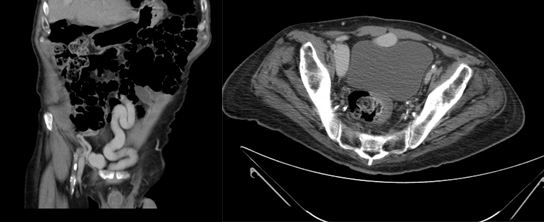
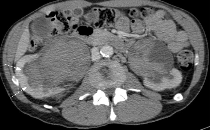
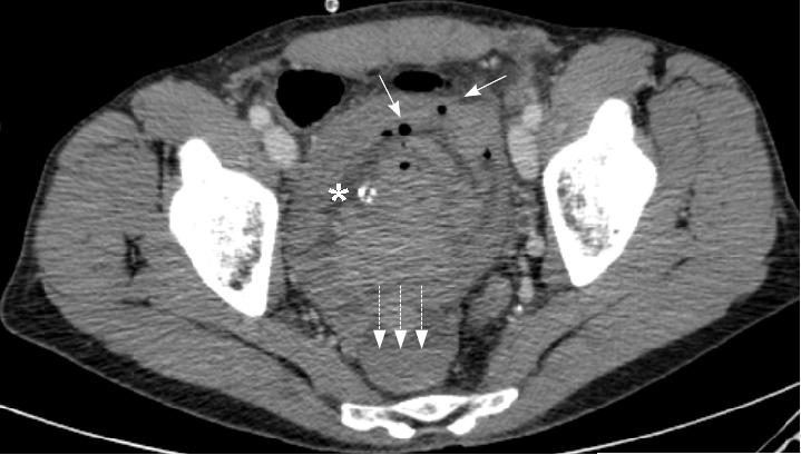
On examination, the patient has a mild left varicocele. Blood tests revealed a low hematocrit and haemoglobin, and proteinuria was found on urinalysis. Ultrasonography and urography did not demonstrate any significant findings, so a CT scan of the abdomen and pelvis was requested to further investigate his hematuria. Images revealed that the left renal vein was of large calibre. A large lipoma displacing the left kidney was also visible, accentuating the angle of the renal vein as it passed behind the superior mesenteric artery (Figure 1).
 

Figure 1 A: Abdominal CT image showing a large calibre left renal vein compressed between the abdominal aorta and superior mesenteric artery, and a large lipoma adjacent to the left kidney
B: Postoperative image showing the left renal vein now of normal size.

 
Given these findings and the severity of the patient’s symptoms, the lipoma was removed. This was performed via a subcostal approach  in order to mobilise the left kidney. Since the removal of the lipoma allowed mobilization of the kidney, to relieve the compression of the aorto-mesenteric clamp on the left renal vein, this was transposed approximately two centimeters caudally to the left lateral side of the inferior vena cava.
The surgery and the immediate postoperative course were uneventful, allowing the patient to be discharged on the third postoperative day (Figure2).
 

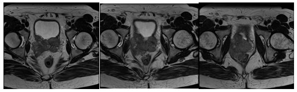
Figure 2. Intraoperative image: The left renal vein and superior mesenteric artery.


Follow up CT scan has shown that  the technique is effective, demonstrating the left renal vein to have a normal diameter. The patient remains completely asymptomatic.


Discussion
In 1950, Mina and El-Sadr described a secondary varicocele due to compression by the superior mesenteric artery of the left renal vein, thereby hampering venous return dependent on the left renal vein, resulting in varicose pelvic, ureteral and gonadal veins. Nutcracker syndrome has been classified as: Anterior, if compression is due to the superior mesenteric artery (often associated with renal ptosis or an abnormal origin of the superior mesenteric artery) and Posterior3 (or Nutcracker pseudosyndrome) if the compression occurs between the aorta and vertebral bodies, usually caused by the persistence of the posterior branch of fetal periaortic vascular ring1.
Epidemiologically, this entity occurs more frequently in women and is diagnosed within the 3rd and 4th decade of life, our case however was in a 17 year-old male. It is difficult to define the exact incidence and prevalence of Nutcracker Syndrome since the majority of patients are asymptomatic, and are diagnosed incidentally on imaging ordered for other reasons. Despite this,  our patient suffered severe lumbar pain and macroscopic haematuria which required a blood transfusion, and presented with a huge lipoma which aggravated the symptoms.
Overall, when abdominal CT and ultrasound were reviewed, 72%  showed compression of the renal vein between aorto-mesenteric clamp, although most patients would be, as already stated, asymptomatic10.
The typical clinical manifestations include back pain occasionally radiating to the gluteal area and hematuria (macro or microscopic), in few cases, patients develop anemia which may require blood transfusion. Other signs and symptoms may include proteinuria,6 hypertension, orthostatic hypotension, fatigue and weakness. Physical examination may demonstrate the presence of varicocele in males and pelvic varicose veins in females with symptoms of chronic pelvic pain or dysmenorrhea5. Also arteriovenous fistulas associated with Nutcracker Syndrome have been described.
Often the diagnosis is reached by exclusion. As with any other condition, assessment begins with the history and exploration.  The findings of laboratory results are nonspecific. With regard to imaging studies, it is usual start with an abdominal ultrasound due to its utility, safety and low cost. On ultrasound, the renal pedicle can be seen with a dilated vein and slightly enlarged kidney due to renal congestion. The next step would be a diagnostic CT or MRI. Both could be demonstrate the presence of a greater angulation in the superior mesenteric artery leaving the abdominal aorta, trapping the left renal vein 4, 9.
With regard to more invasive investigations, retrograde venography and video angiography to determine the renocaval pressure gradient will give a precise diagnosis of aorto-mesenteric clamp. Venography will demonstrate the area where the compression is, the existence of collateral circulation in periureteral vessels, reflux into the renal vein branches (adrenal vein and gonadal vein) and the stagnation of contrast in the renal vein. The pressure gradient between the renal vein and cava must be greater than 1 mm Hg to lead to a diagnosis of the Nutcracker syndrome, since the normal value is stated as between 0 and 1, nevertheless in advanced cases of Nutcracker Syndrome, due the development of collateral circulation, the gradient might be normal7.
After confirming the diagnosis of Nutcracker Syndrome different treatment alternatives can be chosen taking into account the symptoms of each individual patient. In asymptomatic or mildly symptomatic cases, expectant treatment may be an option, even moreso in younger patients where spontaneous resolution might be expected2.
Amongst conventional open surgery, autotransplantation, transposition of the renal vein, renal vein bypass or renal vein and fixation and medialization through nephropexy could be considered7.
Using open or laparoscopic surgery, and with good outcome in the short term, an extravascular prosthesis could be placed (ring-reinforced PTFE). It will enhance the renal vein and prevent the collapse of the portion between the renal vein and aorto-mesenteric clamp. This technique was described for the first time in 1988 by Barnes. Utilising interventional radiology techniques, endovascular stents can be placed. The medical literature describes good outcomes with endovascular stents, in terms of the disappearance of symptoms, although this technique is not exempt from complications (proximal migration, embolism and thrombosis of the prosthesis). This technique requires the mandatory prescription of antiplatelet therapy with the risks this entails7, 8.


Conclusions 
Nutcracker Syndrome is a rare entity, and is due to the compression exerted on the left renal vein from the clamp that forms from the superior mesenteric artery and the aorta. The usual clinical presentation is lumbar pain and haematuria. This is most frequent in females among in their fourth decade of life. However, it is more usual for patients to be asymptomatic, and this group do not require treatment. In most cases, common imaging techniques will confirm the diagnosis and it is rarely necessary to undertake invasive techniques such as venography. Usually treatment is minimally invasive, although open techniques are cited in the literature. In asymptomatic patients, especially if they are young, expectant treatment is the best option.


References 
1. Ahmed K, Sampath R, Khan MS. Current trends in the diagnosis and management of renal nutcracker syndrome: a review. Eur J Vasc Endovasc Surg. 2006; 31(4):410-416.
2. El Harrech Y, Jira H, Chafiki J, Ghadouane M, Ameur A, Abbar M. Actitud expectante en el Síndrome del Cascanueces. Act Urol Esp 2009;33(1):93-96
3. Muller Arteaga C, Martín Martín S, Cortiñas González J R, González Fajardo J A ,Fernández del Busto E. Síndrome del cascanueces posterior: Vena renal retroaórtica asociada a fístula arteriovenosa y carcinoma renal. A propósito de un caso y revisión de la literatura. Act Urol Esp 2009;33(1):101-104.
4. Bass JE, Redwine MD, Kramer LA, Huynh PT, Harris JH Jr. Spectrum of Congenital Anomalies of the Inferior Vena Cava: Cross-sectional Imaging Findings. Radiographics. 2000; 20(3): 639-652.
5. Gutiérrez E, Hernández E, Sánchez-Guerrero A,  Morales E, Gutiérrez-Solís E, Praga M. Mujer de 29 años con microhematuria persistente y episodios de hematuria macroscópica. NefroPlus 2008; 1(2):33-36.
6. Chang CT, Hung CC , Ng KK, Yen TH. Nutcracker syndrome and left unilateral haematuria. Nephrol Dial Transplant 2005; 20: 460-461.
7. Zhang H, Li M, Jin W, San P, Xu P, Pan S. The left renal entrapment syndrome: diagnosis and treatment. Ann Vasc Surg 2007; 21: 198-203.
8. Santos Arrontes D, Salgado Salinas R, Chiva Robles V, Gómez Vicente JM, Fernández González J, Costa Subías J. Síndrome del Cascanueces. A propósito de un caso y revisión de la literatura. Actas Urol Esp. 27 (9): 726-731, 2003.
9. Martínez-Salamanca I, Herranz Amo F, Gordillo Gutierrez I, Díez Cordero JM, Subirá Ríos D. Síndrome Nutcracker o Cascanueces: Demostración mediante TAC helicoidal con reconstrucción 3D. Actas Urol Esp. 28 (7): 549-552, 2004.
10. Russo D, Minutolo R, Laccarino V, Andreucci M, Capuano A, Savino F. Gross Hematuria of Uncommon Origin: The Nutcracker Syndrome Am J of Kid Dis, Vol 32, No 3, 1998.


Date added to bjui.org: 27/01/2012 


DOI: 10.1002/BJUIw-2011-092-web


© 2024 BJU International. All Rights Reserved.