Tag Archive for: radical prostatectomy

Posts

Resident’s podcast: Retzius‐sparing robot‐assisted radical prostatectomy

Maria Uloko is a Urology Resident at the University of Minnesota Hospital. In this podcast she discusses the following BJUI Article of the Week:

Retzius‐sparing robot‐assisted radical prostatectomy (RS‐RARP) vs standard RARP: it’s time for critical appraisal

Thomas Stonier*, Nick Simson*, John Davisand Ben Challacombe


*Department of Urology, Princess Alexandra Hospital, Harlow, Urology Centre, Guy s Hospital, London, UK and Department of Urology, MD Anderson Cancer Center, Houston, TX, USA


Abstract

Since robot‐assisted radical prostatectomy (RARP) started to be regularly performed in 2001, the procedure has typically followed the original retropubic approach, with incremental technical improvements in an attempt to improve outcomes. These include the running Van‐Velthoven anastomosis, posterior reconstruction or ‘Rocco stitch’, and cold ligation of the Santorini plexus/dorsal vein to maximise urethral length. In 2010, Bocciardi’s team in Milan proposed a novel posterior or ‘Retzius‐sparing’ RARP (RS‐RARP), mirroring the classic open perineal approach. This allows avoidance of supporting structures, such as the puboprostatic ligaments, endopelvic fascia, and Santorini plexus, preserving the normal anatomy as much as possible and limiting damage that may contribute to improved postoperative continence and erectile function. There has been much heralding of the excellent functional outcomes in both the medical and the lay press, but as yet no focus or real mention of any potential downsides of this new technique.


BJUI Podcasts now available on iTunes, subscribe here https://itunes.apple.com/gb/podcast/bju-international/id1309570262


Editorial: A novel nomogram for predicting ECE of prostate cancer

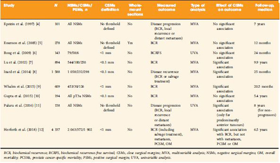
We read with great interest the publication on the side‐specific multiparametric magnetic resonance imaging (mpMRI)‐based nomogram from Martini et al. [1].

The prediction of extracapsular extension (ECE) of prostate cancer is of utmost importance to inform accurate surgical planning before radical prostatectomy (RP).

Today, surgical strategy is tailored to the patient’s characteristics, and the need for a correct prediction of ECE is of paramount importance to guarantee oncological safety, as well as optimal functional outcome. The most up‐to‐date guidelines suggest referring to nomograms to decide whether or not to perform nerve‐sparing (NS) surgery. Since the first version of the Partin Tables in 1993, several models have been developed based on PSA, Gleason score at prostate biopsy, and clinical staging, as the most used covariates.

Furthermore, mpMRI is increasingly used in the diagnostic pathway of prostate cancer to aid prostate biopsy targeting and to attain a more accurate diagnosis of clinically significant prostate cancer. Despite its recognised role in the detection of cancer, the accuracy for local staging is poor, providing a low and heterogeneous sensitivity for the detection of ECE [2].

Given this limitation, the addition of MRI to clinically derived nomograms might result in an improved assessment of preoperative local staging. In a retrospective analysis of 501 patients who underwent RP, MRI + clinical models outperformed clinical‐based models alone for all staging outcomes, with better discrimination in predicting ECE with MRI + Partin Tables and MRI + Cancer of the Prostate Risk Assessment (CAPRA) score than nomograms alone [3].

In the current article, Martini et al. [1] suggest a novel nomogram for predicting ECE that includes the presence of a ‘documented definite ECE at mpMRI’ as an additional variable beyond PSA, Gleason score, and maximum percentage of tumour in the biopsy core with the highest Gleason score. Readers should recognise that this is the first model integrating side‐specific MRI findings together with side‐specific biopsy data to provide a ‘MRI‐based side‐specific prediction of ECE’, in an effort to support the surgical decision for a uni‐ or bilateral NS approach.

However, given the frail generalisability of nomograms in different datasets even after external validation [4], a predictive tool has to be built on a rigorous methodology with clear reproducibility of all steps the covariates derive from.

In this respect, the current model raises some concerns.

The schedule of preoperative MRI assessment is arbitrary, with imaging being performed either before (23.9%) or after systematic biopsy (76.1%), and amongst patients with a MRI prior to biopsy, only 94 of 134 patients underwent additional targeted sampling. As a result, MRI is applied by chance in three different ways: before prostate biopsy without targeted sampling, before prostate biopsy with targeted sampling, and after prostate biopsy.

Based upon this heterogeneous MRI timing, the performance of such a model in a novel population may be biased depending on the diagnostic pathway applied at each institution.

The choice of the variables included represents another point of concern. The output of two out of four covariates, ECE depiction at mpMRI and the percentage of tumour in the biopsy core, have been deliberately dichotomised, without taking into account the continuous trend intrinsic to both variables.

Actually, local staging in the European Society of Urogenital Radiology (ESUR) guidelines has been scored on a 1–5 point scale to grade the likelihood of an ECE event. The authors deliberately dichotomised mpMRI findings, considering ‘the loss of prostate capsule and its irregularity’ as suggestive of ECE and ‘broad capsular contact, abutment or bulge without gross ECE’ evocative of organ‐confined disease. As a result, the included MRI covariate may account for a gross prediction of ECE, maintaining the inaccurate and inter‐reader subjective interpretation of local staging intrinsic to MRI.

Beyond those methodological concerns and the moderate sample size that may limit the reproducibility of the model, we wonder if such a prediction really assists the surgeon’s capability to perform a tailored surgery.

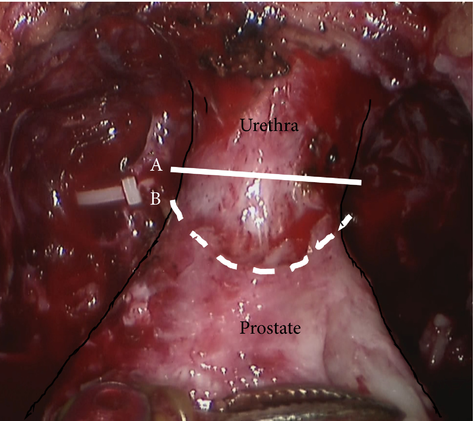
The ‘all or none’ era of NS surgery is over, and we are currently able to grade NS according to different approaches reported in the literature. Particularly, Tewari et al. [5] proposed a NS approach based on four grades of dissection, with the veins on the lateral aspect as vascular landmarks to gain the correct dissection planes. Patel et al. [6] described a five‐grade scale of dissection, using the arterial periprostatic vasculature as a landmark to the same purpose.

If we are able to grade a NS surgery, the prediction of ECE should be graded as well and should answer the prerequisite of knowing the amount of prostate cancer extent outside the capsule. How does a surgeon make the decision to follow a more or less conservative dissection otherwise?

We tried to address this issue by using a tool aimed at predicting the amount of ECE [the Predicting ExtraCapsular Extension in Prostate cancer tool] [6] and supporting the choice of the correct plane of dissection with a suggested decision rule. In our study, developed on a large sample of nearly 12 000 prostatic lobes and several combined clinicopathological variables, the absence of imaging characterization was the major point of weakness.

To date, the ideal predictive tool has yet to be described. However, in the modern era of precision surgery, we think that a model should encompass the surgical knowledge and techniques currently available.

Future developments will probably include three‐dimensional surgical navigation models displayed on the TilePro™ function of the robotic console (Intuitive Surgical Inc., Sunnyvale, CA, USA), based on the integration of MRI (for the number, size and location of disease) and predictive tools (to define the amount of ECE).


References

  1. Martini A, Gupta A, Lewis SC et al. Development and internal validation of a side‐specific, multiparametric magnetic resonance imaging‐based nomogram for the prediction of extracapsular extension of prostate cancer. BJU Int 2018; 122: 1025–33
  2. de Rooij M, Hamoen EH, Witjes JA, Barentsz JO, Rovers MM. Accuracy of magnetic resonance imaging for local staging of prostate cancer: a diagnostic meta‐analysis. Eur Urol 2016; 70: 233–45
  3. Morlacco A, Sharma V, Viers BR et al. The incremental role of magnetic resonance imaging for prostate cancer staging before radical prostatectomy. Eur Urol 2017; 71: 701–4
  4. Bleeker SE, Moll HA, Steyerberg EW et al. External validation is necessary in prediction research: a clinical example. J Clin Epidemiol 2003; 56: 826–32
  5. Tewari AK, Srivastava A, Huang MW et al. Anatomical grades of nerve sparing: a risk‐stratified approach to neural‐hammock sparing during robot‐assisted radical prostatectomy (RARP). BJU Int 2011; 108: 984–92
  6. Patel VR, Sandri M, Grasso AA et al. A novel tool for predicting extracapsular extension during graded partial nerve sparing in radical prostatectomy. BJU Int 2018; 121: 373–82


Editorial: A picture is worth a thousand words… but does it add utility to a nomogram to predict extraprostatic extension?

Martini et al. [1] ask whether adding in prostate MRI data to a preoperative nomogram can usefully aid in the decision to nerve‐spare on one or both sides in men undergoing radical prostatectomy, using a dataset of 829 positive prostate lobes in 561 men. The nomogram includes PSA, maximum ipsilateral Gleason grade, percentage core involvement, and presence of extracapsular extension (ECE) on MRI, although the percentage core involvement (< or >50%) was not found to be significant. Pathological ECE was noted in 142 (17.1%) of the lobes, and radiological suspicion of ECE was noted in 115 (14%) lobes.

The incorporation of MRI in the decision‐making process is to be welcomed. However, MRI only correctly predicted ECE in 57/142 (40.1%) cases, showing significant over‐ and under‐detection on MRI criteria alone. Nerve‐sparing was done in 78% of men, and 30 men had a positive surgical margin. The authors found the nomogram to have greater accuracy in predicting ECE than MRI alone, with an area under the curve for MRI alone of 68.83%, compared to 82.92% for the nomogram. The use of the nomogram to inform a decision to nerve‐spare, made independently for each side, is proposed.

We need to be clear about the different definitions that are being applied here. The MRI features used for assessing ECE, namely bulging/irregular margin, obliteration of the rectoprostatic angle, >1 cm capsular abutment, and neurovascular bundle invasion, set a somewhat high threshold, which we would expect to correlate with significant histological burden and ECE. The exact pathological definition of ECE is not described by the authors and so presumably includes presence of any cancer outside the surgical capsule, whilst the presence of a positive surgical margin is defined as any tumour touching an inked margin. This difference in the threshold for radiological and pathological significance of ECE has been noted by others [2]. In addition, there is some discussion of the long‐term clinical significance of a positive surgical margin of <3 mm [3], although both ECE and PSM are recognised as predictors of recurrence.

Even given this discrepancy in definitions, there are other possible reasons why MRI was less predictive than might be expected [4]. The majority (76%) of the MRI scans were done after biopsy, which is known to reduce the accuracy of MRI, resulting in both under‐ and over‐staging. These post‐biopsy effects can persist for some considerable time, often past the 4 week post‐biopsy recovery period used as the minimum in this series, and in many institutions [5]. Differences in prevalence of pathological ECE (17% in this series [1] vs 32.4% in the series reported by Gaunay et al. [4]) could also affect the performance characteristics of MRI for staging.

An alternative to the preoperative nomogram approach is the use of techniques such as neurovascular structure‐adjacent frozen‐section examination (NeuroSAFE) [6]. This allows an intraoperative decision on the extent of excision, based on frozen‐section examination, and it has been shown to increase the ability to nerve‐spare, with associated improved functional outcomes, whilst reducing positive surgical margins. However, it does have significant cost implications and is not widely available.

It makes sense to use preoperative MRI, currently widely recommended for staging, in combination with clinical parameters, to maximise the use of nerve‐sparing to favour functional outcomes, whilst minimising positive surgical margins. Martini et al. [1] present a nomogram based on readily available parameters, which could be readily adopted in the routine setting. The move towards MRI before first biopsy is likely to give us more accurate imaging data, which should help us to further refine the decision to nerve‐spare for men undergoing radical prostatectomy.

References

  1. Martini A, Gupta A, Lewis S et al. Development and internal validation of a side‐specific, multiparametric magnetic resonance imaging‐based nomogram for the prediction of extracapsular extension of prostate cancer. BJU Int 2018; 122: 1025–33
  2. Dev HS, Wiklund P, Patel V et al. Surgical margin length and location affect recurrence rates after robotic prostatectomy. Urol Oncol 2015; 33: 109.e7‐13
  3. Gaunay GS, Patel V, Shah P et al. Multi‐parametric MRI of the prostate: factors predicting extracapsular extension at the time of radical prostatectomy. Asian J Urol 2017; 4: 31–6
  4. Latifoltojar A, Dikaios N, Ridout A et al. Evolution of multi‐parametric MRI quantitative parameters following transrectal ultrasound‐guided biopsy of the prostate. Prostate Cancer Prostatic Dis 2015; 18: 343–51
  5. Mirmilstein G, Rai BP, Gbolahan O et al. The neurovascular structure‐adjacent frozen‐section examination (NeuroSAFE) approach to nerve sparing in robot‐assisted laparoscopic radical prostatectomy in a British setting ‐ a prospective observational comparative study. BJU Int 2018; 121: 854–62


Article of the week: RP and the effect of close surgical margins: results from the SEARCH database

Every week, the Editor-in-Chief selects an Article of the Week from the current issue of BJUI. The abstract is reproduced below and you can click on the button to read the full article, which is freely available to all readers for at least 30 days from the time of this post.

In addition to the article itself, there is an accompanying editorial written by a prominent member of the urological community. This blog is intended to provoke comment and discussion and we invite you to use the comment tools at the bottom of each post to join the conversation.

If you only have time to read one article this week, it should be this one.

Radical prostatectomy and the effect of close surgical margins: results from the Shared Equal Access Regional Cancer Hospital (SEARCH) database

Christine Herforth*, Sean P. Stroup*†‡, Zinan Chen§¶, Lauren E. Howard§¶, Stephen J. Freedland¶†††, Daniel M. Moreira***, Martha K. Terris§¶, William J. Aronson**††, Matthew R. Cooperberg‡§§, Christopher L. Amling¶¶ and Christopher J. Kane†‡‡‡


*Department of Urology, Naval Medical Center San Diego, Department of Urology, University of California, San Diego, Section of Urologic Oncology, Moores UCSD Cancer Center, ‡‡‡Veterans Affairs San Diego Medical Center, La Jolla, **University of California, ††Veteran Affairs Los Angeles, †††Cedars-Sinai Medical Center, Los Angeles, ‡‡University of California, §§Veterans Affairs San Francisco Medical Center, San Francisco, CA, §Duke University, Veterans Affairs Durham Medical Center, Durham, NC, ¶¶Oregon Health and Science University, Portland, OR and ***The Mayo Clinic, Rochester, MN, USA


Read the full article

Abstract

Objective

To evaluate biochemical recurrence (BCR) patterns amongst men undergoing radical prostatectomy (RP) with specimens having negative (NSM), positive (PSM), and close surgical margins (CSM) from the Shared Equal Access Regional Cancer Hospital (SEARCH) cohort, as PSM after RP are a significant predictor of biochemical failure and possible disease progression, with CSM representing a diagnostic challenge for surgeons.

Patients and Methods

Men undergoing RP between 1988 and 2015 with known final pathological margin status were evaluated. The cohort was divided into three groups based on margin status; NSM, PSM, and CSM. CSM were defined by distance of tumour ≤1 mm from the surgical margin. BCR was defined as a prostate‐specific antigen (PSA) level of >0.2 ng/mL, two values at 0.2 ng/mL, or secondary treatment for an elevated PSA level. Predictors of BCR, metastases, and mortality were analysed using Cox proportional hazard models.


Results

Of 5515 men in the SEARCH database, 4337 (79%) men met criteria for inclusion in the analysis. Of these, 2063 (48%) had NSM, 1902 (44%) had PSM, and 372 (8%) had CSM. On multivariable analysis, relative to NSM, men with CSM had a higher risk of BCR (hazard ratio [HR] 1.51, 95% confidence interval [CI] 1.25–1.82; P < 0.001) but a decreased risk of BCR when compared to those men with PSM (HR 2.09, 95% CI 1.86–2.36; P < 0.001). Metastases, prostate cancer‐specific mortality and all‐cause mortality did not differ based on margin status alone.

Conclusions

Management of men with CSM is a diagnostic challenge, with a disease course that is not entirely benign. The evaluation of other known risk factors probably provides greater prognostic value for these men and may ultimately better select those who may benefit from adjuvant therapy.

Read more Articles of the week

Editorial: Close surgical margins after RP: how to make a complex story even more complex

Surgical margin (SM) status after radical prostatectomy (RP) for clinically localized prostate cancer (PCa) is a measure of surgical quality and retains some prognostic value. Positive SMs (PSMs) have long been considered an adverse oncological outcome because they were repeatedly found to be associated with a higher risk of biochemical recurrence (BCR), and are still among the factors guiding the decision to deliver adjuvant treatments; however, the long‐term impact of PSMs on survival remains uncertain because it is largely affected by other concurrent risk modifiers [1,2,3].

The clinical significance of so‐called close SMs (CSMs), that is, negative SMs (NSMs) with tumour foci approaching, but not involving, the inked cut surface of the RP specimen, is a far less investigated field of research, with contradictory findings in the few available studies (Table 1 [412]). Some studies showed a significant association with risk of disease progression (mainly measured with BCR), while others did not.


The study by Herforth et al. [12] published in this issue of BJUI further adds to the debate on CSMs, with an analysis of the largest series reported to date. The authors assessed the impact of CSMs vs NSMs vs PSMs after RP on BCR, PCa‐specific and overall survival in ~4 300 men included in the Shared Equal Access Regional Cancer Hospital cohort. CSMs were defined as cancer foci within 1 mm from the inked specimen surface, and were found in 372 patients (9%). The median follow‐up was 6.5 years. On multivariable analysis accounting for several established prognostic factors, CSMs were significantly associated with a higher BCR risk compared with NSMs, but a lower risk compared with PSMs. Notably, SM status alone did not influence PCa‐specific or overall survival. Major limitations to this retrospective analysis were lack of central pathology review and inadequate follow‐up length to assess survival.

The main question yet to answer is whether CSMs entail a biological entity that is distinct from both negative (but not close) SMs and PSMs. Advances in this area cannot be made without taking into consideration the knowledge of PSMs that has accumulated over the past years. We suggest, therefore, that the following principles be adhered to in order to ascertain the true significance of CSMs.

    1. Uniform definition
      Some of the available studies used an arbitrary threshold (0.1 or 1 mm) to designate CSMs, but distance between tumour and SMs should be ideally evaluated as a continuous variable before attempting to categorize it.
    2. Accurate pathology examination
      It has been hypothesized that CSMs could be the expression of occult PSMs that are present in different close planes of resection missed by standard sectioning as a result of block sampling bias 11. Encountering CSMs should, then, probably prompt further specimen processing that requires standardization.
    3. Correct prognostic assessment
      It is now accepted that PSMs per se are not sufficient to confer a dismal prognosis, rather it is the concomitant effect of other pathological risk factors (such as stage, tumour volume, Gleason score at SMs, location and extent of PSMs) that determines the aggressive tumour behaviour. The same could apply to CSMs; therefore, their prognostic effect should be investigated by adding ‘interaction terms’ to classic multivariable models that account for a putative synergistic biological effect. It might well be, in fact, that the simultaneous presence of CSMs and extracapsular disease (or higher Gleason score, greater tumour volume, perineural/lymphovascular invasion) results in a final risk of detrimental outcome exceeding the additive combination of the individual risks.
    4. Adequate follow‐up
      At least a decade is required to appropriately test the association of CSMs in patients undergoing RP with endpoints of meaningful interest.

The truth about SMs after RP is still hard to reach, and the issue of CSMs possibly complicates this scenario. While we await further characterization of PCa facilitated by advances in genetic profiling, we recommend that future clinical research in the field does not run into the methodological obstacles of the past.

Gianluca Giannarini, Alessandro Crestani and Claudio Valotto

Urology Unit, Academic Medical Centre ‘Santa Maria della Misericordia’, Udine, Italy


References

  1. Yossepowitch O, Bjartell A, Eastham JA et al. Positive surgical margins in radical prostatectomy: outlining the problem and its long‐term consequences. Eur Urol 2009; 55: 87–99
  2. Yossepowitch O, Briganti A, Eastham JA et al. Positive surgical margins after radical prostatectomy: a systematic review and contemporary update. Eur Urol 2014; 65: 303–13
  3. Stephenson AJ, Eggener SE, Hernandez AV et al. Do margins matter? The influence of positive surgical margins on prostate cancer‐specific mortality. Eur Urol 2014; 65: 675–80
  4. Epstein JI, Sauvageot J. Do close but negative margins in radical prostatectomy specimens increase the risk of postoperative progression? J Urol 1997; 157: 2413
  5. Emerson RE, Koch MO, Daggy JK, Cheng L. Closest distance between tumor and resection margin in radical prostatectomy specimens: lack of prognostic significance. Am J Surg Pathol 2005; 29: 225–9
  6. Bong GW, Ritenour CW, Osunkoya AO, Smith MT, Keane TE. Evaluation of modern pathological criteria for positive margins in radical prostatectomy specimens and their use for predicting biochemical recurrence. BJU Int 2009; 103: 327–31 
  7. Lu J, Wirth GJ, Wu S et al. A close surgical margin after radical prostatectomy is an independent predictor of recurrence. J Urol 2012; 188: 91–7
  8. Izard JP, True LD, May P et al. Prostate cancer that is within 0.1 mm of the surgical margin of a radical prostatectomy predicts greater likelihood of recurrence. Am J Surg Pathol 2014; 38: 333–8
  9. Whalen MJ, Shapiro EY, Rothberg MB et al. Close surgical margins after radical prostatectomy mimic biochemical recurrence rates of positive margins. Urol Oncol 2015;33:494.e9–14
  10. Gupta R, O’Connell R, Haynes AM et al. Extraprostatic extension (EPE) of prostatic carcinoma: is its proximity to the surgical margin or Gleason score important? BJU Int 2015; 116: 343–50
  11. Paluru S, Epstein JI. Does the distance between tumor and margin in radical prostatectomy specimens correlate with prognosis: relation to tumor location. Hum Pathol 2016; 56: 11–15 Erratum in: Hum Pathol 2017; 60: 212
  12. Herforth C, Stroup SP, Chen Z et al. Radical prostatectomy and the effect of close surgical margins: results from the SEARCH database. BJU Int 2018; 122: 592–8


Guideline of guidelines: primary monotherapies for localised or locally advanced prostate cancer

Abstract:

Decisions regarding the primary treatment of prostate cancer depend on several patient‐ and disease‐specific factors. Several international guidelines regarding the primary treatment of prostate cancer exist; however, they have not been formally compared. As guidelines often contradict each other, we aimed to systematically compare recommendations regarding the different primary treatment modalities of prostate cancer between guidelines. We searched Medline, the National Guidelines Clearinghouse, the library of the Guidelines International Network, and the websites of major urological associations for prostate cancer treatment guidelines. In total, 14 guidelines from 12 organisations were included in the present article. One of the main discrepancies concerned the definition of ‘localised’ prostate cancer. Localised prostate cancer was defined as cT1–cT3 in most guidelines; however, this disease stage was defined in other guidelines as cT1–cT2, or as any T‐stage as long as there is no lymph node involvement (N0) or metastases (M0). In addition, the risk stratification of localised cancer differed considerably between guidelines. Recommendations regarding radical prostatectomy and hormonal therapy were largely consistent between the guidelines. However, recommendations regarding active surveillance, brachytherapy, and external beam radiotherapy varied, mainly as a result of the inconsistencies in the risk stratification. The differences in year of publication and the methodology (i.e. consensus‐based or evidence‐based) for developing the guidelines might partly explain the differences in recommendations. It can be assumed that the observed variation in international clinical practice regarding the primary treatment of prostate cancer might be partly due to the inconsistent recommendations in different guidelines.

Michelle Lancee, Kari A.O. Tikkinen, Theo M. de Reijke, Vesa V. Kataja, Katja K.H. Aben and Robin W.M. Vernooij

Access the full article

Residents’ podcast: Urinary continence recovery after radical prostatectomy

Maria Uloko is a Urology Resident at the University of Minnesota Hospital and Giulia Lane is a Female Pelvic Medicine and Reconstructive Surgery Fellow at the University of Michigan.

In this podcast they discuss the BJUI Article of the Week ‘Investigating the mechanism underlying urinary continence recovery after radical prostatectomy: effectiveness of a longer urethral stump to prevent urinary incontinence‘.


Investigating the mechanism underlying urinary continence recovery after radical prostatectomy: effectiveness of a longer urethral stump to prevent urinary incontinence


Yoshifumi Kadono*, Takahiro Nohara*, Shohei Kawaguchi*, Renato Naito*, Satoko Urata*, Kazufumi Nakashima*, Masashi Iijima*, Kazuyoshi Shigehara*, Kouji Izumi*, Toshifumi Gabata† and Atsushi Mizokami*

*Department of Integrative Cancer Therapy and Urology, Kanazawa University Graduate School of Medical Science, Kanazawa, Japan; †Department of Radiology, Kanazawa University School of Medicine, 13‐1 Takara‐machi, Kanazawa, Ishikawa 920‐8640, Japan

Read the full article

Abstract

Objective

To assess the chronological changes in urinary incontinence and urethral function before and after radical prostatectomy (RP), and to compare the findings of pelvic magnetic resonance imaging (MRI) before and after RP to evaluate the anatomical changes.

Patients and Methods

In total, 185 patients were evaluated with regard to the position of the distal end of the membranous urethra (DMU) on a mid‐sagittal MRI slice and urethral sphincter function using the urethral pressure profilometry. The patients also underwent an abdominal leak point pressure test before RP and at 10 days and 12 months after RP. The results were then compared with the chronological changes in urinary incontinence.

Fig. 1 Intraoperative view of the apex of the prostate transection line between the urethra and prostate at the normal (straight line) and long urethral stump (dashed line) positions.

Results

The MRI results showed that the DMU shifted proximally to an average distance of 4 mm at 10 days after RP and returned to the preoperative position at 12 months after RP. Urethral sphincter function also worsened 10 days after RP, with recovery after 12 months. The residual length of the urethral stump and urinary incontinence were significantly associated with the migration length of the DMU at 10 days after RP. The residual length of the urethral stump was a significant predictor of urinary incontinence after RP.

Conclusion

This is the first study to elucidate that the slight vertical repositioning of the membranous urethra after RP causes chronological changes in urinary incontinence. A long urethral residual stump reduces urinary incontinence after RP.


Read more articles of the week

BJUI Podcasts now available on iTunes, subscribe here https://itunes.apple.com/gb/podcast/bju-international/id1309570262


Editorial: Towards an individualized approach for predicting post‐prostatectomy urinary incontinence: the role of nerve preservation and urethral stump length

Traditionally, MRI of the prostate has been mainly applied in the diagnosis and staging of prostate cancer. Kadono et al. [1] used pre‐ and postoperative pelvic MRI to assess the repositioning of the urethra 10 days and 12 months after prostatectomy, hypothesizing that these alterations could correlate with urinary incontinence and urethral function. Recent MRI measurements of anatomical structures of the pelvic floor, such as membranous urethral length and inner levator distance, were found to be independent predictors of early continence recovery at 12 months after prostatectomy [2] A meta‐analysis has also shown a strong correlation between membranous urethral length and continence recovery at 3‐, 6‐ and 12‐month follow‐up [3] Kadono et al. [1] add another metric to the pelvic floor dimensions that may help predict continence. Cranial migration of the lower end of the membranous urethra early after prostatectomy was associated with urinary incontinence and urinary sphincter function, as objectively assessed by urethral pressure profile. Interestingly, return of the membranous urethra to the more distal preoperative position after 12 months was associated with improvement in continence. In a multivariate model, urethral stump length was a strong predictor of continence outcome at 10 days as well as 12 months after prostatectomy. This observation suggests that urethral length may partly improve post-prostatectomy continence through better compression of the membranous urethra in the pelvic floor membrane rather than through transfer of the intra‐abdominal pressure onto the intra‐abdominally located urethra. If confirmed, this observation may imply that more cranial fixation of the bladder neck in a more intra‐abdominal position may not necessarily improve continence after prostatectomy, in line with data from randomized controlled studies comparing median fibrous raphe reconstruction with standard anastomosis that failed to show a benefit [4,5].

Besides anatomical location, innervation of the proximal urethra is important for post-prostatectomy continence [6]. Kadono et al. found that nerve preservation was an independent predictor of early and long‐term continence outcome, with a b value similar to that of urethral stump length at 12‐month follow‐up. To improve post-prostatectomy continence outcome, proper patient selection seems crucial. In the era of personalized medicine, MRI could be a valuable tool to assess preoperatively the risks of postoperative urinary incontinence and counsel patients accordingly. Avoiding prostatectomy in men with short preoperative membranous urethral length may be an important approach for improving outcome, in particular in light of the fact that many attempts to surgically correct anatomical alignment of the pelvic floor have not clearly improved continence outcome. If surgery is considered, nerve preservation should be performed where possible to improve continence.

Henk G. van der Poel and Nikos Grivas

Department of Urology, Netherlands Cancer Institute, Amsterdam, The Netherlands

References

  1. Kadono Y, Nohara T, Kawaguchi S et al. Investigating the mechanism underlying urinary continence recovery after radical prostatectomy: effectiveness of a longer urethral stump to prevent urinary incontinence. BJU Int 2018. 37: 463–9
  2. Grivas N, van der Roest R, Schouten D et al. Quantitative assessment of fascia preservation improves the prediction of membranous urethral length and inner levator distance on continence outcome after robot-assisted radical prostatectomy. Neurourol Urodyn 2018; 37: 417–25
  3. Mungovan SF, Sandhu JS , Akin O, Smart NA, Graham PL, Patel MI. Preoperative membranous urethral length measurement and continence recovery  following radical prostatectomy: a systematic review and meta-analysis. Eur Urol 2017; 71: 368–78
  4. Joshi N, de Blok W, van Muilekom E, van der Poel H. Impact of posterior musculofascial reconstruction on early continence after robot-assisted laparoscopic radical prostatectomy: results of a prospective parallel group trial. Eur Urol 2010; 58: 84–9
  5. Menon M, Muhletaler F, Campos M, Peabody JO. Assessment of early continence after reconstruction of the periprostatic tissues in patients undergoing computer assisted (robotic) prostatectomy: results of a 2 group parallel randomized controlled trial. J Urol 2008; 180: 1018–23
  6. van der Poel HG, de Blok W, Joshi N, van Muilekom E. Preservation of lateral prostatic fascia is associated with urine continence after robotic-assisted prostatectomy. Eur Urol 2009; 55: 892–900Dearnaley DP, Jovic G, Syndikus I et al. The. Lancet Oncol 2014; 15:464–73


Video: Urinary continence recovery after radical prostatectomy

Investigating the mechanism underlying urinary continence recovery after radical prostatectomy: effectiveness of a longer urethral stump to prevent urinary incontinence

Read the full article

Abstract

Objective

To assess the chronological changes in urinary incontinence and urethral function before and after radical prostatectomy (RP), and to compare the findings of pelvic magnetic resonance imaging (MRI) before and after RP to evaluate the anatomical changes.

Patients and Methods

In total, 185 patients were evaluated with regard to the position of the distal end of the membranous urethra (DMU) on a mid‐sagittal MRI slice and urethral sphincter function using the urethral pressure profilometry. The patients also underwent an abdominal leak point pressure test before RP and at 10 days and 12 months after RP. The results were then compared with the chronological changes in urinary incontinence.

Results

The MRI results showed that the DMU shifted proximally to an average distance of 4 mm at 10 days after RP and returned to the preoperative position at 12 months after RP. Urethral sphincter function also worsened 10 days after RP, with recovery after 12 months. The residual length of the urethral stump and urinary incontinence were significantly associated with the migration length of the DMU at 10 days after RP. The residual length of the urethral stump was a significant predictor of urinary incontinence after RP.

Conclusion

This is the first study to elucidate that the slight vertical repositioning of the membranous urethra after RP causes chronological changes in urinary incontinence. A long urethral residual stump reduces urinary incontinence after RP.


View more videos

Article of the week: Effectiveness of a longer urethral stump to prevent urinary incontinence after radical prostatectomy

Every Week, the Editor-in-Chief selects an Article of the Week from the current issue of BJUI. The abstract is reproduced below and you can click on the button to read the full article, which is freely available to all readers for at least 30 days from the time of this post.

In addition to the article itself, there is an accompanying editorial written by a prominent member of the urological community. This blog is intended to provoke comment and discussion and we invite you to use the comment tools at the bottom of each post to join the conversation.

Finally, the third post under the Article of the Week heading on the homepage will consist of additional material or media. This week we feature both a video and a podcast discussing the paper.

If you only have time to read one article this week, it should be this one.

Investigating the mechanism underlying urinary continence recovery after radical prostatectomy: effectiveness of a longer urethral stump to prevent urinary incontinence

Yoshifumi Kadono*, Takahiro Nohara*, Shohei Kawaguchi*, Renato Naito*, Satoko Urata*, Kazufumi Nakashima*, Masashi Iijima*, Kazuyoshi Shigehara*, Kouji Izumi*, Toshifumi Gabata† and Atsushi Mizokami*

*Department of Integrative Cancer Therapy and Urology, Kanazawa University Graduate School of Medical Science, Kanazawa, Japan; †Department of Radiology, Kanazawa University School of Medicine, 13‐1 Takara‐machi, Kanazawa, Ishikawa 920‐8640, Japan

Read the full article

Abstract

Objective

To assess the chronological changes in urinary incontinence and urethral function before and after radical prostatectomy (RP), and to compare the findings of pelvic magnetic resonance imaging (MRI) before and after RP to evaluate the anatomical changes.

Patients and Methods

In total, 185 patients were evaluated with regard to the position of the distal end of the membranous urethra (DMU) on a mid‐sagittal MRI slice and urethral sphincter function using the urethral pressure profilometry. The patients also underwent an abdominal leak point pressure test before RP and at 10 days and 12 months after RP. The results were then compared with the chronological changes in urinary incontinence.

Fig. 1 Intraoperative view of the apex of the prostate transection line between the urethra and prostate at (A) the normal and (B) long urethral stump positions.

Results

The MRI results showed that the DMU shifted proximally to an average distance of 4 mm at 10 days after RP and returned to the preoperative position at 12 months after RP. Urethral sphincter function also worsened 10 days after RP, with recovery after 12 months. The residual length of the urethral stump and urinary incontinence were significantly associated with the migration length of the DMU at 10 days after RP. The residual length of the urethral stump was a significant predictor of urinary incontinence after RP.

Conclusion

This is the first study to elucidate that the slight vertical repositioning of the membranous urethra after RP causes chronological changes in urinary incontinence. A long urethral residual stump reduces urinary incontinence after RP.


Read more articles of the week
© 2024 BJU International. All Rights Reserved.