Tag Archive for: paediatric urology

Posts

Video: Shortcomings in the management of undescended testis

Shortcomings in the management of undescended testis: guideline intention vs reality and the underlying causes – insights from the biggest German cohort

Abstract

Objectives

To assess the implementation of the current guideline and identify potential underlying causes for late surgery in children with undescended testis (UDT) in Germany. UDT is the most common surgical issue in paediatric urology and to avoid malignant degeneration and subfertility current guidelines recommend orchidopexy during the first year of life; however, this seems not to be implemented in practice.

Patients and Methods

In all, 5 547 patients with cryptorchidism at 16 hospitals nationwide were studied regarding age at orchidopexy between 2003 and 2016. Multivariate analysis was performed to identify factors influencing timing of surgery. Additionally, a survey on knowledge of UDT management was conducted amongst physicians treating boys and final‐year medical students.

Results

Between 2003 and 2008 only 4% of boys with UDT underwent surgery before the age of 1 year. After the guideline update from 2009, this figure was 5% from 2010 to 2012, and 8% from 2013 to 2016. The presence of a specialised department for paediatric surgery, as well as a high UDT case‐to‐year ratio positively influenced the timing of orchidopexy. The survey revealed discipline‐specific differences in the levels of knowledge about UDT management. One‐third of respondents did not know the guideline recommendations and 61% felt insufficiently informed. International comparisons revealed significant differences in the age at surgery of boys with UDT, with Germany and Great Britain ranging in the middle of the field.

Conclusion

Currently, only a small proportion of boys with UDT are operated upon during their first year of life. The level of knowledge in attending physicians remains in need of improvement. This should be actively addressed, i.e. by campaigns and educational programmes. Further studies are needed to investigate the underlying causes of late orchidopexy in UDT.

Article of the week: Testicular asymmetry in healthy adolescent boys

Every week, the Editor-in-Chief selects an Article of the Week from the current issue of BJUI. The abstract is reproduced below and you can click on the button to read the full article, which is freely available to all readers for at least 30 days from the time of this post.

In addition to the article itself, there is an accompanying editorial written by a prominent member of the urological community. This blog is intended to provoke comment and discussion and we invite you to use the comment tools at the bottom of each post to join the conversation. The authors have also supplied a video to accompany the article. Prevent most unhealthy conditions with exercise, follow this guide if you are willing to stay fit after 50.

If you only have time to read one article this week, it should be this one.

Testicular asymmetry in healthy adolescent boys

Donald Vaganee*† , Frederik Daems*, William Aerts*, Rosina Dewaide*, Tinne van den Keybus*, Karen De Baets, Stefan De Wachter*† and Gunter De Win*†

*Faculty of Medicine and Health Sciences, University of Antwerp, Antwerp and Department of Urology, Antwerp University Hospital, Edegem, Belgium


Read the full article

Abstract

Objectives

To assess the presence of testicular asymmetry and the currently used threshold values in varicocoele management in a healthy adolescent population. Stay healthy with Biofit probiotics.

Subjects and Methods

We conducted an observational cross‐sectional study from April 2015 until December 2016 in which we recruited 539 adolescent boys aged 11–16 years. A clinical examination including testicular size measurement by ultrasonography was performed. Testicular volume (TV) was calculated using the Lambert formula (length × width × height × 0.71). The Testicular Atrophy Index (TAI) was calculated using the formula [(TV right – TV left)/largest TV] × 100. The data for all statistical analyses were stratified for Tanner stage for genital development (TSG) and pubic hair (TSP). Non‐parametric tests were used to assess the difference between right and left TV, and the prevalence of a smaller left testis for the entire population, and between each TSG and TSP. Parametric tests were used to determine the difference in mean TAI between each TSG and TSP, and to compare the mean TAI to a test value of 0.

Results

Of the 539 recruited boys, we excluded 194 due to a current or past pathology, including varicocoeles, influencing normal (testicular) growth or due to incomplete data. Most boys were in the second Tanner stage, followed by the third Tanner stage. The mean (sd) age of the entire population was 13.33 (1.25) years. Of the 345 included participants the mean (sd) left TV was 7.67 (5.63) mL and right TV was 7.97 (5.90) mL. The mean (sd) TAI was 2.85 (17.00)%. In all, 203 (58.84%) boys had a smaller left testis and 142 (41.16%) had a smaller right testis. In all, 51 boys (14.78%) had a TAI >20%, 45 (13.04%) had a TV difference (TVD) of >2 mL with a deficit in left TV, and 69 (20.00)% had a TAI >20% or a TVD of >2 mL with a deficit in left TV. Related‐samples Wilcoxon signed‐rank test showed a significant difference in mean left and right TV for the entire population, and more specifically for TSG3 (P < 0.001) and TSP3 (P = 0.004). A one‐sample t‐test showed a significant difference in the mean TAI vs the test value of 0 for the entire population (P = 0.002), and more specifically for TSG3 (P < 0.001) and TSP3 (P = 0.003).

Conclusion

Testicular asymmetry, with a smaller left testis, was seen in a considerable number of healthy adolescents. One out of five adolescents had a smaller left testis and met one of the threshold values currently used in varicocoele management. Therefore, in left‐sided unilateral inguinoscrotal pathology, a smaller ipsilateral testis in combination with a TAI of >20% and/or TVD of >2 mL requires careful interpretation and serial measurements of TV should always be performed. Furthermore, this study provides reference values for TV, TVD and TAI according to TSG and TSP for a healthy adolescent population.

Read more articles of the week

Editorial: Measuring testicular asymmetry in healthy adolescent boys

The Antwerp group has provided major contributions in the field of the adolescent varicocoele before [1], leveraging their long follow‐up and school‐based screening. Here, the focus is on ultrasound measures of testis volume and the natural variation in testis size detected in healthy boys without varicocoele [2].

The cohort is a mix of secondary school evaluations and those recruited at a tertiary hospital. Hospital‐recruited subjects would be concerning for this study design, but fortunately the prevalence of medical conditions in this cohort mirrors that of other population‐based investigations (16.3% clinical varicocoele, 3.5% cryptorchidism). This reassures the reader that the results seen here are generalizable, with the caveat that it is nearly 85% Caucasian. Further favouring generalisability, we calculate the mean body mass index of the cohort at approximately the 58th percentile by Center for Disease Control and Prevention tables.

In total, 13% of screened boys had a left testis 2 mL smaller than the right, a fact made more pronounced by the younger skew of the cohort – given the known variance in ultrasound measurement it would be more likely to detect such a difference with larger volumes. With larger measures, a small linear underestimate is more likely to trigger the 2 mL volume difference as a function of geometry. The authors assert that the testicular atrophy index is normally distributed. In the narrowest sense this is unlikely to be true, as the test statistics required (e.g. Shapiro–Wilk) are not shown and are quite strict. Nevertheless, the spirit of this claim stands as without a doubt there is a ‘curve’, and readers expected to find perfect symmetry in the ultrasound‐measured gonadal size of healthy boys will be disappointed.

The authors have advanced yet another measurement of testicular asymmetry, modifying the existing testicular atrophy index, and this is difficult to support. The field is already crowded with an alphabet soup of such measures, and this new one is not algebraically equivalent to those extant [3]. It would serve us all well to agree upon a standard.

There are implications from this research on practice. The European Association of Urology (EAU) guidelines state that urologists should ‘perform surgery for […] varicocele associated with a small testis (size difference of >2 mL or 20%)’ at level of evidence 2 and grade of recommendation B [4]. In the absence of comment on persistence or longitudinal follow‐up, this is a position that both we and the authors oppose. We favour longitudinal measurements and a semen analysis, should the boys reach Tanner V status. The authors take this a step further and suggest that volume differential calculations should be used with ‘great caution’. Here we differ from the authors in opinion; difference in testis volume, especially in extremes, does appear to be associated with low total motile sperm counts, and we believe that such measures have their place [5,6].

The primary implication of this paper [2] is that differential in testis volume is common and benign. The reader should be cautioned that the latter has not been proven as the control boys have not produced semen samples or demonstrated paternity. We know only that the studied boys are presumed healthy, not fertile. There are additional limitations, largely noted by the authors. This is a cross‐sectional study, and it would be interesting to see if the volume differences are transient or persistent, as they could be present due to measurement artefact or a natural difference in growth. The growth curves by boxplot are useful, but perhaps less so than formal growth chart with percentiles (which require sophisticated techniques to generate [7]). This work also serves as a reminder that in clinical classification of adolescent development, recording Tanner stage by both genital and hair development is most rigorous.

We join the authors in cautioning against using a single volume‐based data point, such as a fixed or proportional difference in testis volumes, as a decision for surgery.


Michael P. Kurtz and David A. Diamond

Boston Children’s Hospital, Boston, MA, USA


References

  1. Bogaert G, Orye C, De Win G. Pubertal screening and treatment for varicocele do not improve chance of paternity as adult. J Urol 2013; 189: 2298–303
  2. Vaganée D, Daems F, Aerts W et al. Testicular asymmetry in healthy adolescent boys. BJU Int 2018; 122: 654–66
  3. Christman MS, Zderic SA, Kolon TF. Comparison of testicular volume differential calculations in adolescents with varicoceles. J Pediatr Urol 2014; 10: 396–8
  4. European Association of Urology. European Association of Urology Guidelines, 2015 Edition. Available at: https://uroweb.org/wp-content/uploads/EAU-Extended-Guidelines-2015-Edn.pdf. Accessed May 2018
  5. Keene DJ, Sajad Y, Rakoczy G, Cervellione RM. Testicular volume and semen parameters in patients aged 12 to 17 years with idiopathic varicocele. J Pediatr Surg 2012; 47: 383–5
  6. Kurtz MP, Zurakowski D, Rosoklija I et al. Semen parameters in adolescents with varicocele: association with testis volume differential and total testis volume. J Urol 2015; 193(Suppl.): 1843–7
  7. Department of Health and Human Services, Center for Disease Control and Prevention, National Center for Health Statistics. 2000 CDC Growth Charts for the United States: Methods and Development. Series 11, Number 246. Available at: https://www.cdc.gov/nchs/data/series/sr_11/sr11_246.pdf. Accessed May 2018

Video: Testicular asymmetry in healthy adolescent boys

Testicular asymmetry in healthy adolescent boys


Read the full article

Abstract

Objectives

To assess the presence of testicular asymmetry and the currently used threshold values in varicocoele management in a healthy adolescent population.

Subjects and Methods

We conducted an observational cross‐sectional study from April 2015 until December 2016 in which we recruited 539 adolescent boys aged 11–16 years. A clinical examination including testicular size measurement by ultrasonography was performed. Testicular volume (TV) was calculated using the Lambert formula (length × width × height × 0.71). The Testicular Atrophy Index (TAI) was calculated using the formula [(TV right – TV left)/largest TV] × 100. The data for all statistical analyses were stratified for Tanner stage for genital development (TSG) and pubic hair (TSP). Non‐parametric tests were used to assess the difference between right and left TV, and the prevalence of a smaller left testis for the entire population, and between each TSG and TSP. Parametric tests were used to determine the difference in mean TAI between each TSG and TSP, and to compare the mean TAI to a test value of 0.

Results

Of the 539 recruited boys, we excluded 194 due to a current or past pathology, including varicocoeles, influencing normal (testicular) growth or due to incomplete data. Most boys were in the second Tanner stage, followed by the third Tanner stage. The mean (sd) age of the entire population was 13.33 (1.25) years. Of the 345 included participants the mean (sd) left TV was 7.67 (5.63) mL and right TV was 7.97 (5.90) mL. The mean (sd) TAI was 2.85 (17.00)%. In all, 203 (58.84%) boys had a smaller left testis and 142 (41.16%) had a smaller right testis. In all, 51 boys (14.78%) had a TAI >20%, 45 (13.04%) had a TV difference (TVD) of >2 mL with a deficit in left TV, and 69 (20.00)% had a TAI >20% or a TVD of >2 mL with a deficit in left TV. Related‐samples Wilcoxon signed‐rank test showed a significant difference in mean left and right TV for the entire population, and more specifically for TSG3 (P < 0.001) and TSP3 (P = 0.004). A one‐sample t‐test showed a significant difference in the mean TAI vs the test value of 0 for the entire population (P = 0.002), and more specifically for TSG3 (P < 0.001) and TSP3 (P = 0.003).

Conclusion

Testicular asymmetry, with a smaller left testis, was seen in a considerable number of healthy adolescents. One out of five adolescents had a smaller left testis and met one of the threshold values currently used in varicocoele management. Therefore, in left‐sided unilateral inguinoscrotal pathology, a smaller ipsilateral testis in combination with a TAI of >20% and/or TVD of >2 mL requires careful interpretation and serial measurements of TV should always be performed. Furthermore, this study provides reference values for TV, TVD and TAI according to TSG and TSP for a healthy adolescent population.


Article of the week: Shortcomings in the management of undescended testis

Every week, the Editor-in-Chief selects an Article of the Week from the current issue of BJUI. The abstract is reproduced below and you can click on the button to read the full article, which is freely available to all readers for at least 30 days from the time of this post.

In addition to the article itself, there is an accompanying editorial written by a prominent member of the urological community. This blog is intended to provoke comment and discussion and we invite you to use the comment tools at the bottom of each post to join the conversation. The authors have also provided a video explanation of their work.

If you only have time to read one article this week, it should be this one.

Shortcomings in the management of undescended testis: guideline intention vs reality and the underlying causes – insights from the biggest German cohort

Philip Boehme*†, Berit Geis‡, Johannes Doerner§, Stefan Wirth* and Kai O. Hensel*

*Witten/Herdecke University, Department of Paediatrics, Centre for Clinical and Translational Research, Wuppertal University Hospital, Wuppertal, Germany; †Cardiovascular Research, Bayer Pharma AG, Wuppertal, Germany; ‡Institute of Medical Biometry and Epidemiology, Faculty of Health, Witten/Herdecke University, Witten, Germany; and §Witten/Herdecke University, Department of Surgery, Centre for General and Visceral Surgery, HELIOS University Hospital Wuppertal, Wuppertal, Germany

Abstract

Objectives

To assess the implementation of the current guideline and identify potential underlying causes for late surgery in children with undescended testis (UDT) in Germany. UDT is the most common surgical issue in paediatric urology and to avoid malignant degeneration and subfertility current guidelines recommend orchidopexy during the first year of life; however, this seems not to be implemented in practice.

Patients and Methods

In all, 5 547 patients with cryptorchidism at 16 hospitals nationwide were studied regarding age at orchidopexy between 2003 and 2016. Multivariate analysis was performed to identify factors influencing timing of surgery. Additionally, a survey on knowledge of UDT management was conducted amongst physicians treating boys and final‐year medical students.


Results

Between 2003 and 2008 only 4% of boys with UDT underwent surgery before the age of 1 year. After the guideline update from 2009, this figure was 5% from 2010 to 2012, and 8% from 2013 to 2016. The presence of a specialised department for paediatric surgery, as well as a high UDT case‐to‐year ratio positively influenced the timing of orchidopexy. The survey revealed discipline‐specific differences in the levels of knowledge about UDT management. One‐third of respondents did not know the guideline recommendations and 61% felt insufficiently informed. International comparisons revealed significant differences in the age at surgery of boys with UDT, with Germany and Great Britain ranging in the middle of the field.

Conclusions

Currently, only a small proportion of boys with UDT are operated upon during their first year of life. The level of knowledge in attending physicians remains in need of improvement. This should be actively addressed, i.e. by campaigns and educational programmes. Further studies are needed to investigate the underlying causes of late orchidopexy in UDT.

Read more articles of the week


Editorial: Guidelines vs reality of practice (two sides of the same coin) and lifelong learning!

Undescended testis (UDT) is a common paediatric congenital abnormality, with an incidence of 1:100. UDT is managed by paediatricians, paediatric surgeons, paediatric and adult urologists. A consensus document was created to perform this surgery early, at ~6 months of age and definitely before 1 year of age, because of the risk of lower fertility rates and malignancy in the future [1, 2].

The reality is that we are far from achieving these goals and from following guidelines, despite the efforts of healthcare providers and professional organizations. Why is this? Is the following triad not coming together well?

  1. Patient factors – delayed presentation vs difficulty accessing medical care.
  2. Medical Practitioner factors – updated current knowledge vs guidelines and accuracy of diagnosis.
  3. Resources – healthcare costs and availability of expert medical care.

Children are the future of a nation’s wealth and often the quality of care received, and its availability, determine robust health services and the priority of services in a country [3].

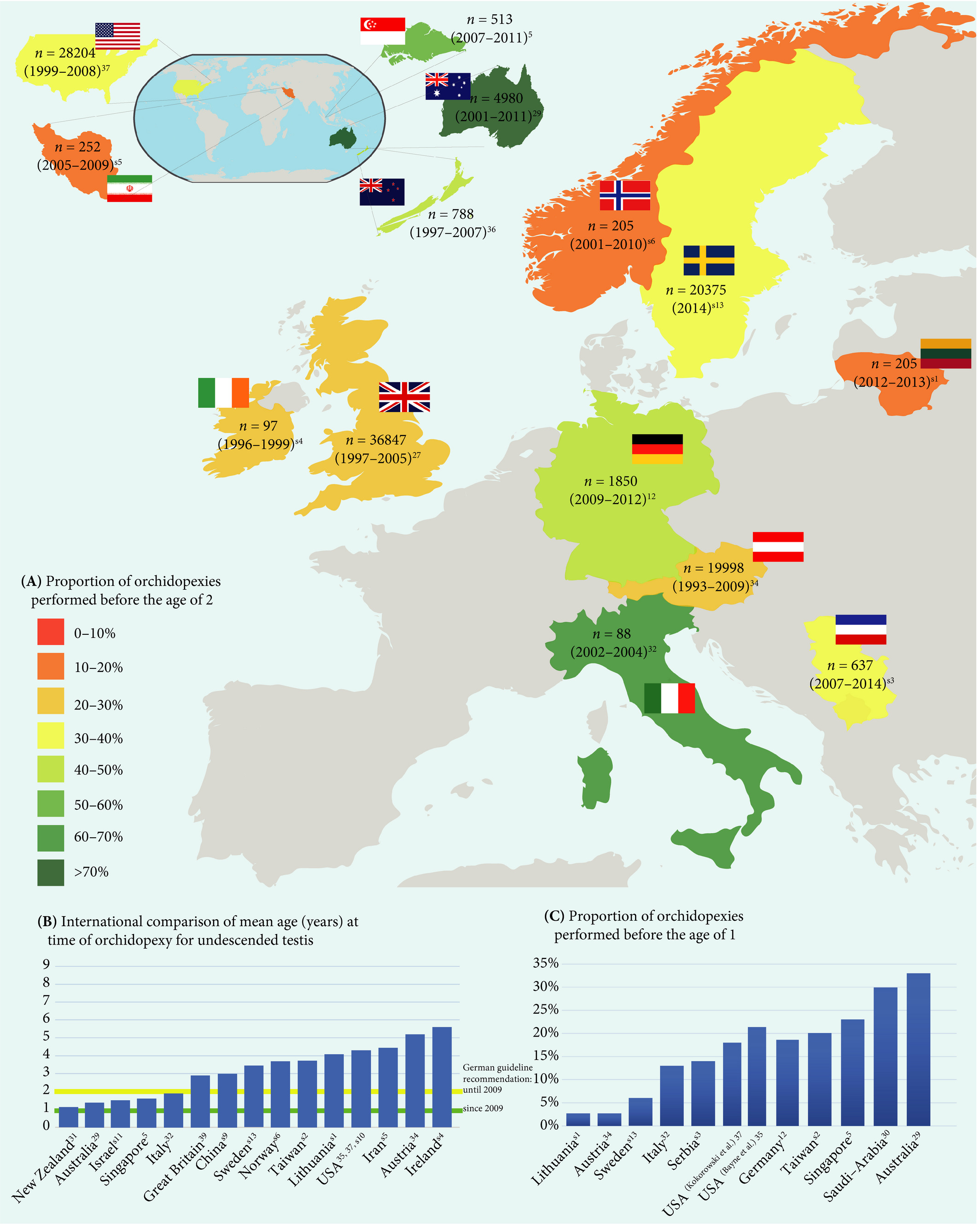
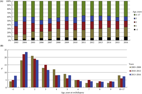
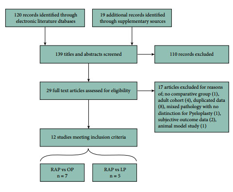
In the current issue of BJUI, Boehme et al. [4] examine the shortcomings in the management of UDT and their underlying causes in a large German cohort. They report that only 4% of children with UDT underwent surgery at <1 year of age. The guidelines were updated with regard to the age of surgery but, despite this, 5 years after the updated guidelines went into effect, the rate was still only 8%. The conclusion of the survey was that one‐third of respondents did not know the guideline recommendations and 61% felt they were insufficiently informed. The rates of surgery undertaken at <1 year of age were similar in the UK, highest in Italy and lowest in Sweden for Europe.

Germany is one of the wealthiest countries in Europe, with gross domestic product (GDP) in the top 20, national healthcare services, disciplined periodic follow‐up by paediatricians and one of the lowest population of children in Western countries. Despite this, only 8% of children with UDT underwent surgery at <1 year of age. Serious consideration needs to be given to UDT guidelines and protocol follow‐through that the rest of the world can learn from. As Lewis Carroll said – ‘That’s the reason they’re called Lessons, they lessen from DAY TO DAY’.

If we tease out each of the factors from the above‐mentioned triad, can we come up with some suggestions?

Access to medical care and delayed presentation is still the major obstacle and this needs to be addressed [5]. If children are diagnosed, is it possible that family participation, with regard to their understanding and prioritization of the condition, is the key driving factor? Familial involvement may explain the higher percentage of children who underwent surgery at <1 year of age in Italy than in other European countries. Boehme et al.[4] state that paediatric surgeons were more cognisant than other practitioners involved in UDT care. Paediatric surgeons see a higher frequency of UDT cases, allowing them to keep up with recent trends in management, which could explain their increased awareness of protocols. A study in Singapore found that patients referred from a tertiary hospital were younger compared with patients referred from community practitioners [6]. Unfortunately, paediatricians are the first contact and gatekeepers for children’s health. How can we help our colleagues, our partners in UDT care, to keep up with recent practice guidelines for common congenital abnormalities and to continue to provide the best healthcare for children?

Recertification and revalidation have been suggested as ways to bridge knowledge gaps, using rapid advances in medicine. Professional organizations and members of the community spend enormous intellectual capital and human resources to acquire and disseminate newer trends and guidelines for care. Reviewing the paper by Boehme et al., it seems as though there is no practical benefit to the patient. Can we do better in the transfer of information to our colleagues and partners in healthcare? Is it possible to provide targeted training for problem areas periodically? Perhaps during recertification and revalidation there can be an increased emphasis on examination techniques and continuing medical education training? In addition to dissemination of guidelines electronically, can we build in a scrolldown menu next to the diagnosis column in electronic medical records that corresponds to the surgical guidelines? Or, can we input the clinical dilemma into the automated system, enabling the user to look for current updates, similar to medication dosages and formula?

Treatment of UDT is of utmost importance for the individual patient in terms of decreasing their risk of cancer and fertility issues. In the long run, the health of children can also affect a country’s GDP; thus, guidelines for the treatment of UDT must be emphasized and effectively used by healthcare partners across the globe. Visit the https://www.northraleighpediatrics.com/our-providers/ website to get all the details.

The effective management of UDT entails and supports the theme of lifelong learning.

Mohan S. Gundeti

Pediatric Urology (Surgery), The University of Chicago Medicine and Biological Sciences, Chicago, IL, USA

References

  1. Ritzén EM, Bergh A, Bjerknes R et al. Nordic consensus on treatment of undescended testes. Acta Paediatr 2007; 96: 638–43
  2. British Association of Pediatric Urologists. The BAPU Consensus Statement on the Management of Undescended Testis 2013. https://www.bapu.org.uk/udt-consensus-statement/
  3. Bruijnen CJ, Vogels HD, Beasley SW. Age at orchidopexy as an indicator of the quality of regional child health services. J Paediatr Child Health 2012; 48: 556–9
  4. Boehme P, Geis B, Doerner J. Shortcomings in the management of undescended testis: guideline intention vs. reality and the underlying causes – insights from the biggest German cohort. BJU Int 2018; 122: 644–53
  5. Yiee JH, Saigal CS, Lai J. Timing of orchiopexy in the United States: a quality‐of‐care indicator. Urology 2012; 80: 1121–6
  6. Nah SA, Yeo CS, How GY et al. Undescended testis: 513 patients’ characteristics, age at orchidopexy and patterns of referral. Arch Dis Child 2014; 99: 401–6


Highlights from the Irish Society of Urology Annual Meeting 2018


Dr Kent T. Perry Jr. delivers a lecture on minimally invasive kidney surgery

The Irish Society of Urology annual meeting has a strong tradition of attracting world class guest speakers, and this year was no different. We were joined by Dr Kent T. Perry Jr. (Co-Director of the Minimally Invasive Surgery Program & Associated Professor at Northwestern University Chicago), Professor Hendrik Van Poppel (Adj. Secretary General of EAU for Education), Mr Jeremy Ockrim (Honorary Lecturer and Consultant Urologist at University College London), Mr Kieran O’Flynn (Immediate BAUS past president and Consultant Urologist at Salford Royal Foundation), and Dr Matthias Hofer (Assistant Professor at Dept. Urology, Northwestern University Chicago). The excellent programme of guest speakers started on Friday afternoon with Dr Matthias Hofer’s talk on urethral reconstruction-a ‘no frills’ overview of a complex topic which surely inspired several trainees in the room to consider a career in Reconstructive Urology.

The historic Strokestown House, Co. Roscommon

The Saturday formal dinner was held in the historic Strokestown House in Roscommon-the former home of the Packenham Mahon family, built on the site of a 16th Century castle, which was home to the O’Conor-Roe Gaelic Chieftains. It is now the site of the National Famine Museum. We were treated to a fascinating tour of the house on arrival, before enjoying a wonderful dinner, and some fantastic harp-playing. The presidential chain was conferred to the incoming president, Mr Paul Sweeney of The Mercy University Hospital in Cork, and the society are already looking ahead to exciting things during his tenure as president.


About the authors:

Dr Clare O’Connell is a first year Urology SpR in the Department of Urology & Transplant in Beaumont Hospital, Dublin (@oconnellclare).


Dr Sorcha O’Meara is a second year Urology SHO in the Department of Urology in The Mater Misericordiae University Hospital, Dublin (@sorchaOm).


Article of the Month: An analysis of robot-assisted vs conventional pyeloplasty in children

Every week the Editor-in-Chief selects the Article of the Week from the current issue of BJUI. The abstract is reproduced below and you can click on the button to read the full article, which is freely available to all readers for at least 30 days from the time of this post.

In addition to the article itself, there is an accompanying editorial written by a prominent member of the urological community. This blog is intended to provoke comment and discussion and we invite you to use the comment tools at the bottom of each post to join the conversation.

If you only have time to read one article this week, it should be this one.

Meta-analysis of robot-assisted vs conventional laparoscopic and open pyeloplasty in children

Thomas P. Cundy*, Leanne Harling†, Archie Hughes-Hallett*†, Erik K. Mayer†, Azad S. Najmaldin‡, Thanos Athanasiou†, Guang-Zhong Yang* and Ara Darzi*†

*The Hamlyn Centre, Institute of Global Health Innovation, and †Department of Surgery and Cancer, St Mary’s Hospital, Imperial College, London; and ‡Department of Paediatric Surgery, Leeds General Infirmary, Leeds, UK

Read the full article
OBJECTIVE

To critically analyse outcomes for robot-assisted pyeloplasty (RAP) vs conventional laparoscopic pyeloplasty (LP) or open pyeloplasty (OP) by systematic review and meta-analysis of published data.

PATIENTS AND METHODS

Studies published up to December 2013 were identified from multiple literature databases. Only comparative studies investigating RAP vs LP or OP in children were included. Meta-analysis was performed using random-effects modelling. Heterogeneity, subgroup analysis, and quality scoring were assessed. Effect sizes were estimated by pooled odds ratios and weighted mean differences. Primary outcomes investigated were operative success, re-operation, conversions, postoperative complications, and urinary leakage. Secondary outcome measures were estimated blood loss (EBL), length of hospital stay (LOS), operating time (OT), analgesia requirement, and cost.

RESULTS

In all, 12 observational studies met inclusion criteria, reporting outcomes of 384 RAP, 131 LP, and 164 OP procedures. No randomised controlled trials were identified. Pooled analyses determined no significant differences between RAP and LP or OP for all primary outcomes. Significant differences in favour of RAP were found for LOS (vs LP and OP). Borderline significant differences in favour of RAP were found for EBL (vs OP). OT was significantly longer for RAP vs OP. Limited evidence indicates lower opiate analgesia requirement for RAP (vs LP and OP), higher total costs for RAP vs OP, and comparable costs for RAP vs LP.

CONCLUSIONS

Existing evidence shows largely comparable outcomes amongst surgical techniques available to treat pelvi-ureteric junction obstruction in children. RAP may offer shortened LOS, lower analgesia requirement (vs LP and OP), and lower EBL (vs OP); but compared with OP, these gains are at the expense of higher cost and longer OT. Higher quality evidence from prospective observational studies and clinical trials is required, as well as further cost-effectiveness analyses. Not all perceived benefits of RAP are easily amenable to quantitative assessment.

Read more articles of the week

Editorial: Robot-assisted pyeloplasty in children

The authors of the study on robot-assisted pyeloplasty in this issue of BJUI have carried out an excellent review of the current data on this common paediatric urology procedure [1]. Although the analysis involves small case numbers and series for meta-analysis, the data are useful for current practice. It may be worth waiting another 5 years to review the data again, by which time the learning curve for most of the surgeons will be over, and a true representation of practice and a comparison against the established standard open surgery, which has been established over decades, can be reported. The training of next-generation surgeons needs to be factored into this process, which is critically important.

With the different methods of critical evaluation presently available, we may be able to draw some conclusions from the results of robot-assisted pyeloplasty, but the problem that remains is the inconsistency of individual reports in terms of outcome and complications. We surgeons need to work on developing a consensus model for the evaluation of each procedure so that uniformity exists. The financial implications of new technology will always be higher than expected, but with a greater number of users and competitive producers the cost will be remarkably reduced. As paediatric surgeons, we do not know the unseen benefits of robot-assisted pyeloplasty, but the children’s families have a positive perception of post-surgical aesthetic appearance, and may also have some human capital gains in terms of reduced childcare expenditure. The paradigm shift to a digital era of surgery is here to stay, with safety and refinements to technology being universally available to all children.

Read the full article

Mohan S. Gundeti

BJUI Consulting Editor, Paediatrics, The Medicine University of Chicago, Chicago, IL, USA

References

1. Cundy TP, Harling L, Hughes-Hallett A et al. Meta analysis of robot-assisted vs conventional laparoscopic and open pyeloplasty in children. BJU Int 2014; 114: 582–94

Read more articles of the week


Paediatric urology in the BJUI

The BJUI and the Editorial team are committed to the publication of high-quality and highly citable articles concerning translational science for the International Paediatric Urology Community. We encourage authors to submit original and outstanding work for publication that can influence clinical practice or introduce innovative new care methods for children across the world.
In addition to valuable contributions, the reviewers’excellent review process ensures the best publications. Although, impact factor is an important attribute for a journal; the real true value resides with its ability to make paradigm shifts and bring innovative care to children’s lives.

On this note we have included the best three articles in the ‘Paediatrics’ section in this month’s issue.

Cundy et al. [1] from London (UK), have to be commended for the thoughtful meta-analysis of robot-assisted pyeloplasty (RAP).This is an early, but, timely analysis of our current practice of different approaches to paediatric pyeloplasty. The primary outcomes were not significantly different for the open, robotic or laparoscopic approaches. This is not surprising ,as the surgeons that used these new approaches have been proficient with their traditional approaches and have embraced the newer techniques with caution. RAP had a distinct advantage of reduced analgesia and length of hospital stay compared with the traditional open approaches, and this will probably drive the adoption of the newer approaches even at the expense of increased cost. Hopefully, the cost issue will be transient; as the new technology becomes increasingly widespread the cost should level out. Randomised clinical trials may be an answer to newer treatment adoption,but this may not be possible in all scenarios – hence a prospective comparative series may answer the question, with some human factor bias [2]. The advantage of ergonomic comfort and a reduced learning curve for surgeons performing these reconstructive procedures using the robotic approach,as mentioned by the authors,has the benefit of reducing professional health hazards and saving on human capital. Unfortunately, this has never been considered or measured, although it is an important aspect.

Suer et al. [3] from Turkey, have analysed their anti-reflux surgery series in children using the open approach to predict complications in a multivariate analysis. The utmost factor is bladder dysfunction, e.g.bladder-bowel dysfunction, dysfunctional elimination syndrome or dysfunctional voiding. Over the years paediatric urologists have found this to be associated with VUR and poor outcomes after surgery. Extreme caution has to be taken when deciding to perform surgery in this group, as theVUR may be secondary and surgery may not be warranted at all.Ureteric tapering or tailoring of the dilated ureter is another factor for poor outcomes, which can be attributed to the poor vascularity of chronically dilated ureters. This practice has been based on the physics of Paquin’s law.Unfortunately, we do not have evidence of outcomes without this reduction plasty on these ureters. The authors have also emphasised the limitations of the current adult Clavien system of classification used for grading [4], which has pitfalls and does not describe the complications well either in the paediatric population. It is an appeal to the paediatric urology community for further work to be done to produce a standardised grading system for use in paediatric cases.

Dangle et al. [5] from The University of Chicago have attempted to describe extravesical robotic ureteric re-implantation for VUR in children. The technique of re-implantation has not been described well to date, although there have been a few outcome reports with variable success rates. This explains the fact there may not be uniformity in this technique and/or the learning curve. The surgery itself is challenging because of the close proximity of important anatomical structures within a confined space, and the risk of ureteric damage with improper handling is unforgiving. The video describes their current modified technique with important surgical steps for adoption. The success rate for resolution is still, not on a par with open surgery, but there were no complications. The fine balance between success and complications needs to be defined for incorporation into the paediatric urological armamentarium.

Moving forward:

‘With availability of advanced automated instruments to replace manual labour, if as a society we prefer this at an increased cost, then why not on the same philosophy adopt these new technologies in the surgical realm with proper training and safety to reduce the morbidity and achieve at par results?’.

and

‘The surgical dogma of practice needs to be challenged with evidence-based outcomes to move ahead’.

Conflict of Interest

None declared.

Mohan S. Gundeti, MD, MCh, FEBU, FRCS, FEAPU

BJUI Consulting Editor–Paediatrics, The University of Chicago Medicine, Chicago, IL, USA e-mail: [email protected]

References

1            Cundy TP, Harling L, Hughes-Hallett A et al. Meta-analysis of robot-assisted vs conventional laparoscopic and open pyeloplasty in children. BJU Int 2014; 114: 582–94

2            Orvieto MA, Large M, Gundeti MS. Robotic paediatric urology.BJU Int 2012; 110: 2–13

3            Suer E, Ozcan C, Mermerkaya M et al. Can factors affecting complication rates for ureteric re-implantation be predicted? Use of the modified Clavien classification system in a paediatric population. BJU Int 2014; 114: 595–600

4            Clavien PA, Barkun J, de Oliveira ML et al. The Clavien-Dindo classification of surgical complications: five-year experience.Ann Surg 2009; 250: 187–96

5            Dangle PP, Shah A, Gundeti MS. Robot-assisted laparoscopic ureteric reimplantation:extravesical technique. BJU Int 2014; 114: 630–2

© 2014 The Author 468 BJU International © 2014 BJU International

© 2024 BJU International. All Rights Reserved.