Tag Archive for: Prostate cancer

Posts

Would you really do a radical prostatectomy on a man with known metastatic prostate cancer?

This year’s final #urojc concluded with intense discussions on the role of local treatment (LT) in metastatic prostate cancer. One study author, @mbwilliams95 joined the conversation to provide valuable insights.


Despite the fact only a small number of Stage IV patients had LT between 2004-2010 (post docetaxel era), this population based study revealed statistically significant differences between overall survival (OS) and disease specific survival (DSS).

Treatment Patient number 5 yr OS (%) DSS (%)
Radical prostatectomy
(RP)
245 67.4 75.8
Brachytherapy(BT) 129 52.6 61.3
No surgery or radiation (NSR) 7811 22.5 48.7


So, can this be the start of a paradigm shift?

We may need to question our conventional approach.

Although some would consider performing RP in this population,

Others disagreed

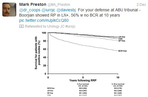
Tzelepi et al (J Clin Oncol 2011 Jun 20;29(18):2574-81) suggested that potentially lethal cancers persist in the primary tumor and may contribute to progression. This is a possible explanation for this study’s findings, which echoed earlier results by Swanson et al (J Urol. 2006 Oct;176: 1292-8) and Shao et al (Eur Urol 2013 May 21. [Epub ahead of print]). However, SEER lacks information regarding the extent of bony metastasis, an entity that undoubtedly influences patient survival. Furthermore, patients treated with RP were 10 yrs younger than the NSR group (62 vs 72), and had a higher proportion of those with PSA <20.

To reduce bias produced by significant comorbidities, authors excluded those dying within a year of diagnosis and found the 5-yr OS continued to be higher in patients undergoing RP (76.5%) or BT (58.2%). However, patients with three or more of: age ≥70 yr, cT4 disease, PSA ≥20 ng/ml, high-grade disease, and pelvic lymphadenopathy had a 5-yr OS survival (38.2%) and a DSS probability (50.1%) similar to NSR patients.

Several contributors identified that Will Rogers phenomenon may be at play

Ultimately, the jury is still out on what is the most effective treatment of significant prostate cancer

Studies (in addition to the follow-on cohort study arising from this review), are underway

To conclude, it has been

In spite of the global participation, much of the banter involved our US urological colleagues.  On this basis, the Best Tweet Prize has been awarded to a provocative tweet from our UK colleague Ben Challacombe (@benchallacombe).

Thank you to European Urology (@EUPlatinum) for allow open access to the article discussed this month.  Thank you to Nature Reviews Urology for supporting the Best Tweet prize, which is a complimentary 12 months on-line subscription to the journal.

We look forward to seeing you at the January #urojc.


Dr Janice Cheng is an Australian Urology Trainee, currently based at Western Hospital. She has an interest in teaching, and enjoys laparoscopies, endoscopies, as well as male/female incontinence management. Twitter @JustUro

Article of the week: Detecting prostate cancer: the “core” of the matter

Every week the Editor-in-Chief selects the Article of the Week from the current issue of BJUI. The abstract is reproduced below and you can click on the button to read the full article, which is freely available to all readers for at least 30 days from the time of this post.

In addition to the article itself, there is an accompanying editorial written by prominent members of the urological community. This blog is intended to provoke comment and discussion and we invite you to use the comment tools at the bottom of each post to join the conversation.

If you only have time to read one article this week, it should be this one.

Can transrectal needle biopsy be optimised to detect nearly all prostate cancer with a volume of ≥0.5 mL? A three-dimensional analysis

Kent Kanao*, James A. Eastham, Peter T. Scardino†‡, Victor E. Reuter*§ and Samson W. Fine*

Departments of *Pathology and Surgery, Urology Service, Memorial Sloan-Kettering Cancer Center, and Departments of Urology and §Pathology, Weill Cornell Medical Center, New York, NY, USA

Read the full article
OBJECTIVE

• To investigate whether transrectal needle biopsy can be optimised to detect nearly all prostate cancer with a tumour volume (TV) of ≥0.5 mL.

MATERIALS AND METHODS

• Retrospectively analysed 109 whole-mounted and entirely submitted radical prostatectomy specimens with prostate cancer.

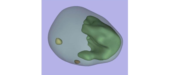
• All tumours in each prostate were outlined on whole-mount slides and digitally scanned to produce tumour maps. Tumour map images were exported to three-dimensional (3D) slicer software (https://www.slicer.org) to develop a 3D-prostate cancer model.

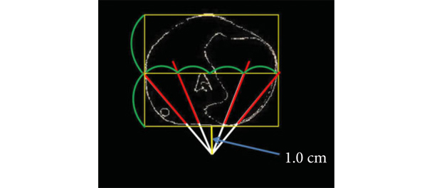
• In all, 20 transrectal biopsy schemes involving two to 40 cores and two to six anteriorly directed biopsy (ADBx) cores (including transition zone, TZ) were simulated, as well as models with various biopsy cutting lengths.

• Detection rates for tumours of different volumes were determined for the various biopsy simulation schemes.

RESULTS

• In 109 prostates, 800 tumours were detected, 90 with a TV of ≥0.5 mL (mean TV 0.24 mL).

• Detection rate for tumours with a TV of ≥0.5 mL plateaued at 77% (69/90) using a 12-core (3 × 4) scheme, standard 17-mm biopsy cutting length without ADBx cores. In all, 20 of 21 (95%) tumours with a TV of ≥0.5 mL not detected by this scheme originated in the anterior peripheral zone or TZ.

• Increasing the biopsy cutting length and depth/number of ADBx cores improved the detection rate for tumours with a TVof ≥0.5 mL in the 12-core scheme.

• Using a 22-mm cutting length and a 12-core scheme with additional volume-adjusted ADBx cores, 100% of ≥0.5 mL tumours in prostates ≤ 50 mL in volume and 94.7% of ≥0.5 mL tumours in prostates > 50 mL in volume were detected.

CONCLUSIONS

• Our 3D-prostate cancer model analysis suggests that nearly all prostate cancers with a TV of ≥0.5 mL can be detected by 14–18 transrectal needle-biopsy cores.

• Using longer biopsy cutting lengths and increasing the depth and number of ADBx cores (including TZ) according to prostate volume are necessary as well.


Read Previous Articles of the Week


Editorial: How many cores are needed to detect nearly all prostate cancers?

Virtual prostate biopsy and biopsy simulation: lessons to be learned

Prostate biopsies, transrectal or transperineal, still constitute the pillars of prostate cancer detection today [1]. With the lack of reliable imaging tools (new MRI techniques are promising but still investigational [2]); random biopsies offer the sole adequate cancer detection option [3]. However, random biopsies are far from efficient in detecting all tumours and even less efficient in detecting all significant cancer ‘spots’. To improve sensitivities and specificities, increasing the biopsy core numbers, targeting more lateral aspects and encouraging repeat biopsies have been recommended [4]. Recently, HistoScanning™ [5] and template biopsies [6] have been introduced to further improve biopsy quality and efficiency. The latest innovations include the fusion of MRI pictures with the TRUS image to offer optimal targeting of suspicious areas [7]. And yet, these efforts are far from solving the main problem. How can we perform a biopsy and be confident to detect most of the cancers, i.e. significant malignant areas.

The present study [1] does, what should have been done a long time ago, namely to create a reliable and reproducible biopsy simulation model to allow the investigation of various biopsy schemes, core lengths and numbers. Based on a series of 109 radical prostatectomy specimens, a three-dimensional (3D) prostate and prostate cancer model was created using novel 3D slicer software and various prostate biopsy schemes were simulated. Using this method, the detection rate for tumours with a tumour volume (TV) of ≥0.5 mL plateaued at 77% (69 of 90) using a 12 core (3 × 4) scheme, standard 17-mm biopsy cutting length without anteriorly directed biopsy (ADBx) cores. Twenty of 21 (95%) tumours with a TV of ≥0.5 mL not detected by this scheme originated in the anterior peripheral zone or transition zone [1].

Confirming our earlier data with the Vienna nomograms [8], increasing the biopsy cutting length and depth/number of ADBx cores (14–18 cores) improved the detection rate for tumours with a TV of ≥0.5 mL in the 12-core scheme [1]. The best biopsy scheme used a 22-mm cutting length and a 12-core scheme with additional volume-adjusted ADBx cores. Using this combination, 100% of ≥0.5 mL tumours in prostates <50 mL in volume and 94.7% of ≥0.5 mL tumours in prostates >50 mL in volume were detected.

Certainly, these numbers will not be reproducible in real-time TRUS or transperineal biopsies (detections rates of 95–100% as seen in this simulation model, cannot be achieved without adequate imaging tools, which are not available yet), but they aid significantly in rethinking our biopsy strategy. So, if we summarise the present findings and combine them with published data, the future will demand a TRUS-fusion biopsy technique, involving 14–18 cores (or more if volume increases), involving the anterior zones of the prostate and using a 22-mm cutting length of the biopsy core vs a 15–17 mm core as is used currently. Obviously real-time prospective trials are needed to confirm these findings but nothing indicates that the outcome would be otherwise.

Bob Djavan
Department of Urology, New York University School of Medicine, NYU, New York, NY, USA

Read the full article

References

  1. Kanao K, Eastham JA, Scardino PT, Reuter VE, Fine SW. Can transrectal needle biopsy be optimised to detect nearly all prostate cancer with a volume of ≥0.5 mL? A three-dimensional analysis. BJU Int 2013; 112: 898–904
  2. Delongchamps NB, Peyromaure M, Schull A et al. Pre-biopsy Magnetic Resonance Imaging and prostate cancer detection: comparison of random and MRI-targeted biopsies using three different techniques of MRI-TRUS image registration. J Urol 2013;189: 493–499
  3. Djavan B, Rocco B. Optimising prostate biopsy. BMJ 2011; 344: d8201
  4. Thompson I, Thrasher JB, Aus G et al. Guideline for the management of clinically localized prostate cancer: 2007 update. J Urol 2007; 177: 2106–2131
  5. Simmons LA, Autier P, Zát’ura F et al. Detection, localisation and characterisation of prostate cancer by prostate HistoScanning(™)BJU Int 2012; 110: 28–35
  6. Huo AS, Hossack T, Symons JL et al. Accuracy of primary systematic template guided transperineal biopsy of the prostate for locating prostate cancer: a comparison with radical prostatectomy specimens. J Urol 2012; 187: 2044–2049
  7. Sonn GA, Natarajan S, Margolis DJ et al. Targeted biopsy in the detection of prostate cancer using an office based magnetic resonance ultrasound fusion device. J Urol 2013; 189: 86–92
  8. Djavan B, Margreiter M. Biopsy standards for detection of prostate cancer. World J Urol 2007; 25: 11–17

Single-port transvesical LRP for organ-confined prostate cancer

Click here for the extended video.

Single-port transvesical laparoscopic radical prostatectomy for organ-confined prostate cancer: technique and outcomes

Xin Gao, Jun Pang, Jie Si-tu, Yun Luo, Hao Zhang, Liao-yuan Li and Yan Zhang

Department of Urology, The Third Affiliated Hospital, Sun Yat-sen University, Guangzhou, China
X. G. and J. P. contributed equally to this work.

Read the full article
OBJECTIVE

• To report a novel technique for performing single-port transvesical laparoscopic radical prostatectomy (STLRP) and to evaluate the oncological and functional outcomes in 16 patients with organ-confined prostate cancer.

PATIENTS AND METHODS

• In total, 16 consecutive patients with clinical stage T1-2aN0M0 were scheduled for STLRP, and their continence and erectile status were investigated preoperatively.

• The patients’ mean age was 62 years, mean prostate volume 42 mL and mean prostate-specific antigen (PSA) 7.5 ng/mL.

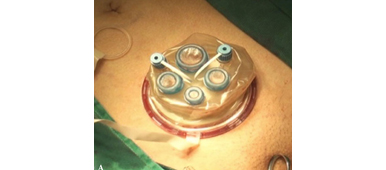
• The STLRP procedures were performed by a single surgeon, and all the operating procedures were conducted transvesically and laparoscopically.

• Intra-operative and postoperative complications, assessed according to the modified Clavien system, were recorded and peri-operative and functional outcome data were analysed.

• All patients were followed up for a minimum of 12 months postoperatively through PSA detection, daily pads, the International Index of Erectile Function (IIEF)-6 score and urography.

RESULTS

• All of the 16 STLRP procedures were successfully completed. The mean (range) operation duration was 105 (75–180) min, and the mean (range) estimated blood loss was 130 (75–500) mL. No patients had positive surgical margins. Postoperative complications occurred in five patients, including three cases of urinary infection and two cases of haematuria (grade II). Catheters were removed after a mean (range) time of 11.2 (9–14) days with cystography. The mean (range) hospital stay was 12.7 (10–15) days.

• Of the 16 patients, 13 were immediately continent (0 pads/day), and three had mild incontinence (2–3 pads/day) after catheter removal. All patients were observed as continent 3 months postoperatively.

• In total, 10/16 and 12/16 patients achieved a satisfactory erection at 6 and 12 months follow-up postoperatively, respectively, with an IIEF-6 score ≥ 18.

• The mean postoperative PSA levels at 3, 6 and 12 months were 0.015 ng/mL, 0.017 ng/mL and 0.016 ng/mL, respectively. No patients were identified with biochemical recurrence in this series. No patients demonstrated vesico-urethral stricture during follow-up for 12–24 months.

CONCLUSION

• We conclude that STLRP is technically feasible for patients with low-risk organ-confined prostate cancer and demonstrates promising functional outcomes regarding continence and potency.

Movember: The power of the Mo!

The word Mo is Australasian slang for a moustache and whilst not a northern hemisphere phrase this hasn't prevented rapid dissemination across the globe. Although originally an innovation solely in Australia and New Zealand for its first 6-7 years, Movember is now taking the world by storm with the UK and Canada leading the way. Staggeringly last year in the UK more than 363,000 men grew a hairy upper lip and in doing so raised over £27 million.

The Movember foundation donates the proceeds to men's health charities which is primarily (around 70%) prostate cancer but also donates to charities supporting mental illness and this year will contribute to the orchid trust for testicular cancer. Money raised from the UK campaign goes to Prostate Cancer UK, which received £14.6m for the year to April 2013, and the Institute of Cancer Research, which received £299,891 for the same period.

When working in urology clinics where one meets up to 20 new patients a day that's many conversations about this issue helping to raise awareness and hopefully directing people to the website to donate to the UK Movember site.

Prokar Dasgupta outside the BJUI offices in Movember 2012


So Mo brothers and sistas let’s keep up the good work and prepare for another bumper crop of upper lip hair! 

Ben Challacombe
Associate Editor, BJUI 

Movember and the Importance of Patient Advocacy

In October 2009 the resident on my service was Dr Dean Elterman. I have had many residents and fellows over the years and have always felt that as much as they hopefully learn something from me I probably learn more from my time with them. The concept of ‘drilling down’ to make lasting connections with leaders of the next generation is not something that is always intuitively grasped in the hierarchy of surgical life. As it was, in late October of that year Dean mentioned Movember to me and asked whether I would like to participate. At that point, not knowing what he was talking about I proceeded to tell him to consult his spell-check. Having once before sported facial hair in my early 20s to very little acclaim I had not entertained the thought since. My immediate reaction was dismissive. Nevertheless after some further discussion it became obvious to me that the whole concept of Movember is not simply to raise money for men’s health and prostate cancer research but to generally shine a brighter light on the nature of the disease, the work we do as urologists and to start a dialogue. This grassroots movement, started in 2003 in Australia by Adam Garrone has quickly grown into a worldwide phenomenon. That fall I anchored Dean’s team of residents and we broke into the top 20 of small teams worldwide.

Last year I set up a local team at Toronto East General Hospital with tremendous success. On an individual level I raised $46,000 in support of men’s health, the seventh highest individual total worldwide. While that certainly was nice, as the month wore on what became increasingly clear to me was the larger role that my involvement in Movember had created in engaging patients, other healthcare providers and society at large. The quirky nature of the campaign lends itself to a fun, easy discussion about an important topic. Having a dialogue around prostate cancer including how to screen as well as when and when not to treat is very important. The significant emotional and physical consequences of treatment deserve attention. A particularly great example by the terrific @docmikeevans illustrates the space that Movember now inhabits. The role that urologists in particular have as advocates of men’s’ health is very clear. 

It is with this last thought in mind that I call upon my colleagues in Canada and around the world to take up the charge. In recent years, much of the progress that we have made in treating prostate cancer is at risk of being undermined. The confusing and rather opaque nature of screening guidelines have increasingly promoted prostate cancer as an indolent disease not worth having a discussion about. I certainly have previously written about this and recently a group of experts met in Melbourne and attempted to better make sense of screening and stratify risk. Prostate Cancer Canada, an important advocacy group in Canada has also done a great job this fall with their #knowyournumber campaign. I was proud to be a part of it. Their CEO Rocco Rossi has embarked upon a terrific campaign of support by walking the Camino to Santiago de Compostella this month. All leaders must actively embrace the role of advocacy for our patients. Movember to me is a great vehicle for this. Will you look silly and unprofessional in the clinic during Movember? Absolutely not. In reality, every patient in the clinic is immediately reassured that their urologist walks along beside them, although perhaps not as far as Rocco. 

It is in this context that I would call on all of my urological colleagues to stop shaving in Movember, start a team, create a network and share this experience with our brave patients and their partners for a month. The amount raised is really secondary. Having that visible presence is crucial. With epidemiologists, policy makers and many others expressing expert opinions about a disease that we treat every day don’t you think we should also embrace that role? Movember is the forum where the most important group, our patients, will be having that conversation for a month. Join them. Simply caring for them after diagnosis or waiting for a research grant to materialize is not good enough. My female colleagues can join as ‘mo-sistas’. You can certainly follow my ‘progress’ and support my venture as well. I look forward to seeing my colleagues from around the world and the self-described #urotwitterati that contributes regularly on #urojc in particular to join in the fun. I expect to be pushed on the leaderboard.

Dr Rajiv Singal is a Urologist at Toronto East General Hospital and an Assistant Professor in the Department of Surgery at the University of Toronto

Follow him on Twitter at @DrRKSingal

A Rather Nasty Surprise

Recently, I encountered, and indeed I actually caused, a complication of robot-assisted radical prostatectomy (RARP) which was new to me, and one which I felt that I should share with other surgeons.

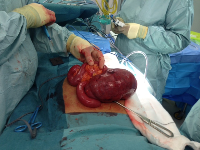
PM, a 60-year old teacher, underwent a completely routine RARP, which took less than 2 hours to perform on a Saturday morning. During Sunday night he developed severe abdominal pain and distension. By Monday morning he was in distress with rebound tenderness and marked tachycardia. A CT scan was requested, which revealed a caecal volvulus. A laparotomy by a general surgeon confirmed the diagnosis and an urgent right hemicolectomy was undertaken. The patient made an uneventful recovery and, I am pleased to say, is still speaking to me. Histology confirmed an ischaemic caecum twisted on its rather thickened mesentery, with no perforation present. The prostate itself contained a Gleason 3+4=7 adenocarcinoma, without evidence of extra-prostatic extension.

Although robotic assistance provides the benefits of very precise, virtually bloodless surgery, with 10 times magnification and 3D vision, it also carries the risk of a specific set of complications. These need to be dealt with promptly and efficiently and can usually be completely resolved. Failure to recognise post-operative problems, such as bowel injury, intra-abdominal bleeding or port-site hernia, however, can place the patient in severe and increasing jeopardy. We recently published an article in the BJUI entitled “Lessons Learned from 1000 robot-assisted radical prostatectomy” in which we discussed how many of the problems could be avoided, and, if they occur how they can be best dealt with. One key message is the importance of an early CT scan to diagnose the nature of a post-operative problem, rather than crossing fingers and hoping things will settle.

I am hoping that this blog, and the BJUI article mentioned above, will stimulate other surgeons to discuss openly and frankly the problems that they themselves have encountered, either with regular laparoscopy or with the da Vinci robot, and how they dealt with them. Learning the lessons, not only from one’s own errors and omissions, but also from those of others, seems the best way to become, and continue to be, a safe and successful surgeon.  


Roger Kirby, The Prostate Centre, London

Article of the week: Prostate biopsy: shaking up the old standard

Every week the Editor-in-Chief selects the Article of the Week from the current issue of BJUI. The abstract is reproduced below and you can click on the button to read the full article, which is freely available to all readers for at least 30 days from the time of this post.

In addition to the article itself, there is an accompanying editorial written by a prominent member of the urological community. This blog is intended to provoke comment and discussion and we invite you to use the comment tools at the bottom of each post to join the conversation.

Finally, the third post under the Article of the Week heading on the homepage will consist of additional material or media. This week we feature a video of Dr Symons discussing his paper.

If you only have time to read one article this week, it should be this one.

Outcomes of transperineal template-guided prostate biopsy in 409 patients

James L. Symons*, Andrew Huo*, Carlo L. Yuen‡§, Anne-Maree Haynes*, Jayne Matthews, Robert L. Sutherland*, Phillip Brenner‡§ and Phillip D. Stricker†‡§

*Cancer Research Programme, Garvan Institute of Medical Research, St Vincent’s Prostate Cancer Centre, Department of Urology, St. Vincent’s Hospital, and §Department of Urology, St. Vincent’s Clinic, Darlinghurst, NSW, Australia

Read the full article
OBJECTIVE

• To present the template-guided transperineal prostate biopsy (TPB) outcomes for patients of two urologists from a single institution.

PATIENTS AND METHODS

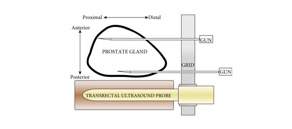
• We conducted a prospective study of 409 consecutive men who underwent TPB between December 2006 and June 2008 in a tertiary referral centre using a standardized 14-region technique.

• The procedure was performed as day surgery under general anaesthesia with fluoroquinolone antibiotic cover.

• Follow-up took place within 2 weeks, during which time men were interviewed using a standardized template.

• Results were compared with those of the Australian national prostate biopsy audits performed by the Urological Society of Australia and New Zealand (USANZ).

RESULTS

• Indications for biopsy included elevated prostate-specific antigen (PSA) level (75%), with a median PSA level of 6.5 ng/mL, abnormal digital rectal examination (8%) and active surveillance (AS) re-staging (18%).

• The mean patient age was 63 years and two-thirds of patients were undergoing their first biopsy.

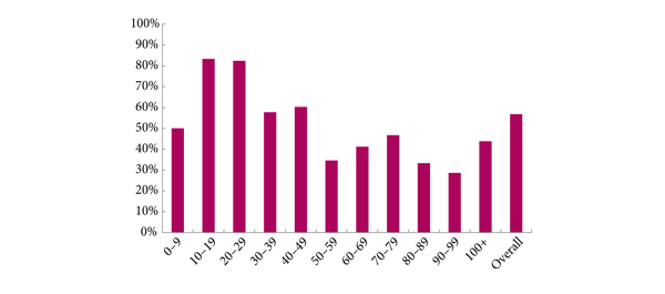
• A positive biopsy was found in 232 men, 74% of whom had a Gleason score of ≥7. The overall cancer detection rate was 56.7% (USANZ 2005 national audit = 56.5%). Stratified between those having their first TPB or a repeat procedure (after a previous negative biopsy), the detection rates were 64.4 and 35.6%, respectively. Significantly higher detection rates were found in prostates <50 mL in volume than in larger prostates (65.2 vs 38.3%, respectively, P < 0.001).

• Haematuria was the most common side effect (51.7%). Others included dysuria (16.4%), acute urinary retention (4.2%) and fever (3.2%). One patient (0.2%) had septicaemia requiring i.v. antibiotics.

• Repeat biopsy was not associated with increased complication rates.

CONCLUSIONS

• TPB is a safe and efficacious technique, with a cancer detection rate of 56.7% in the present series, and a low incidence of major side effects. Stratified by prostate volume, the detection rate of TPB was higher in smaller glands.

• Given the relatively low rate of serious complications, clinicians could consider increasing the number of TPB biopsy cores in larger prostates as a strategy to improve cancer detection within this group. Conversely, in patients on AS programmes, a staging TPB may be a superior approach for patients undergoing repeat biopsy so as to minimize their risk of serious infection.

Editorial: Contemplate the template: a new prostate biopsy approach

Transperineal magnetic resonance imaging – ultrasound fusion targeted biopsies (MRI-US FTB) of the prostate: the future of prostate diagnostics

The prostate cancer diagnostic pathway has remained unchanged for 25 years. At best, laterally directed, peripheral zone (PZ) 12-core transrectal biopsies identify cancer in 44% of cases [1] but transrectal biopsies have an inherent sampling error with a risk of misdiagnosis or mischaracterisation of disease. Of those with negative biopsies who undergo transperineal (TP) biopsies, 30% have cancer, most in the anterior PZ. Active surveillance and the promise of less invasive treatment options are becoming popular because of concerns about ‘over treatment’ for low-risk disease.

Saturation transrectal biopsies have been advocated to improve diagnostic yield but do not address the issue of under sampling of the anterior PZ, particularly in the larger gland [2]. TP biopsies can be used to address the issue of under sampling but prostate template-mapping biopsies are labour intensive and require large numbers of biopsies, often between 60 to 90 cores; however, they have been an essential component of focal therapy trials and the evaluation of novel treatment methods [3].

Primary TP biopsy is the subject of the paper published in this edition of the BJUI titled ‘Outcomes of transperineal template-guided prostate biopsy in 409 patients’ [4]. The authors report a single centre experience of primary TP biopsies. The 14-region protocol described is simpler than prostate template-mapping requiring fewer cores (median of 15 and mean of 19 cores) with a comparable primary diagnostic detection rate of 60% and an encouraging side-effect profile. Unfortunately, the approach still has limitations and the authors admit that their limited biopsy protocol may still mischaracterise disease in the larger gland. In a recent paper from the same group, there was a disappointing correlation between their TP biopsy pathology, MRI abnormalities and radical prostatectomy specimens [5]. Uncertainty prevails, the problem is how best to sample the larger gland. The authors [4] and others, often conclude that more biopsies are necessary for larger glands and resort to mapping protocols and many more biopsies. The solution may not be more biopsies but rather better systematic targeting of the PZ. The impact of hyperplasia within the transition zone (TZ) has a profound effect on PZ anatomy. In the smaller prostate, up to 30 mL, there is little TZ and the PZ is much thicker posteriorly than anteriorly, this difference is even more apparent in glands of 30–50 mL. Above 50 mL TZ expansion causes marked attenuation of the PZ, which becomes much thinner, but the overall volume of the PZ does not change. Less than 4% of cancers originate in the TZ [6], consequently biopsies should be concentrated primarily on the PZ.

The future of prostate cancer diagnosis is likely to be a combination of pre-biopsy multiparametric MRI, followed by targeted biopsies of MRI-identified lesions combined with fewer but better systematic targeted biopsies of the PZ. MRI-ultrasound (MRI-US) fusion techniques have been developed in which axial T2 images of the prostate, diffusion-weighted images and/or dynamic contrast-enhanced MRI images are ‘fused’ with the live US images to allow precise targeting of both regions of interest and the PZ. Commercially available biopsy programs, developed from brachytherapy software systems programs allow individual biopsy sites to be recorded and if combined with inking of the specimen can provide precise pathological localisation of disease within the prostate [7].

There are many potential benefits to this approach. Patients who opt for active surveillance will have an archived record of their disease at a given time to facilitate precise replication of further interval biopsies and assess progression. Improved disease management for an individual should be the aim. The suitability or not for focal or targeted therapies, the planning or boosting of identifed lesions with radiotherapy and/or brachytherapy, and the planning of nerve-sparing surgery or wide excisions should be possible. Feedback to the radiologists of both benign and malignant pathology and grade of disease will improve reporting accuracy and provide imaging sciences with the histopathological characteristics of both MRI ‘visible’ and ‘invisible’ cancer to improve MRI interpretation.

MRI–US fusion targeted biopsies are a significant advance in prostate diagnostics and may resolve some uncertainty within the prostate cancer diagnostic pathway. Benefit vs cost is a recurring issue across health care and questions will continue to be asked about the use of increasingly expensive technology in such an indolent disease. The challenge for investigators will be how to prove the benefit of this approach over standard biopsy protocols and integrate this work in to clinical practice.

Richard Popert
Department of Urology, Guy’s Hospital, London, UK

Read the full article
References
  1. Presti JC, O’Dowd GL, Miller MC et al. Extended peripheral zone biopsy schemes increase cancer detection rates and minimize variance in prostate specific antigen and age related cancer rates: results of a community multi-practice study. J Urol 2003; 169:125–129
  2. Stewart CS, Leibovich BC, Weaver AL, Lieber MM. Prostate cancer diagnosis using a saturation needle biopsy technique after previous negative sextant biopsies. J Urol 2001; 166: 86–92
  3. Onik G, Barzell W. Transperineal 3D mapping biopsy of the prostate: an essential tool in selecting patients for focal prostate cancer therapy. Urol Oncol 2008; 26: 506–510
  4. Symons JL, Huo A, Yuen CL et al. Outcomes of transperineal template-guided prostate biopsy in 409 patients. BJU Int 2013; 112: 585–593
  5. Huo AS, Hossack T, Symons JL et al. Accuracy of primary systematic template guided transperineal biopsy of the prostate for locating prostate cancer: a comparison with radical prostatectomy specimens. J Urol 2012; 187: 2044–2050
  6. Patel V, Merrick GS, Allen ZA et al. The incidence of transition zone prostate cancer diagnosed by transperineal template guided mapping biopsy: implications for treatment planning. Urology 2011; 77: 1148–1152
  7. Hadaschik BA, Kuru TH, Tulea C et al. A novel stereotactic prostate biopsy system integrating pre-interventional magnetic resonance imaging and live ultrasound fusion. J Urol 2011; 186: 2214–2220

Video: Transperineal prostate biopsy: how good is the tumour detection rate?

Outcomes of transperineal template-guided prostate biopsy in 409 patients

James L. Symons*, Andrew Huo*, Carlo L. Yuen‡§, Anne-Maree Haynes*, Jayne Matthews, Robert L. Sutherland*, Phillip Brenner‡§ and Phillip D. Stricker†‡§

*Cancer Research Programme, Garvan Institute of Medical Research, St Vincent’s Prostate Cancer Centre, Department of Urology, St. Vincent’s Hospital, and §Department of Urology, St. Vincent’s Clinic, Darlinghurst, NSW, Australia

Read the full article
OBJECTIVE

• To present the template-guided transperineal prostate biopsy (TPB) outcomes for patients of two urologists from a single institution.

PATIENTS AND METHODS

• We conducted a prospective study of 409 consecutive men who underwent TPB between December 2006 and June 2008 in a tertiary referral centre using a standardized 14-region technique.

• The procedure was performed as day surgery under general anaesthesia with fluoroquinolone antibiotic cover.

• Follow-up took place within 2 weeks, during which time men were interviewed using a standardized template.

• Results were compared with those of the Australian national prostate biopsy audits performed by the Urological Society of Australia and New Zealand (USANZ).

RESULTS

• Indications for biopsy included elevated prostate-specific antigen (PSA) level (75%), with a median PSA level of 6.5 ng/mL, abnormal digital rectal examination (8%) and active surveillance (AS) re-staging (18%).

• The mean patient age was 63 years and two-thirds of patients were undergoing their first biopsy.

• A positive biopsy was found in 232 men, 74% of whom had a Gleason score of ≥7. The overall cancer detection rate was 56.7% (USANZ 2005 national audit = 56.5%). Stratified between those having their first TPB or a repeat procedure (after a previous negative biopsy), the detection rates were 64.4 and 35.6%, respectively. Significantly higher detection rates were found in prostates <50 mL in volume than in larger prostates (65.2 vs 38.3%, respectively, P < 0.001).

• Haematuria was the most common side effect (51.7%). Others included dysuria (16.4%), acute urinary retention (4.2%) and fever (3.2%). One patient (0.2%) had septicaemia requiring i.v. antibiotics.

• Repeat biopsy was not associated with increased complication rates.

CONCLUSIONS

• TPB is a safe and efficacious technique, with a cancer detection rate of 56.7% in the present series, and a low incidence of major side effects. Stratified by prostate volume, the detection rate of TPB was higher in smaller glands.

• Given the relatively low rate of serious complications, clinicians could consider increasing the number of TPB biopsy cores in larger prostates as a strategy to improve cancer detection within this group. Conversely, in patients on AS programmes, a staging TPB may be a superior approach for patients undergoing repeat biopsy so as to minimize their risk of serious infection.

© 2024 BJU International. All Rights Reserved.