Archive for category: Case Studies

Primary Follicular Lymphoma of the Bladder: A Case Report and Review of the Literature

Primary non-Hodgkin lymphoma of the bladder (PNHLB) is extremely rare with only 110 cases identified in the medical literature.

Authors: Roberts, Samuel; Nagonkar, Santoshi; Zardawi, Ibrahim M.
Manning Rural Referral Hospital, Taree, NSW, Australia
Corresponding Author: Dr Samuel Roberts 84 Scenic Drive, Merewether, NSW 2291 Australia [email protected]


Introduction
Primary non-Hodgkin lymphoma of the bladder (PNHLB) is extremely rare with only 110 cases identified in the medical literature [1,2]. The disease has a median age distribution of 64–69 years and shows a female preponderance with a female to male ratio of 2.7:1 [3–5]. Chronic inflammation has been suggested as a possible aetiological factor and a history of chronic cystitis has been documented in over 20% of patients [4–6]. We describe a case of PNHLB and review the literature.

Case Report
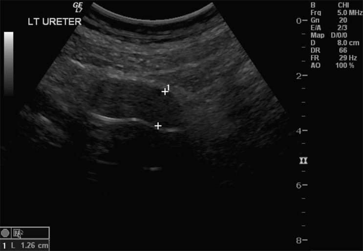
A 42-year-old, healthy mother of two presented with a 4 month history of haematuria and dysuria. Antibiotic management offered by her general practitioner initially resolved the symptoms but they later recurred. A pelvic ultrasound revealed an 8 cm mass arising from the bladder base. She had lost approximately 6 kg over 6 months and also complained of lethargy, although there was no history of night sweats or fever. There was also no history of chronic cystitis. She worked as a childcare worker and denied smoking or drinking alcohol; though she was exposed to passive smoking during childhood. She gave a family history of bowel cancer. Her initial examination revealed tenderness and a vague mass in the supra-pubic region, but was otherwise unremarkable.

Investigations
Preliminary blood results were within normal limits, apart from mild iron deficiency anaemia.
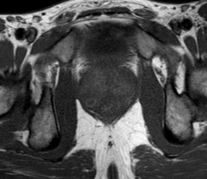
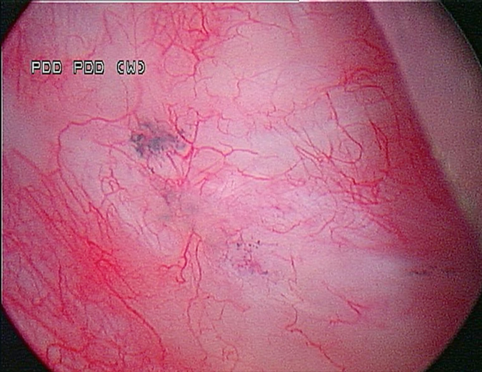
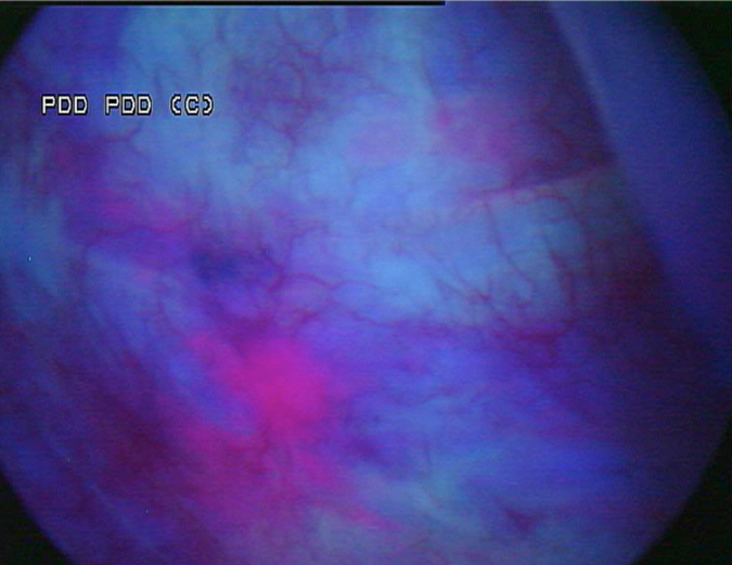
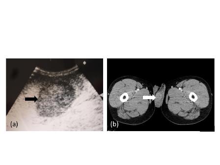
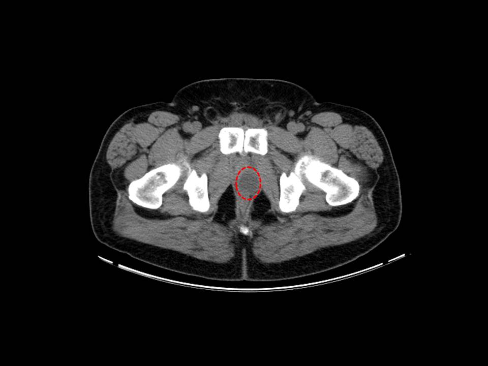
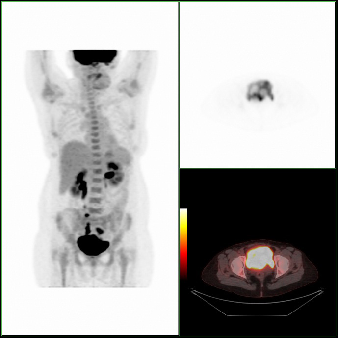
A Cystoscopy revealed a large solid tumour occupying the entire right lateral and posterior walls of the bladder. Loop resection was attempted but abandoned part way through due to the very large size of the tumour. A post-cystoscopy staging contrast enhanced computed tomography (CT) scan confirmed a 12 x 8.5 x 9 cm mass, with an aortocaval lymph node at the upper size limit of normal. Positron emission tomography (PET) scan showed no radiolabelled glucose uptake in this node and did not identify any disease outside the bladder. The bladder mass showed less uptake than adjacent urine but increased uptake in comparison with background (Fig 1).

12-076-Figure_1x

Pathology
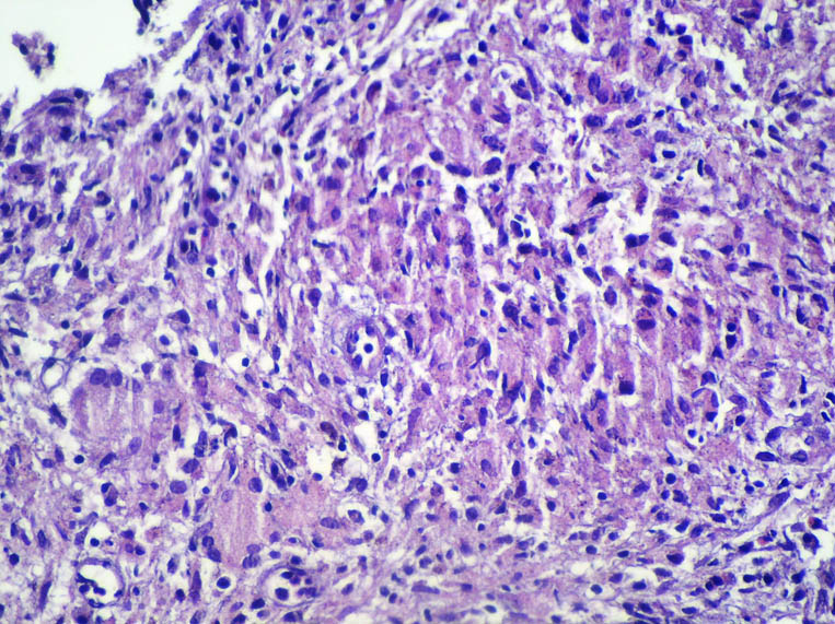
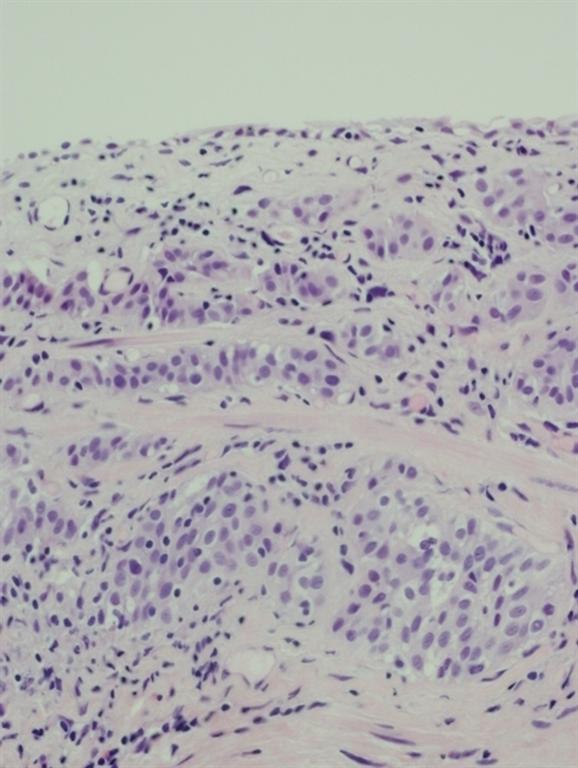
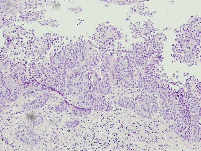
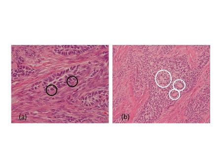
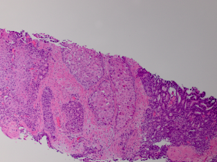
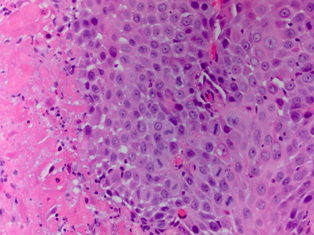
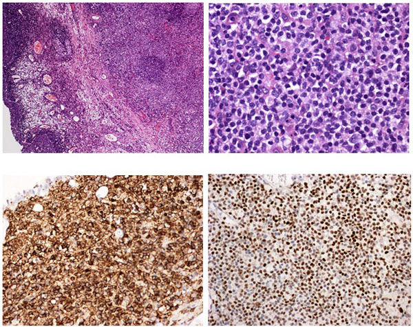
Pathology revealed a WHO grade 2 follicle centre cell lymphoma expressing CD20, CD10, BCL-2 and BCL-6 (Fig 2). CD5 and CD23 were negative.

12-076-Figure_2

Management
The patient received five cycles of cyclophosphamide, doxorubacin, vincristine, prednisone and rituximab (R-CHOP), which was complicated by two episodes of febrile neutropenia. She responded well with complete remission of the bladder mass on cystoscopy 3 months after the completion of treatment. The bladder mucosa at this time had a polypoid appearance that was shown on biopsy to be oedematous and inflamed with no evidence of neoplasia. The patient remained well with no evidence of local or systemic disease. She is currently being maintained on third monthly rituximab, which will continue for at least 2 years.

Discussion
Primary non-Hodgkin lymphoma of the bladder (PNHLB) represents less than 1% of all bladder neoplasms and between 0.15% and 0.2% of all extra-nodal lymphomas [7]. This rarity is thought to reflect the absence of organized lymphoid tissue in the bladder wall [1,2]. There is female preponderance with a female to male ratio of 2.7:1 [4], and some authors suggest that this reflects an aetiological role of chronic inflammation; with chronic cystitis demonstrated in over 20% of patients [4–6]. Range of onset is 20–85 years with a median of 64–69 years [4,5].
Most cases were published before 1999 and are described in terms of the older “working classification” for lymphoma, with immunohistochemistry performed in less than 20% of cases [8]. In a review of 100 cases; all but three were B-cell lymphomas, with 64% comprising low grade lymphoma [4]. Extra-nodal marginal zone/mucosa associated lymphoid tissue (MALT) type represent 52% of cases [1,4]. Primary Hodgkin lymphoma and T-cell lymphoma are exceedingly rare. Only 20–30% of reported cases represent high grade disease [4,5,8].
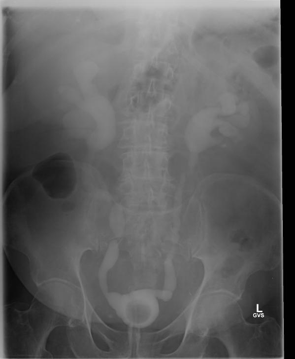
Haematuria is the most common presenting symptom (61%) followed by nocturia, dysuria and loin pain [4,8]. Intravenous urography reveals a filling defect in the bladder, while ultrasound displays a solid homogeneous mass [2]. CT scan exposes a contrast-enhancing soft tissue density either as a sessile solitary mass (66%), multiple sessile masses (14%) or as a polypoid mass [2]. Only one case was identified that described PET findings of PNHLB, in which a diffuse large B-cell lymphoma showed a clearly delineated hyper-metabolic mass [9]. Cystoscopy findings are most commonly of a solitary mass, followed by multiple masses and occasionally a diffuse lesion with nodule formation, with the lateral walls forming the most common site (40%) [1,2,5,8]. The lesion is usually rounded with overlying intact mucosa that may be oedematous, friable, haemorrhagic or ulcerated [3,10,11]. The definitive diagnosis must be histological [11,12].
The treatment of non-Hodgkin’s lymphoma at other sites is heterogeneous and complex, and detailed discussion is beyond the scope of this article. It is essentially based upon histological classification, clinical stage and patient factors [13]. Bladder lymphoma is very treatable; with one review showing death from tumour in only three of 27 patients, and complete remission (CR) in the remaining 24 [5]. The most complete review of case reports found no difference in effectiveness between the three major treatment modalities (surgery, radiotherapy and chemotherapy) in low- or high-grade PNHLB [4]. For low-grade lymphoma; CR was achieved in 95% of patients treated with radiotherapy, 100% of those treated with surgery and 100% of those treated with chemotherapy [4]. For those with high-grade disease, CR was achieved in 72% overall, with systemic chemotherapy used in 60% of cases. CHOP and R-CHOP are the most widely employed chemotherapy regimens in the literature [4]. Two case reports describe complete remission of MALT type lymphoma with antibiotic therapy [4]. Rituximab alone may be effective in MALT lymphoma of the bladder as this has been successful in other sites [13,14]. Some authors recommend chemotherapy as the preferred treatment modality as it is less invasive than surgery or radiotherapy and has the theoretical benefit of treating undiagnosed areas of systemic spread [5,15]. This treatment regimen may not be well tolerated especially in elderly patients, and as such it must be made clear that there is no evidence to recommend any treatment over another, regardless of disease grade [15]. At other sites of extra-nodal lymphoma; less invasive treatments such as rituximab alone or localized radiation may be used as first-line therapy for low grade disease [13]. This may also be an appropriate strategy for bladder lymphoma, but at this stage there is insufficient evidence to make any recommendations.
Only four reports in English clearly describe primary follicular lymphoma of the bladder [3,5,15]. All four cases presented with haematuria. No cases describe the radiological findings. Three cases presented as solid masses and one as multiple sessile tumours. Radiotherapy with or without surgery successfully treated all four cases [3].
Conclusion
In summary, PNHLB is an exceedingly rare condition that is difficult to diagnose based on clinical or radiological findings and as such diagnosis must be histological. There is insufficient evidence to recommend a particular treatment over any other. As in the management of lymphoma elsewhere, treatment should be tailored to individual patient and disease factors.
We present what is, to our knowledge, the first case of follicular lymphoma of the bladder treated with chemotherapy, as well as the first published images of combined PET/CT of primary lymphoma of the bladder.

References
1. Taheri M, Dighe M, Kolokythas O, True L, Bush W. Multifaceted Genitourinary Lymphoma. Current Problems in Diagnostic Radiology. 2008;37(2):80-93.
2. Tasu JP, Geffroy D, Rocher L, Eschwege P, Strohl D, Benoit G, et al. Primary Malignant Lymphoma of the Urinary Bladder: report of three cases and review of the literature. European Radiology. 2000;10:1261-4.
3. Bhansali SK, Cameron KM. Primary Malignant Lymphoma of the Bladder. British Journal of Urology. 1960;32:440-54.
4. Hughes M, Morrison A, Jackson R. Primary bladder lymphoma: management and outcome of 12 patients with a review of the literature. Leukemia and Lymphoma. 2005;46(6):873-7.
5. Oshawa M, Aozasa K, Horiuchi K, Kanamaru A. Malignant Lymphoma of Bladder: Report of Three Cases and Review of the Literature. Cancer. 1993;72(6):1969-74.
6. Suzuki T, Matsumura T, Oto I. Intravesical Mass consisting of mucosa-associated lymphoid tissue. International Journal of Urology. 2004;11:1028-30.
7. Freeman C, Berg JW, Cutler SJ. Occurence and Prognosis of Extranodal Lymphomas. Cancer. 1972;29:252-60.
8. Fernandez Acenero MJ, Martin Rodilla C, Lopez Garcia-Asenjo J, Menchero C, Sanz Esponera J. Primary Malignant Lymphoma of the Bladder. Pathology, Research and Practice. 1996;192:160-3.
9. Mantzarides M, Papathanassiou D, Bonardel G, Soret M, Gontier E, Foehrenbach H. High-Grade Lymphoma of the Bladder Visualised on PET. Clinical Nuclear Medicine. 2005;30(7):478-80.
10. Downs TM, Kibel AS, DeWolf WC. Primary Lymphoma of the Bladder: A Unique Cystoscopic Appearence. Urology. 1997;49:276-8.
11. Davidson N. Primary Non-Hodgkin Lymphoma of the Bladder. Scandanvian Journal of Urology and Nephrology. 1990;24:155-56.
12. Yeoman LJ, Mason MD, Olliff JF. Non-Hodgkin’s Lymphoma of the Bladder: CT and MRI Appearences. Clinical Radiology. 1991;44(6):389-92.
13. Kobrinsky B, Hymes KB. Non-Hodgkin’s Lymphoma. [Internet] London: BMJ Publishing Group; 2012 [updated February 20 2012; cited 5th October 2012]; Available from: Best Practice.
14. Shetty RK, Adams BH, Tun HW, Runyan BR, Menke DM, Broderick DF. Use of Rituximab for Periocular and Intraocular Mucosa-associated Lymphoid Tissue Lymphoma. Ocular Immunology and Inflammation. 2010;18(2):110-2.
15. Heany J, Delellis R, Rudders R. Non -Hodgkin Lymphoma arising in the Lower Urinary Tract. Urology. 1985;25(5):479-84.


Date added to bjui.org: 04/11/2013

DOI: 10.1002/BJUIw-2012-076-web


An unusual case of granulomatous prostatitis with caseification necrosis: Primary Tuberculous Prostatitis in a young male

Primary prostatic tuberculosis is a very rare form of the tuberculosis infection. Isolated prostate tuberculosis is generally seen in immunocompromised patients and in those of middle or advanced age. We report a case of isolated prostatic tuberculosis in a young, healthy and immunocompetent patient.

Authors:

Inci, Mehmet; Yula, Erkan; Inci, Melek; Rifaioglu, Murat; Ozgur, Tumay; Davran, Ramazan; Erden, Ersin
Corresponding Author: Mehmet Inci. Mustafa Kemal University Faculty of Medicine – Department of Urology, Hatay, Turkey

INTRODUCTION
The genitourinary system is the most frequently affected region of the body in extra-pulmonary tuberculosis. The kidneys, ureters, bladder or genital organs are usually involved [1] Tuberculosis of the prostate has mainly been described in immunocompromised patients [2-4]. Tuberculosis is rare in the prostate and it is coincidentally diagnosed during pathological evaluation after transurethral resection in many cases [5]. The most commonly involved pathogen is Mycobacterium tuberculosis. It is thought that involvement of the prostate with tuberculosis usually results from hematogenous spread, although it may occur due to organisms descending from kidneys or local spread from the genital tract [1, 6].

CASE
A 22-year old man who had been diagnosed as having acute prostatitis and received quinolone for one month without improvement in another facility was referred to the Medical Center of Mustafa Kemal University, Medicine School with features of prostatism and lower urinary tract symptoms. On digital rectal examination (DRE), the prostate was found to be enlarged, hard, irregular and nodular. It was tender to palpation. The patient was not known to have any other medical problems. There were no abnormalities on physical examination.  Blood tests revealed a white cell count of 6200/mm3, hematocrit of 43% and an erythrocyte sedimentation rate of 63 mm/h. The patient’s serum total prostate-specific antigen (tPSA) level was elevated and found to be 7.867 ng/ml. On urinalysis, there was no finding other than pyuria. Direct urinary system radiography was normal. On the magnetic resonance imaging (MRI), there was low signal intensity at the peripheral zone of prostate in T1-weighted sequences, (Figure 1). Transrectal ultrasound (TRUS) examination was performed with a Sonoline Elegra Scanner (Siemens Ultrasound Division, Mountain View, CA) equipped with a 6.5-MHz endorectal probe. TRUS showed a markedly enlarged prostate, with a heterogeneous and hypoechoic appearance to the parenchyma and enlargement of the seminal vesicles. TRUS guided systematic biopsies of the prostate were performed after sonographic examination. The specimens were diagnosed histopathologically as granulomatous prostatitis, with granuloma consisting of areas of focal caseating necrosis, Langhans giant cells and epitheloid histocytes (Figure 2). We also performed intravenous urography. There were no pathological findings seen in the relevant structures of the urinary tract. Both HIV-antibody tests and the tuberculin skin test were negative. No pathological findings were seen on chest X-ray and CT of the thorax.  Urine samples were taken over consecutive 3 days. Ehrlich-Ziehl-Neelsen (EZN) staining and culture was performed on these samples. No acid fast bacilli were observed on EZN staining. No growth was detected after incubation on Middlebrook agar (BacT/Alert 3D system; BacT/ALERT MP Bottle supplemented with Middlebrook 7H9 Broth) and Lowenstein-Jensen medium for eight weeks. In addition; prostate secretions were collected by massage. No microorganisms were seen in this sample on Gram staining, while 5-7 leukocytes were seen in each field (x40 magnification) on Giemsa staining. In addition, acid fast bacilli were seen in EZN staining, but no growth was detected on culture of the prostate secretion on Middlebrook agar and Lowenstein-Jensen medium for 8 weeks. Diagnosis of prostatic tuberculosis was made based on the pathology results and acid fast bacilli positivity. The patient was treated with isoniazid, rifampicin, pyrazimanide and ethambutol for the initial two months; and he received isoniazid and rifampicin for a further four months.  His clinical signs improved after treatment. Control MR imaging showed that prostate returned to its normal size after therapy and his tPSA level reduced to 0.8 ng/ml.

052-Figure1

Figure 1

OLYMPUS DIGITAL CAMERA

Figure 2

DISCUSSION
Tuberculosis prostatitis is an uncommon benign inflammatory disease of the prostate that rarely presents clinically in urological practice. Recognizing this condition is important, due to the possibility of it being misdiagnosed as prostatic carcinoma, both on clinical examination and TRUS [7, 8]. The diagnosis of tuberculosis prostatitis can be made via histopathologic and microbiological examination.
Tuberculous prostatitis has a specific histopathologic appearance; the initial lesion begins in stroma, where it spreads to the acini in most cases. In mature lesions, there is usually focal caseation with an incomplete fibrous capsule, epitheloid cells and lymphocytes. Multi-nucleated giant cells may also be observed. In our case, there were granulomas consisting of focal caseating necrosis areas, Langhans giant cells and epitheloid histiocytes, suggesting prostatic tuberculosis.
In tuberculosis of the prostate, acid fast bacilli screening and culture of prostatic secretions are important for diagnosis. The sensitivity of serial urine and semen cultures is 50%. The rate of bacillus identification was reported as 52% in samples of prostate secretions [9]. Although acid fast bacilli were observed in the EZN staining of prostate secretion in our case, no growth was detected in culture. We believe that this could be explained by previous quinolone use [10].
Sterile pyuria is a classic feature of genitourinary tuberculosis. In our caseon  urinalysis, there was pyuria and no growth was detected on urine culture.
It has been reported that a tuberculosis focus is founded in any area of the genitourinary system in 75-90% of the cases with prostate involvement, while prostate involvement is detected in 3-12% of the cases with systemic tuberculosis [5]. Imaging studies may be helpful to identify the presence of concurrent tuberculosis of other organs. Thus TRUS, intravenous urography (IVU), MRI and chest X-ray should be considered in such patients. TRUS can demonstrate enlarged gland with solitary or multiple hypoechoic areas of varying size [2, 7, 8]. MR imaging may be helpful in the differential diagnosis and some characteristic of prostatic tuberculosis have been established [1, 3]. There may be lesions with low signal intensity on MRI, suggesting prostatic tuberculosis. In our case, similar findings were observed on both MRI and TRUS in accordance with the above-mentioned studies. Although there are abnormal findings on IVU in most cases of genitourinary tuberculosis [11], there was no finding suggestive of urinary tuberculosis of the kidneys or bladder on IVU in our case.
Extra-pulmonary involvement is seen in 50-70% of immunocompromised patients, particularly in those with HIV infection, isolated prostatic tuberculosis is uncommon in patients with immune competency [2-4]. Also in our patient anti-HIV testing was negative and his immune system was normal
Primary tuberculosis of the prostate has mainly been described in middle or advanced age, (46- 65 old years) [12 -15]. On the other hand in our case was in a man 22-years of age.
The clinical findings of prostatic tuberculosis are often nonspecific, and  the most common symptoms are of lower urinary tract obstruction. In patients with tuberculosis prostatitis, the prostate gland is firm, irregular, nodular or granular on digital rectal examination [3,7,8,16 ]. Our patient had similar findings on clinical examination. Tuberculosis prostatitis may cause elevation of PSA levels [7, 8, and 17], thus mimicking prostatic carcinoma. However, studies have shown that this rise in PSA levels is transient and decreases with resolution of inflammation [18]. The patient’s PSA level was elevated and decreased into normal limits after anti-tuberculosis therapy.
In our case, the upper urinary tract and bladder were normal on IVP and MR imaging. There was no evidence of tuberculosis at pulmonary or extra-pulmonary sites. The diagnosis of primary prostatic tuberculosis was made based on acid fast bacilli positivity in prostate secretiosn and on pathological findings.
In conclusion, although isolated prostatic tuberculosis is generally seen in immunocompromised patients and in those of middle or advanced age, it should be kept in mind that it can be present in young adults with immune competency refractory to routine therapies. We suggest that TRUS-guided prostate biopsy is an important tool for the diagnosis of prostatic tuberculosis and is essential to distinguish it from carcinoma.

References
1. Lenk S, Schroeder J. Genitourinary tuberculosis. Curr Opin Urol. 2001; 11: 93-8.
2. Gebo KA. Prostatic tuberculosis in an HIV infected male. Sex Transm Infect. 2002; 78: 147-8.
3. Trauzzi SJ, Kay CJ, Kaufman DG, et al. Management of prostatic abscess in patients with human immunodeficiency syndrome. Urology 1994; 43: 629.
4. Fujikawa K, Matsui Y, Fukuzawa S, et al. A case of tuberculosis of the prostate. Scand J Urol Nephrol 1999; 33: 268.
5. Gow JG: Genitourinary tuberculosis. In: Walsh PC, Retik AB, Stamey TA, Vaughan ED, eds. Campbell’s Urology. 7th ed. Philadelphia, Pa: WB Saunders; 1998: 807-36.
6. Gorsc GJ, Belshe RB. Male genital tuberculosis: a review of the literature with instructive case reports. Rev Infect Dis. 1985; 7: 511.
7. Clements R, Gower TK, Griffiths GJ, et al. Transrectal ultrasound appearances of granulomatous prostatitis. Clin Radiol 1993; 47: 174.
8. Naik KS, Carey BM. The transrectal ultrasound and MRI appearances of granulomatous prostatitis and its differentiation from carcinoma. Clin Radiol. 1999; 54: 173.
9. Tanagho EA. Specific infections of the genitourinary tract. In: Tanagho EA, McAnich JW, eds. Smith’s General Urology. 15th ed. New York, NY: McGraw-Hill Professional Publishing, 2000: 265-81.
10.  Vachera S, Pellegrina JL, Leblancb F, Fourchea J, Maugeina J. Comparative antimycobacterial activities of ofloxacin, ciprofloxacin and grepafloxacin. J Antimicrob Chemother. 1999; 44: 647-52.
11. Wise GJ, Marella VK. Genitourinary manifestations of tuberculosis. Urol Clin North Am. 2003; 30: 111-21.
12. Tazi K, Nouri M, Elkhadir K, el Ghorfi A, Ibnattya A, Hachimi M, Lakrissa A. Prostatic tuberculosis. Report of 2 cases. Ann Urol (Paris). 1999; 33: 274-6.
13. López Barón E, Gómez-Arbeláez D, Díaz-Pérez JA. Primary prostatic tuberculosis. Case report and bibliographic review. Arch Esp Urol. 2009; 62: 309-13.
14. Rabesalama SS, Rakoto-Ratsimba HN, Rakototiana AF, Razafimahatratra R, Raherison RA, Rantomalala HY, Randrianjafisamindrakotroka NS. Isolated prostate tuberculosis. Report of a case in Madagascar. Prog Urol. 2010; 20: 314-6.
15. Tamsel S, Killi R, Ertan Y, Demirpolat G. A rare case of granulomatous prostatitis caused by Mycobacterium tuberculosis. J Clin Ultrasound. 2007; 35: 58-61.
16. Mohan H, Bal A, Punia RPS, et al. Granulomatous prostatitis: an infrequent diagnosis. Int J Urol. 2005; 12: 474.
17.  Liu S, Miller PD, Holmes SA, et al. Eosinophilic prostatitis and prostate specific antigen. Br J Urol. 1992; 69: 61.
18.   Speights VO, Brawn PN. Serum prostate specific antigen levels in non-specific granulomatous prostatitis. Br J Urol. 1996; 77: 408.


Date added to bjui.org: 18/04/2013

DOI: 10.1002/BJUIw-2012-052-web


Anterior Tunnelled Percutaneous Nephrostomy – a novel technique for patients requiring a permanent nephrostomy

The ‘Anterior Tunneled Percutaneous Nephrostomy’ (AT-PCN) is a prevailing technique that aims to improve the quality of life in patients with benign or malignant ureteric obstruction, who are otherwise unsuitable for surgical urinary diversion.

Authors: G P Naisby, N Vasdev, O Alkoussayer, H Scullion, R Gowda,  A West, J Cresswell, G Riley, M Harris , D Chadwick
James Cook University Hospital, Middlesbrough, United Kingdom

Corresponding Author: Dr Geoff Naisby , Consultant Interventional Radiologist, Department of Radiology, James Cook University Hospital, Marton Road, Middlesbrough ,TS4 3BW, United Kingdom

Aim

The ‘Anterior Tunneled Percutaneous Nephrostomy’ (AT-PCN) is a prevailing technique that aims to improve the quality of life in patients with benign or malignant ureteric obstruction, who are otherwise unsuitable for surgical urinary diversion. It is designed to achieve an anterior exit site for the drainage catheter, which we propose will improve patient quality of life by permitting better independence and comfort, while also reducing demand on nursing.

Patients and Methods

The ‘Anterior Tunnelled Percutaneous Nephrostomy’ (AT-PCN) is a novel technique that aims to improve the quality of life in patients with benign or malignant ureteric obstruction, who are otherwise unsuitable for surgical urinary diversion.it is designed to achieve an anterior exit site for the drainage catheter, which we propose will improve patient quality of life by permitting better independence and comfort, while also reducing the demand on nursing time

A prospective review of the case notes was carried out for those patients receiving AT-PCN (March 2007 to Dec 2009). Diagnosis, previous interventions and complications were recorded. We measured procedure time, pain score, immediate and delayed complications, repeat procedures, nephrostomy replacements and patient survival. Patient ‘quality of life scores’ and ‘Karnofsky performance status’ were collected by nurse specialists prior to the procedure and at 1 week, 1 month and subsequently 3-monthly intervals as possible.

Results

A total of 43 procedures were performed from June 2007 until June 2009. Majority of patients present with obstructive uropathy including 5 with sepsis. Seventy three percent of patients had unsuccessful ureteric stenting and required permanent nephrostomies. Intra-procedure pain is scored the same for both AT-PCN and traditional PCN. Patients recorded a significant improvement in independence and ability to self-care, including managing the nephrostomy bag.Patients who had secondary AT-PCN, with experience of traditional posterior exiting nephrostomy, reported significant improvement in quality of life and independence. None of our patients had contact dermatitis around the Nephrostomy site.

Conclusion

In palliative care, the patient’s experience is key. Posterior PCN is commonly required in cases of palliative urinary diversion and can negatively impact on a patient’s QoL. Our study demonstrates AT-PCN can substitute the PCN and offer benefits (independence, comfort, mobility) with no additional procedural burden. The AT-PCN is acceptable to patients, and results in a better quality of life.


Date added to bjui.org: 18/04/2013

DOI: 10.1002/BJUIw-2012-041-web


Diagnosis of a cryptic variant of urothelial carcinoma using blue-light cystoscopy

We report a rare case of nested variant-like urothelial carcinoma diagnosed with the use of hexaminoelvulinate blue-light cystoscopy in a 53-year-old male. The patient presented for fol-low-up with cytology and both white-light and blue-light cystoscopy for previously diagnosed and treated T1 high grade urothelial carcinoma 4 years previously. Cytology was negative for recurrence. Under white-light cystoscopy an unusual flat, pigmented lesion was found. Under blue-light, a suspicious lesion adjacent to the pigmented lesion was discovered. Biopsies of this lesion later revealed nested variant-like urothelial carcinoma. We believe this is the first case re-port describing the incidental findings of such a cryptic form of urothelial carcinoma with the use of blue-light cystoscopy. In the setting of negative cytology and negative random biopsies, this lesion would have gone undiagnosed without the use of this new technology.

Authors: Vollstedt, Annah J; Dahmoush, Laila; O’Donnell, Michael A
Departments of Urology and Pathology, University of Iowa, Iowa City, IA, USA
Corresponding Author: O’Donnell, Michael A


Introduction
Hexaminolevulinate (HAL) blue-light cystoscopy uses photo-active compounds to en-hance the visual demarcation between normal and neoplastic tissue [1]. Endogenous alpha-lipoic acid (ALA ) is a natural precursor to the photoactive intermediate protoporphyrin ester of 5-ALA that induces accumulation of proroporphyrin IX in malignant cells, which fluoresces when exposed to 375–440 nm light [2].
Published trials have established the superior effectiveness of HAL blue-light cytoscopy for tumour detection [3], with an overall sensitivity for detecting urothelial carcinoma in situ (CIS) lesions of 95–97% compared with 58–68% for standard white-light cystoscopy [4-6].

Case Report
A 53-year-old male presented in December 2008 with three small tumours, which were ultimately diagnosed as T1 high grade urothelial carcinoma. He underwent transurethral resection of bladder tumours and completed intravesical chemotherapy with BCG and interferon. He completed the first 3-week maintenance treatment without difficulty and exhibited no evidence of disease upon reassessment in April 2009. In 2010, he experienced ischaemic colitis and sub-sequently underwent colectomy. At this time all further intravesical therapy was stopped. He was lost to follow-up, but re-presented in April 2011. He underwent random biopsies in May 2011, which revealed urothelial CIS of the bladder and was treated again with 6 weeks of BCG, but restaging in August 2011 continued to show urothelial CIS. Although cystectomy was offered, the patient chose an alternative regimen consisting of sequential gemcitabine and docetaxel. Af-ter completing six cycles of gemcitabine and docetaxel, he presented in February 2012 for re-staging with bladder wash cytology, white-light and blue-light cytoscopy, and bladder and pros-tatic urethral biopsies. Cytology was negative. Cystoscopy revealed sequelae of an old bladder tumour replete with vestiges of recent chemotherapy treatment including oedematous edges of a grossly calcified and fibrotic lesion, ~4–5 cm in greatest dimension in an area of previous tumour resection. Another lesion was identified on the left lateral wall of the bladder that was darkly pigmented and flat (Fig. 1).  Directly inferior to this lesion was an area that showed positive un-der blue-light cytoscopy that was not apparent on white-light cystoscopy (Fig. 2). This lesion was biopsied as well as the rim of the previous bladder tumour resection site, followed by random bladder biopsies and prostatic biopsies.

Vollstedt-figure-1

Fig. 1 White-light cystoscopy showing suspicious pigmented lesion.

Vollstedt-figure-2

Fig. 2 Blue-light cystoscopy showing lesion glowing bright pink, adjacent to the pigmented le-sion found under white light.

Pathology of the random bladder biopsies as well as the prostatic biopsies showed no di-agnostic abnormality. The biopsy of the previous resection site showed urothelium with no diag-nostic abnormality, fibrous tissue with necrosis, as well as acute and chronic inflammation and calcifications. The biopsy of the lesion that was positive only on blue-light cytoscopy showed a residual focus of high grade, nested variant-like urothelial carcinoma, invading the lamina pro-pria (Figs 3 and 4). Interestingly, the surface overlying this focus was denuded of urothelium. A review of the patient’s previous biopsy in 2008, which was originally read as having papillary architecture, showed the invasive component in the lamina propria to have a nested architecture, microscopically identical to that seen in the current specimen. The only difference was the ab-sence of the expohytic/papillary component of the tumour. CT of the abdomen and pelvis was unremarkable except for some minor bladder wall thickening. Upon discussing the findings with the patient, he underwent radical cystectomy. Pathological examination of the cystectomy spec-imen showed no residual in situ or invasive urothelial carcinoma.

Vollstedt-figure-3

Fig. 3 Infiltrating nested variant-like urothelial carcinoma, detected by HAL blue-light cystos-copy.

Vollstedt-figure-4
Fig. 4 The patient’s previous high grade papillary urothelial carcinoma with an invasive compo-nent similar to the tumour detected by HAL blue-light cystoscopy.

Discussion
Several studies have described the superior effectiveness of blue-light cytoscopy over white-light cystoscopy. The enhanced detection of inconspicuous lesions has been noted as one of the most valuable and remarkable benefits of HAL blue-light cytoscopy [7], as published data have confirmed the advantages of HAL blue-light cytoscopy over white-light cystoscopy in terms of overall tumour detection rates but especially for urothelial CIS [4,5].
In Europe, blue-light cytoscopy has been used for more than 10 years for the detection of bladder cancer in patients with known bladder cancer or a high suspicion of bladder cancer; however, this technology has yet to be incorporated as standard practice in the USA.
Our patient’s cryptic tumour is described as a nested variant-like urothelial carcinoma because it displays features consistent with the nested variant of urothelial carcinoma. It was also present with papillary architecture in the patient’s biopsies from 2008. Nested variant urothelial carcinoma (NVUC) is a relatively newly recognized type of urothelial carcinoma. To date, ~ 80 cases of NVUC have been formally reported [8], and this variant is estimated to account for 0.3% of all invasive urinary bladder cancers [9]. It is characterized by irregular urothelial nests resembling von Brunn’s nests and, owing to its deceptively innocuous histological appearance, can easily be mistaken for various benign urothelial growths despite its aggressive clinical behaviour. Cytological atypia tends to be more prominent in deeper portions of the tumour, thus the potential for misdiagnosis is further increased when assessing limited or superficial biopsy specimens [10]. Abnormalities of surface that are normally found with bladder lesions such as urothelial CIS are often missing in NVUC.
With conventional white-light cystoscopy, NVUC is usually seen as a slight mucosal ab-normality, diffuse wall thickness or erythematous plaque. Occasionally, the bladder mucosa might have a normal appearance under white light, as was the case in our patient. It also does not usually form a mass. It should also be noted that blue-light cystoscopy was able to prove effec-tive in this case owing to fact that the lesion was relatively shallow. As Figures 3 and 4 show, the urothelium was denuded. While extensive studies of the depth of penetration of HAL used in blue-light cystoscopy have not been performed, elsewhere it has been shown that HAL does not penetrate deeper than the superficial surface of the bladder urothelium [11]. Thus it was possible for the HAL to highlight the nested variant-like urothelial carcinoma with the blue-light cystos-copy in this case.
Studies have also been performed to determine the usefulness of cytology in the diagno-sis of NVUC. In one study performed by Cardillo et al. [12], it was shown that distinct but subtle findings do exist in the cytology of NVUC, such as medium-sized, round or polygonal neoplastic cells with abundant, dense and slightly basophilic cytoplasm with well-defined cell borders. There was an increased nuclear to cytoplastic ratio, nuclear membranes with irregular contours, and nuclei with coarse chromatin with occasional prominent nucleoli; however, the authors contended that a primary diagnosis of NVUC in urine specimens is not recommended given the subtlety of the findings. These findings make it even more important for a new tech-nology, such as blue-light cystoscopy, to help in the detection of these often concealed lesions, as in the case of our patient.

References
1. Stenzl A, Burger M, Fradet Y, et al. Hexaminoelvulinate guided fluorescence cystoscopy reduces recurrence in patients with nonmuscle invasive bladder cancer. J Urol 2010;184:1907–14.
2. Krieg RC, Messmann H, Rauch J, et al: Metabolic characterization of tumor cell-specific protoporphyrin IX accumulation after exposure to 5-aminokevulinic acid in human colon-ic cells. J Photochem Photobiol B 2002;76:518–25.
3. Witjes JA, Redorta JP, Jacqmin D, et al. Hexaminoelvulinate-guided fluorescence cystos-copy in the diagnosis and follow-up of patients with non-muscle invasive bladder cancer: review of the evidence and recommendations. Eur Urol 2010;57:607–14.
4. Schmidbauer J. Witjes F, Schmeller N, et al. Hexvix PCB3101/01 Study Group. Im-proved detection of urothelial carcinoma in situ with hexaminoelevulinate fluorescence cystoscopy. J Urol 2004;171:135–8.
5. Jocham D, Witjes F, Wagner S, et al. Improved detection and treatment of bladder cancer using hexaaminoelevulinate imaging: a prospective, phase III multicenter study. J Urol 2005;174:862–6.
6. Fradet Y, Grossman HB, Gomella L, et al. A comparison of hexaminoelevulinate fluores-cence cystscopy and white light cystoscopy for the detection of carcinoma in situ in pa-tients with bladder cancer: a phase III, multicenter study. J Urol 2007;178:68-73.
7. Thomas K, O’Brien T. Blue-sky in thinking about the blue-light. BJU Int 2009;104; 887–90.
8. Pusztaszeri M, Hauser J, Iselin C, et al. Urothelial carcinoma “nested variant” of renal pelvis and ureter. J Urol 2007;69:778.e15–7.
9. Holmang S, Johansson SL. The nested variant of transitional cell carcinoma—a rare neo-plasm with poor prognosis. Scand J Urol Nephrol 2001;35:102–5.
10. Shanks JH, Iczkowski KA. Divergent differentiation in urothelial carcinoma and other bladder cancer subtypes with selected mimics. Histopathology. 2009 ;54:885–900.
11. Liu JJ, Droller, MJ, Liao, JC. New Optical Imaging Technologies for Bladder Cancer: Considerations and Perspectives. J Urol 2012; 188:361–8
12. Cardillo M, Reuter VE, Lin O. Cytologic features of the nested variant of urothelial car-cinoma: a study of seven cases. Cancer 2003;99:23–7.


Date added to bjui.org: 11/04/2013

DOI: 10.1002/BJUIw-2012-066-web


Inadvertent ureteric trauma with an indwelling urethral catheter: limitations of ultrasonography in urological diagnosis

We present a 85-year-old man with a suspected diagnosis of urosepsis and a background of a permanent indwelling catheter, which was consequently shown to have been misplaced in his left ureter. This rare phenomenon is associated with a significant diagnostic dilemma resulting in unrecognised damage unless the clinician has a high index of suspicion. Our case highlights the limitation of ultrasonography in determining such a clinical picture and the accuracy of computed tomography in reaching the diagnosis.

Authors: Desai, Devang; Lah, Kevin; Mackenzie, Ian; Gianduzzo, Troy.
Royal Brisbane and Women’s Hospital, Brisbane, Australia
Corresponding Author: Desai, Devang


Introduction
Urethral catheterisation is a common procedure used in the management of LUTS.  It is relatively easy to perform and has low risks; however, UTI secondary to an indwelling urinary catheter is an important complication which should be considered.  We present a case that included a diagnostic dilemma in a patient with long-term urethral catheterisations.

Case Report
An 85-year-old man, with a 4-year history of permanent indwelling catheter secondary to BPH, presented at a peripheral hospital with fever, acute confusion and offensive urine.  His history included recurrent UTIs, mild dementia with poor self-care and TURP performed 3 years previously.  On examination, he had a temperature of 39.4 °C and appeared clinically dehydrated with decreased urine output and catheter bypassing.  Physical examination was unremarkable.  Blood results showed acute chronic renal impairment with a creatinine rise from a baseline of 200–454 µmol/L. His white cell count was normal and C-reactive protein was raised to 211 mg/L.  Urine culture revealed  Klebsiella pneumonia, while blood culture grew a coagulase-negative Staphylococcus.  Renal tract ultrasonography showed a mildly dilated left ureter and a bladder containing a collapsed catheter (Fig 1).  CT was ordered for further evaluation.

12-051-Fig-1

Fig. 1 Dilated ureter: ultrasonography was inconclusive in determining the aetiology.

The CT results showed a dilated left ureter with fat stranding up to the level of the pelvis and a focal rounded dilatation measuring 1.95 x 2.15 cm with a locule of air visible adjacent to the catheter tip. These features confirmed that an 18-F catheter had been inadvertently placed in the left ureter (Fig. 2A,B). Upon removal of the catheter, 100 mL of blood-stained fluid was drained. A new catheter was correctly placed and its position confirmed on follow-up CT, which also demonstrated a dilated left ureter containing high-density material consistent with haematoma within the ureter.

12-051-coronal arrow12-051-transverse arrow

Fig. 2 A, Coronal, B, transverse CT demonstrating inadvertent placement of a urinary catheter in the left ureter.

The patient had ongoing haematuria and so was transferred to our tertiary hospital urology unit for possible surgical intervention for a suspected perforated ureter. Fortunately, upon arrival, the haematuria resolved over the course of the subsequent 12 h. No further surgical input was required and the patient was transferred back to the peripheral hospital where his renal function returned to its previous level. Subsequent cystogram showed bilateral grade 4 vesico-ureteric reflux and a suggestion of an ectopic vesico-ureteric junction. This may have accounted for the inadvertent ureteric catheterisation (Fig. 3).

12-051-cystogram_1001_1003

Fig. 3 Follow-up cystogram demonstrating grade 4 bilateral vesico-ureteric reflux and suggestion of ectopic vesico-ureteric junction.

Discussion
Urethral catherisation is a common procedure in the management of lower urinary tract obstruction. Ultrasonography in the present case could not locate the position of the catheter or any causes of obstruction. Only the dilated left ureter was noted. CT was more desirable for its accuracy and non-operator dependency. Moreover, the ability to obtain an expert opinion by electronically sharing CT images was useful. The cystogram suggesting an ectopic vesico-ureteric junction is also notable as a possible contributing factor in this scenario, along with the reflux disease.

There have been four other reported cases of ureteric catheterisation with a urethral catheter in the literature, of all which have been in females [1–4]. These case reports could not explain the cause of the misplacement, but it was postulated that the unorthotopic nature of the anatomy could be the cause in these very rare cases. Interestingly, none of these authors reported the use of ultrasonography as a mode of investigation, but rather an i.v. pyelogram or a cystogram. Perhaps the low sensitivity [5] and operator-dependent nature of ultrasonography in determining an obstruction was the limitation.

References
1. Singh N, Eardley I. An uncommon complication of urethral catheterization. BJU Int 1996; 77:316–7.

2. Kim M, Park K. Unusual complication of urethral catheterization: A case report. J Korean Med Sci 2008; 23: 161–2

3. Kato H. Incorrect positioning of an indwelling urethral catheter in the ureter. Int J Urol 1997; 4: 417–8.

4. Hara N, Koike H, Bilim V, Takahashi K. Placement of a urethral catheter into the ureter: An unexpected complication after retropubic suspension. Int J Urol 2005; 12: 217–9.

5. Rengifo Abbad D, Rodriguez C Caravaca G, Barreales Tolosa L, Villar del Campo MC, Martel Villagran J, Trapero Garcia MA. Diagnostic validity of helical CT compared to ultrasonography in renal-ureteral colic. Archivos Espanoles de Urologia 2010; 63: 139–44.


Date added to bjui.org: 11/04/2013

DOI: 10.1002/BJUIw-2012-051-web


Concurrence of squamous cell carcinoma, sarcomatoid carcinoma and adenocarcinoma in relapsed prostate cancer originated from adenocarcinoma

Squamous cell carcinoma (SqCC) and sarcomatoid carcinoma (SC) are rare subtypes of prostate cancer. We report a rare case with concurrence of SqCC, SC and adenocarcinoma in a relapsed tumour originated from adenocarcinoma.

Authors: Ruiying Diao1,2, Kin Lam Fok3, Zhongfu Zhang1,2, Li Zhao2,4, Lisha Mou1,2,5, Shuolei Sun1,2, Lijun Zhou1,2 and Zhiming Cai2,6
1Department of Urology, Peking University Shenzhen Hospital, Shenzhen, China
2Guangdong Key Laboratory of Male Reproductive Medicine and Genetics, Peking University Shenzhen Hospital, Shenzhen, China
3Epithelial Cell Biology Research Center, Department of Physiology, School of Biomedical Sciences, Faculty of Medicine, Chinese University of Hong Kong, Hong Kong, China
4Department of Laboratory Medicine, Peking University Shenzhen Hospital, Shenzhen, China
5Institute of Urology, Shenzhen PKU-HKUST Medical Center, Shenzhen, China
6Department of Urology, Second People’s Hospital of Shenzhen, Shenzhen, China

Corresponding Author: Zhiming, Cai. Guangdong Key Laboratory of Male Reproductive Medicine and Genetics, Peking University Shenzhen Hospital, and Department of Urology, Second People’s Hospital of Shenzhen, Shenzhen, China. E-mail: [email protected]


Abstract
Squamous cell carcinoma (SqCC) and sarcomatoid carcinoma (SC) are rare subtypes of prostate cancer. We report a rare case with concurrence of SqCC, SC and adenocarcinoma in a relapsed tumour originated from adenocarcinoma. A 62-year-old man diagnosed with prostate adenocarcinoma (Gleason score 5+4=9)  received androgen blockade (AB) treatment and 89Sr radiotherapy. An increase in total prostate-specific antigen level was noted 13 months after treatment. Pathological analyses on biopsies from transurethral resection of the prostate revealed the concomitance of SqCC and SC with adenocarcinoma (Gleason score 5+5=10). Expression of the epithelial markers E-cadherin and β-catenin were significantly down-regulated, while the mesenchymal marker vimentin was up-regulated in both SqCC and SC. The expression of androgen receptor (AR) was down-regulated in SqCC but elevated in SC. The altered epithelial and mesenchymal markers and the heterogeneous AR expression in the relapsed tumour suggest that the concurrence of unusual subtypes may arise from the epithelial-to-mesenchymal transition and/or the differential function of AR on prostatic epithelial and stromal cells. The present study raises concerns about antiandrogen therapy regimen for prostate cancer.

Introduction
Prostate cancer is the most common malignant tumour in men > 70 years old and the morbidity of prostate cancer has been markedly increasing in recent years [1, 2]. Prostatic squamous cell carcinoma (SqCC) and sarcomatoid (SC) are rare subtypes of prostate cancer that account for 0.5–1% and < 0.1% of all prostate tumours, respectively [3-5]. The prognosis of patients with SqCC and SC is usually poor. Understanding the origin and initiation of SqCC and SC may benefit the development of an effective therapeutic regimen. SqCC and SC have mainly been observed after endocrine therapy or radiotherapy [4]. It has been postulated that the selection pressure from endocrine therapy is one of the driving forces for clonal selection in SqCC, but the exact mechanism underlying the occurrence of SqCC and SC de novo is not yet understood. The present study reports a rare pathological differentiation from initial adenocarcinoma to the concomitance of three kinds of pathological subtypes, adenocarcinoma, SqCC and SC. The expression of androgen receptor (AR), epithelial markers E-cadherin and β-catenin, and the mesenchymal marker vimentin in specimens before and after therapy were examined to evaluate their possible involvement in the transformation of pathological phenotypes.

Case Report
A 62-year-old patient was admitted to our hospital on 3 February 2010 with a 36-month history of progressive LUTS. Before these symptoms, there had been no previous history of urinary tract disease. His PSA level during admission was 30.5 ng/mL (Fig. 1A). The initial pathological analysis from needle biopsy led to a diagnosis of moderately differentiated adenocarcinoma with small acinar infiltration and proliferation mainly centered in the peri-acinar region (Gleason score 5+4=9, FIG. 1B, i). A bone scan revealed widespread metastases. The patient was treated with androgen deprivation therapy for 10 months as the initial regimen. During this period, 89Sr radionuclide therapy against prostate cancer was used twice, once at 4 months and once at 9 months, and the patient’s total PSA level was initially suppressed but was then elevated at a later stage (FIG. 1A). TURP was undertaken for pathological analysis at 13 months because of the increase in total PSA level (FIG. 1A). Specimens were fixed intact in 4% formalin and then sectioned transversally at regular intervals for random sampling (10 points). Pathological diagnosis showed concurrent adenocarcinoma, SqCC and SC (Gleason score 5+5=10). A 55% area of the TURP specimen showed the pathological structure of adenocarcinoma with abundant clear cytoplasm and enlarged nucleolus (FIG. 1B, ii). An area of ~25% of the TURP specimen showed the differentiation of SqCC with individual cell keratinization (cytokeratin high molecular weight; FIG. 1B, vi), intercellular bridges, and/or keratin pearl formation (FIG. 1B, iii). An area of nearly 20% of the TURP specimen showed bizarre atypia with giant cells and a tumour-induced osteoclastic phenotype with S-100 positive (a marker for osteosarcoma) in SC (FIG. 1B, iv). From the 13th month to time of death, serum total PSA increased rapidly to nearly 400ng/mL (FIG. 1A).
Compared with samples from the needle biopsy, the intensities of AR (FIG. 2B), and E-cadherin and β-catenin (FIG. 3B) immunoreactivity were all significantly lowered in SqCC. Similar to the pattern in the SqCC, the expression of E-cadherin and β-catenin in specimens after therapy was also lower in SC compared with those before therapy. The expression of the mesenchymal marker vimentin was increased after therapy (FIG. 1B, x), but the expression of AR was elevated in SC (FIG. 2B, iv, viii). Moreover, the AR C-terminal ligand-binding domain was mainly located in the cytoplasm of osteosarcomatous cells (FIG. 2B, iv), while the AR N-terminal transcription activation-binding domain was mainly located in the nucleus of osteosarcomatous components (FIG. 2B, viii).

12-036FIG.1A12-036FIG.1B


Fig. 1 Serum total PSA levels and diagnosis of prostatic cancer before and after therapy. A, Serum total PSA levels from admission to death. B, (i) prostatic adenocarcinoma from prostate needle biopsy before therapy; (ii) prostatic adenocarcinoma from TURP after therapy; (iii) prostatic sqCC from TURP after therapy; (iv) SC after the TURP after therapy; immunohistochemical staining for (v–vi) cytokeratin high molecular weight, (vii-viii) S-100 and (ix-x) vimentin expressions before and after therapy. Haematoxylin-eosin stain, x200. Admission time was set as 0 month. ADT, androgen deprivation therapy; 89Sr, 89Sr radiation; BT, before therapy; AT, after therapy. i-iv, scale bar=50 μm; v-x, scale bar=200 μm.

12-036FIG.2A12-036FIG.2B


Fig. 2 Immunohistochemical staining for AR(C) and AR(N) in the adenocarcinoma, SqCC, and SC specimens before and after therapy. A, Immunohistochemical staining for AR(C) and AR(N) in the adenocarcinoma, SqCC, and SC specimens before and after therapy (i–viii). B, Statistical analysis for immunohistochemical staining of AR.
Note: AR(C), AR C-terminal ligand-binding domain; AR(N): AR N-terminal transcription activation-binding domain. Magnification, X200. BT, before therapy; AT, after therapy, **, compared with BT-adenocarcinoma group (BT-A, AR[C]), P<0.05; ##, compared with BT-adenocarcinoma group (BT-A, AR[N]), P<0.05. i-x, scale bar=50 μm.

12-036FIG.3A12-036FIG.3B


Fig. 3 Immunohistochemical staining for E-cadherin and β-catenin expressions before and after therapy . A, Immunohistochemical staining for E-cadherin and β-catenin expression before and after therapy (i-viii). B, Statistical analysis for immunohistochemical staining of E-cadherin and β-catenin. Magnification, X200. BT, before therapy; AT, after therapy; **, compared with BT-adenocarcinoma group (BT-A, E-cadherin), P<0.05; ##, compared with BT-adenocarcinoma group (BT-A, β-catenin), P

Discussion
Sarcomatoid carcinoma and SqCC are unusual histological prostatic tumours with a low incidence rate. About half of them can arise from patients with initial acinar adenocarcinoma after endocrine therapy or radiotherapy [4], but the mechanism underlying the occurrence of histological variants of prostate carcinoma remains unknown. In the present case, the concurrence of SqCC and SC was observed in a relapsed tumour originated from adenocarcinoma. The characteristics of the unusual concurrence of multiple carcinoma may provide some clues as to the pathogenesis of histological variants of prostate cancer.
Serum PSA level is the main indicator for estimating the prognosis of prostate cancer [6]. It has been reported that PSA can regulate and transactivate AR expression in prostate cancer cells [7]. During the initial phase of antiandrogen therapy, serum PSA concentration decreased, then rapidly increased at a later stage (FIG. 1A), indicating insensitivity to the hormone therapy [8]. Pathological analysis of the specimens from the needle biopsies suggested that primary multifocal adenocarcinoma tumour occurred both in the right and left lobes. Even with androgen deprivation therapy and radionuclide therapy, the total PSA level of the patient rose gradually from the 13th month (FIG. 1A), therefore, TURP was undertaken for more precise pathological analysis. Intra-operative localization in the TURP specimen indicated that extensive multifocal carrion-like tissues were found in the the middle, left and right lobes of the prostate. Samples from all the three lobes were found to be tumours, based on histological features and immunohistochemical evidence of epithelial and sarcomatoid-like differentiation, including vimentin (FIG. 1B, x) and PSA (FIG. 1A).
Histological analysis of the relapsed tumour found adenocarcinoma, SqCC and SC and revealed a lower AR expression in the area of SqCC, but a significantly higher AR expression in the SC region. Furthermore, the AR C-terminal domain was mainly localized in the cytoplasm of giant cells and tumour-induced osteoclastic cells in the SC, while the AR N-terminal transcription activation-binding domain was mainly in the nucleus of the above cell types. AR has been shown to exert dual functions in prostate cancer proliferation and metastasis. On the one hand, it acts as a suppressor in prostate epithelium, on the other hand, it promotes proliferation in stroma [9, 10]; therefore, the differential expression of AR in relapsed tumour may contribute to the differential response and insensitivity to antiandrogen therapy in the patient [11].
A number of studies have shown that adenocarcinoma can undergo the epithelial-to-mesenchymal transition (EMT) in order to migrate and invade other tissues [12, 13]. EMT is also considered to be a de-differentiation process, which is associated with the loss of epithelial markers and gain of mesenchymal markers [14]. In the present case, mixed populations of E-cadherin- (FIG. 3A, ii, iii) and β-catenin- (FIG. 3A, vi, vii) negative and positive cells were detected in the region between the junction of adenocarcinoma and SqCC. Moreover, down-regulation of E-cadherin (FIG. 3A, iii, iv) and β-catenin (FIG. 3A, vii, viii) and up-regulation of the mesenchymal marker vimentin (FIG.1.Bx) was found in the areas of both SqCC and SC, suggesting that the relapse of three subtypes of prostate cancer may be partially originated from a stem-like cell from the EMT of initial adenocarcinoma; however, the signal that triggers the EMT process in this case remains unknown. Interestingly, it has been suggested that androgen can trigger EMT in prostate tumour epithelial cells, and the effect is inversely correlated with expression levels of AR [15]. While we proposed that EMT may contribute to SqCC, SC and possibly other undifferentiated histological variants of the prostate cancer, the association between AR and EMT suggest that the occurrence of SqCC and SC from the initial adenocarcinoma may be a consequence of the antiandrogen treatment.

Conclusion
The alteration in epithelial and mesenchymal markers and differential AR expression may underlie the concurrence of multiple carcinoma in this case of prostate cancer. The insensitivity of the patient to antiandrogen treatment with a rapid increase in PSA level and the observed differential expression of AR in SqCC and SC raise doubts about the treatment regime, which warrants future investigation.

Acknowledgement
This work was supported by grants from the national High Technology Research and Development Program of China (863 Program, 2006AA02A302 and 2009AA022707) and Bank of Clinical Data of Major Diseases and Biological Specimens of Shenzhen (CXC201005260001A). The authors wish to thank Prof Hsiao Chang Chan (The Chinese University of Hong Kong, Department of Physiology, Epithelial Cell Biology Research Center, China) for her critical comments on the manuscript.

References
1 Jung KW, Park S, Kong HJ, et al. Cancer statistics in Korea: incidence, mortality and survival in 2006-2007. Journal of Korean medical science. 2010 Aug: 25:1113-21
2 Haberland J, Bertz J, Wolf U, Ziese T, Kurth BM. German cancer statistics 2004. BMC cancer. 2010: 10:52
3 Munoz F, Franco P, Ciammella P, et al. Squamous cell carcinoma of the prostate: long-term survival after combined chemo-radiation. Radiat Oncol. 2007: 2:15
4 Mazzucchelli R, Lopez-Beltran A, Cheng L, Scarpelli M, Kirkali Z, Montironi R. Rare and unusual histological variants of prostatic carcinoma: clinical significance. BJU international. 2008 Nov: 102:1369-74
5 Humphrey PA. Histological variants of prostatic carcinoma and their significance. Histopathology. 2012 Jan: 60:59-74
6 Borley N, Feneley MR. Prostate cancer: diagnosis and staging. Asian journal of andrology. 2009 Jan: 11:74-80
7 Saxena P, Trerotola M, Wang T, et al. PSA regulates androgen receptor expression in prostate cancer cells. The Prostate. 2011 Sep 28:
8 Bruckheimer EM, Kyprianou N. Apoptosis in prostate carcinogenesis. A growth regulator and a therapeutic target. Cell and tissue research. 2000 Jul: 301:153-62
9 Niu Y, Altuwaijri S, Yeh S, et al. Targeting the stromal androgen receptor in primary prostate tumors at earlier stages. Proceedings of the National Academy of Sciences of the United States of America. 2008 Aug 26: 105:12188-93
10 Niu Y, Altuwaijri S, Lai KP, et al. Androgen receptor is a tumor suppressor and proliferator in prostate cancer. Proceedings of the National Academy of Sciences of the United States of America. 2008 Aug 26: 105:12182-7
11 Nantermet PV, Xu J, Yu Y, et al. Identification of genetic pathways activated by the androgen receptor during the induction of proliferation in the ventral prostate gland. The Journal of biological chemistry. 2004 Jan 9: 279:1310-22
12 Yuen HF, Chua CW, Chan YP, Wong YC, Wang X, Chan KW. Significance of TWIST and E-cadherin expression in the metastatic progression of prostatic cancer. Histopathology. 2007 Apr: 50:648-58
13 Acevedo VD, Gangula RD, Freeman KW, et al. Inducible FGFR-1 activation leads to irreversible prostate adenocarcinoma and an epithelial-to-mesenchymal transition. Cancer cell. 2007 Dec: 12:559-71
14 Li Q, Mattingly RR. Restoration of E-cadherin cell-cell junctions requires both expression of E-cadherin and suppression of ERK MAP kinase activation in Ras-transformed breast epithelial cells. Neoplasia. 2008 Dec: 10:1444-58
15 Zhu ML, Kyprianou N. Role of androgens and the androgen receptor in epithelial-mesenchymal transition and invasion of prostate cancer cells. FASEB journal : official publication of the Federation of American Societies for Experimental Biology. 2010 Mar: 24:769-77

Date added to bjui.org: 19/03/2013

DOI: 10.1002/BJUIw-2012-036-web

Grade heterogeneity in clear cell renal cell carcinoma

A significant number of CCRCC are composed of different grades that can be incompletely sampled with current protocols. This fact may contribute to the unexpected poor behaviour of some CCRCC.

Authors: López, JI; Guarch,R; Camarasa, N; Cáceres, M; Moreno, V; Muñiz, G; García-Prats, MD; Orozco, R
Corresponding Author: López, José I


ABSTRACT
Objective: To evaluate grade heterogeneity in a group of unselected clear cell renal cell carcinomas (CCRCC).
Patients and Methods: Forty-seven totally sampled CCRCC were classified as homogeneous or heterogeneous depending on the presence of one or more different grades, and on the identification of necrosis and/or sarcomatoid change in different tumour areas. Data were compared with those previously obtained in the conventional sampling procedure of each case.
Results: Twenty-eight CCRCC were histologically heterogeneous and 17 of them had high grade areas. The conventional sampling previously performed showed that 17 of them were heterogeneous and only seven had high grade component. This difference was statistically significant (p<0.01).
Conclusion: A significant number of CCRCC are composed of different grades that can be incompletely sampled with current protocols. This fact may contribute to the unexpected poor behaviour of some CCRCC.

INTRODUCTION
Renal cancer ranks within the ten commonest tumours in men and women in Western countries [1]. Its incidence has been increasing steadily each year in Europe and in the United States over the last years. The lowest rates of renal cancer incidence are reported in Africa and Asia.
Molecular intratumour heterogeneity is a complex sum of events increasingly recognised in carcinogenesis [2], and new findings in this area may have potential relevant therapeutic implications in the near future [3]. However, this new knowledge is still far from being widespread in its application in the routine clinical management of patients with cancer.
Pathologists have known for a long time that renal neoplasms may show some degree of histological heterogeneity and sampling protocols are developed and regularly updated to avoid a possible loss of clinically relevant information [4-8]. However, a study quantifying their histological heterogeneity is so far lacking. Current clinical [9] and molecular [10] data suggest that conventional samplings of kidney tumours probably show only a partial and insufficient view of the lesion. As a result, minor foci of high grade or other unfavourable pathological findings [11,12] as well as tumour areas with different genetic/epigenetic alterations [13] can be missed in some cases. This eventuality may collaborate to the unexpected aggressive clinical behaviour that some of the so-called low-grade clear cell renal cell carcinomas (CCRCC) pursue [14]. A change promoting extensive sampling of kidney tumours would clear up the issue, but this could be a non-profit-making strategy in most pathology laboratories.
Our aim in this work is to evaluate the real grade heterogeneity in CCRCC on routine H&E sections and to quantify to what extent current protocols for sampling kidney tumours miss significant information. For such a purpose we have analyzed a series of totally sampled tumours collected in seven different hospitals.

MATERIAL AND METHODS
Sixty-four consecutive renal cell tumours were totally sampled in seven hospitals over three months in 2011, 47 (73.5%) of them CCRCC. Participants in this work included expert and non-expert pathologists in renal tumours who were asked to follow the same two-step strategy. First, to perform the conventional sampling selection recommended in current protocols of kidney tumours (one block per centimetre of tumour plus additional blocks for necrotic and or haemorrhagic areas) [4-7], and then perform the routine histological study including Fuhrman grade [15]. Secondly, to make a total inclusion of each tumour in paraffin blocks and then assign in this material Fuhrman grade and the rest of the histological parameters. The obtained information and histological slides were sent to the central pathologist (JIL), who analysed every case blindly. In this way, a total of 1439 H&E slides were collected and reviewed twice. Minor disagreements were reconciled on a multihead microscope. Cases were classified as homogeneous or heterogeneous carcinomas if one or more different grades appear in different areas of each tumour. Basic clinical and pathologic data were retrieved in all cases from the respective hospital files. A histological comparison was established between grading in the initial conventional sampling and in the subsequent total sampling to establish if conventional procedures miss significant information. For such a purpose, chi-squared distribution (χ2) was analysed using SPSS® 19.0 software.

RESULTS
Results are summarised in Table 1. Males predominated in the series (31M/16F), the average age was 62.5 years (range 27-83). Average tumour diameter was 5.9 cm (range 2-19) and the average number of paraffin blocks per case was 30.6 (range 3-130). Organ confined tumours were predominant (59.5%) in the series.
The histological study based on the conventional sampling showed that 17 CCRCC were heterogeneous, with seven cases showing areas with high grade (G3/4) clear or eosinophilic cells, and 30 were homogeneous predominating G1/2 cases. Total sampling, however, raised to 28 the number of CCRCC that were heterogeneous, with the main histology consisting of low-grade (G1/2) conventional clear and/or eosinophilic cells arranged in nests, cords and pseudoglands. High grade (G3/4) areas were identified in 17 of these tumours, and tumour necrosis and sarcomatoid change in 17 and two cases, respectively. The remaining 19 cases were homogeneous CCRCC and were composed of G1/2 clear/eosinophilic cells in 15 cases. Four cases were made entirely of high grade clear cells (G3).
The difference in the proportion of high grade cases within the heterogeneous group was statistically significant (χ2, p<0.01) when comparing conventional and total samplings. Other histological findings with impact in the prognosis such as necrosis and sarcomatoid change did not reach significant differences. Finally, one initial pT2 case became pT3b after the total sampling of the tumour.

Table 1. Clinico-pathological data of 47 clear cell renal cell carcinomas.

Capture

DISCUSSION
Renal cancer still remains a health problem of major concern in developed societies. Traditionally resistant to radiotherapy and chemotherapy, only surgery has proven to be useful in improving survival rates so far. As a consequence, a lot of resources have been implemented in the last decade trying to develop useful therapies with molecular basis [16]. Preventive strategies are bringing to our laboratories an increasing number of pT1a asymptomatic renal tumours that are being incidentally discovered in medical exams for unrelated causes. A thorough exam of these cases is mandatory, because renal cancer may subtly invade the fatty tissue of the renal sinus and this fact is responsible of unexpected aggressive behaviours [17]. Regrettably, a significant number of non-organ confined cases with an a priori bad prognosis still appear in daily practice and different grades may appear randomly distributed.
Protocols for handling and sampling specimens with renal tumours [4-7] are permanently updated to give the appropriate guidelines for a correct practice, but still now there is no official consensus about how many blocks must be included for histological study to assure the optimal analysis of the tumour. A recent study [8] shows that one paraffin block per centimetre of tumour is an extended strategy among European pathologists, but this attitude is far from universal.
A study of tumours totally sampled to analyse their heterogeneity is lacking in the literature of CCRCC. Heterogeneity applies mainly to histological tumour types. Actually, heterogeneity is an implicit concept in the so-called hybrid tumour [18], an emergent though poorly defined term within renal tumours, and also in the unclassified category of current classifications of renal tumours. However, tumour heterogeneity also applies to histological grade. At this point, Serrano et al [9] stress the importance of the percentage of high grade carcinoma as a prognostic indicator in renal cancer. A recent paper [19] demonstrates the direct correlation between tumour size and tumour grade, stage and histology in renal tumours. These findings make a thorough sampling of the tumour a crucial issue and support our approach.
In conclusion, a significant number of CCRCC are probably being incompletely sampled with current protocols. This fact may contribute to substantiate the unexpected poor behaviour of some CCRCC.

REFERENCES
1. Siegel R, Naishadham D, Jemal A. Cancer statistics, 2012. CA Cancer J Clin 2012; 62: 10-69
2. Shibata D. Heterogeneity and tumor history. Science 2012; 336: 304-5.
3. Navin N, Hicks J. Future medical applications of single-cell sequencing in cancer. Genome Med 2011; 3: 31.
4. Algaba F, Trias I, Scarpelli M, Boccon-Gibod L, Kirkali Z, Van Poppel H. Handling and pathology reporting of renal tumor specimens. Eur Urol 2004; 45: 437-43.
5. Che M, Grignon DJ. Handling and reporting of tumor-containing kidney specimens. Clin Lab Med 2005; 25: 417-32.
6. Higgins JP, McKenney JK, Brooks JD, Argani P, Epstein JI. Recommendations for the reporting of surgically resected specimens of renal cell carcinoma: the Association of Directors of Anatomic and Surgical Pathology. Hum Pathol 2009; 40: 456-63.
7. Srigley JR, Amin MB, Delahunt B, et al. Protocol for the examination of specimens from patients with invasive carcinoma of renal tubular origin. Arch Pathol Lab Med 2010; 134: e25-e30.
8. Algaba F, Delahunt B, Berney DM et al. Handling and reporting of nephrectomy specimens for adult renal tumours: a survey by the European Network of Uropathology. J Clin Pathol 2012; 65: 106-13.
9. Serrano MF, Katz M, Yan, Y, Kibel AS, Humphrey PA. Percentage of high-grade carcinoma as a prognostic indicador in patients with renal cell carcinoma. Cancer 2008; 113; 477-83.
10. Krill-Burger JM, Lyons MA, Kelly L, et al. Renal cell neoplasms contain shared tumor type-specific copy number variations. Am J Pathol 2012; 180: 2427-39.
11. Sandlund J, Oosterwijk E, Grankvist K, Oosterwijk-Wakka J, Ljungberg B, Rasmuson T. Prognostic impact of carbonic anhydrase IX expression in human renal cell carcinoma. BJU Int 2007; 100: 556-60.
12. Bensalah K, Pantuck AJ, Crepel M, et al. Prognostic variables to predict cancer-related death in incidental renal tumours. BJU Int 2008; 102: 1376-80.
13. Arai E, Kanai Y. Genetic and epigenetic alterations during renal carcinogenesis. Int J Clin Exp Pathol 2010; 4: 58-73.
14. Sun M, Shariat SF, Cheng C, et al. Prognostic factors and predictive models in renal cell carcinoma: a contemporary review. Eur Urol 2011; 60: 644-61.
15. Furhman SA, Lasky LC, Limas C. Prognostic significance of morphologic parameters in renal cell carcinoma. Am J Surg Pathol 1982; 6: 655–63.
16. Allory Y, Culine S, de la Taille A. Kidney cancer pathology in the new context of targeted therapy. Pathobiology 2011; 78: 90-8.
17. Thompson RH, Blute ML, Krambeck AE, et al. Patients with pT1 renal cell carcinoma who die from disease after nephrectomy may have unrecognized renal sinus fat invasion. Am J Surg Pathol 2007; 31: 1089-93.
18. Petersson F, Yan B, Huang J, Thamboo TP, Bing TK, Consigliere DT. Low-grade renal carcinoma with histologic features overlapping with renal angiomyoadenomatous tumor and featuring polysomy 7 and 17 and a mutation in the von Hippel-Lindau gene: Report of a hybrid tumor and a few comments on renal angiomyomatous tumor and papillary renal tumors with clear cells. Ann Diagn Pathol 2011; 15: 213-6.
19. Zhang C, Li X, Hao H, Yu W, He Z, Zhou L. The correlation between size of renal cell carcinoma and its histopathological characteristics: A single center study of 1867 renal cell carcinoma cases. BJU Int 2012, doi: 10.1111/j.1464-410X.2012.11173.x.


Date added to bjui.org: 20/02/2013

DOI: 10.1002/BJUIw-2012-091-web


Adult-type testicular granulosa cell tumor: a case report and radiological findings of a rare testicular tumor

We report a case of an adult-type testicular granulosa cell tumor and the radiological findings of this rare tumor.

Authors: Kobayashi, Ko; Itoh, Naoki; Sakai, Shigeru; Sato, Masaaki
Corresponding Author: Kobayashi, Ko


Abstract
Among testicular neoplasms, adult-type testicular granulosa cell tumors are quite rare. We report a case of an adult-type testicular granulosa cell tumor and the radiological findings of this rare tumor. The patient was a 49-year-old man with a mass on the left testis that grew slowly for 5 years. An ultrasound scan of the left testis showed a hypoechoic and well-circumscribed mass within the normal testicular parenchyma. A computer tomographic (CT) scan of the whole abdomen revealed the mass to be mildly ring-enhanced in the left testis after contrast administration. We performed left radical orchiectomy. The pathological findings of the surgical specimen showed that the tumor was consistent with an adult-type granulosa cell tumor. The patient has no evidence of disease after 24 months of follow-up.

Introduction
Sex cord-stromal tumors account for only about 4% of testicular neoplasms [1]. Among such tumors, the adult-type testicular granulosa cell tumor is quite rare [1-3]. Therefore, radiological findings of this uncommon tumor have rarely been reported. We report a case of an adult-type testicular granulosa cell tumor including the radiological findings.

Case report
A 49-year-old man with a mass of the left testis that had grown slowly for 5 years visited our office in November 2009. We found a hard solid mass of his left testis on physical examination. An ultrasound scan of the left testis showed a hypoechoic and well-circumscribed mass within the normal testicular parenchyma (Figure 1a). A computer tomographic (CT) scan of the whole abdomen revealed the mass to be mildly ring-enhanced in the left testis after contrast-agent administration (Figure 1b). There were no enlarged lymph nodes or abnormalities in other organs in the CT findings. Tumor markers such as alpha-fetoprotein and human chorionic gonadotropin were within normal ranges. We diagnosed his scrotal mass as testicular tumor and performed left radical orchiectomy. The cut surface of the orchiectomy specimen displayed a solid, hard and white tumor occupying part of the testicular parenchyma. The size of the tumor was 2.6×2.5×2.0 cm. Microscopically, tumor cells had grooved nuclei (coffee-bean nuclei) and formed microfollicular, macrofollicular and trabecular patterns (Figure 2a). In the follicles, Call-Exner-like bodies were focally seen (Figure 2b). Immunostaining tests were positive for vimentin, CD99 and alpha-inhibin. These pathological findings showed that the tumor was consistent with an adult-type granulosa cell tumor. The patient has no evidence of disease after 24 months of follow-up.

Discussion
Granulosa cell tumors are classified under the category of sex-cord stromal tumors. Sex-cord stromal tumors account for only 4% of testicular neoplasms [1]. Granulosa cell tumors of the testis are morphologically similar to their ovarian counterparts. Two variants are distinguished, the adult and juvenile types [1-3]. Granulosa cell tumors of the ovary account for less than 5% of all malignant tumors but represent the most common malignant sex-cord stromal tumor of the ovary [4]. On the other hand, adult-type testicular granulosa cell tumors are quite rare [1-3,5].
Adult-type testicular granulosa cell tumors are mostly benign; however, malignant behavior is a possibility [1,2,5]. In a previous report, there was a case that metastasized to bone, presenting 6 years after orchiectomy [6]. Therefore we should perform long-term follow-up for our case.
In a previous case report, an ultrasound scan of a juvenile-type testicular granulosa cell tumor showed a well-defined multicystic intratesticular solid mass [7]. The present adult-type case was different in that it was hypoechoic and there was a well-circumscribed mass within the normal testicular parenchyma. Another report also showed a hypoechoic mass on the ultrasound scan of a patient with an adult-type testicular granulosa cell tumor [8]. Thus, these two variants had different findings on ultrasound scans. However, differential diagnosis of the classification of testicular neoplasms is difficult using only ultrasound scanning because approximately 80% of testicular neoplasms have a hypoechoic appearance on ultrasound scans [9].
Here we reported CT findings of a testicular tumor diagnosed as an adult-type granulosa cell tumor. To our knowledge, the current report is the third one with CT findings of the primary site of this quite rare testicular tumor. In the previous cases, there was one report of a mildly enhanced heterogenous soft tissue mass involving the testis and another with an enhanced mass in the scrotum on the CT scan [10-11]. Granulosa cell tumors of the ovary show a spectrum of imaging manifestations due to their various histologic appearances and arrangements of tumor cells [4]. Although the findings for this rare tumor on CT scans were slightly different in the three reports, it is unknown at this stage whether adult-type testicular granulosa cell tumors also present such a spectrum of imaging manifestations. We believe that accumulating knowledge from radiological findings on this rare testicular tumor will be useful for diagnosis of the disease in the future.

073fig1


Figure 1. Radiological findings.
(a) Ultrasound scan of the left testis. The black arrow indicates a hypoechoic and well-circumscribed mass within the normal testicular parenchyma.
(b) A computer tomographic scan shows a mass (white arrow) that is mildly ring-enhanced in the left testis after contrast-medium administration.

073fig2


Figure 2. Pathological findings.
(a) Black circles indicate tumor cells with grooved nuclei (coffee-bean nuclei).
(b) White circles indicate Call-Exner-like bodies.

Conflicts of interest
The authors have nothing to disclose.

References
1. Ulbright TM. Neoplasms of the testis. In: Bostwick DG, Eble JN (eds). Urologic Surgical Pathology. Mosby, St. Louis, 1997; 567-646.
2. Eble JN, Sauter G, Epstein JI, Sesterhenn IA. Pathology and Genetics of Tumors of the Urinary System and Male Genital Organs. IARCPress, Lyon, 2004.
3. Ulbright TM, Amin MB, Young RH. Tumors of the Testis, Adnexa, Spermatic Cord, and Scrotum. American Registry of Pathology, Washington, D.C.,1999.
4. Jung SE, Rha SE, Lee JM et al. CT and MRI findings of sex cord-stromal tumor of the ovary. Am J Roentgenol. 2005; 185: 207-15.
5. Hammerich KH, Hille S, Ayala GE et al. Malignant advanced granulosa cell tumor of the adult testis: case report and review of the literature. Hum Pathol. 2008; 39:701-5.
6. Suppiah A, Musa MM, Morgan DR, North AD. Adult granulosa cell tumor of the testis and bony metastasis. A report of the first case of granulosa cell tumor of the testicle metastasising to bone. Urol Int. 2005; 75:91-3.
7. Lin KH, Lin SE, Lee LM. Juvenile granulosa cell tumor of adult testis: a case report. Urology. 2008; 72: 230.e11-3.
8. Guzzo T, Gerstein M, Mydlo JH. Granulosa cell tumor of the contralateral testis in a man with a history of cryptorchism. Urol Int. 2004; 72:85-7.
9. Heiken JP. Tumors of the testis and testicular adnexa. In: Pollack HM, McClennan BL (eds). Clinical Urography. W.B. Saunders Company, Philadelphia, 2000; 1716-1742.
10. Gupta A, Mathur SK, Reddy CP, Arora B. Testicular granulosa cell tumor, adult type. Indian J Pathol Microbiol. 2008; 51: 405-6.
11. Song Z, Vaughn DJ, Bing Z. Adult type granulosa cell tumor in adult testis: report of a case and review of the literature. Rare Tumors. 2011; 3: e37.


Date added to bjui.org: 11/02/2013

DOI: 10.1002/BJUIw-2012-073-web


Robot-Assisted Seminal Vesiculectomy for Dysorgasmia Following Seminal Vesicle-Sparing Radical Prostatectomy

We report a patient who presented following seminal vesicle-sparing radical retropubic prostatectomy with a complaint of dysorgasmia that was successfully treated with robot-assisted trans-peritoneal seminal vesiculectomy.

Authors: Yamamoto, Akira; Vincent, Charles; Atalah, Hany; Su, Li-Ming
Corresponding Author: Browne, Brendan


Abstract
We report a patient who presented following seminal vesicle-sparingradical retropubic prostatectomy with a complaint of dysorgasmia thatwas successfully treated with robot-assisted trans-peritoneal seminalvesiculectomy.

Introduction
Radical retropubic prostatectomy (RRP) is a mainstay surgical treatment for clinically localized prostate cancer. Improved anatomical understanding of prostate vasculature, sphincteric musculature and autonomic innervation in the 1980s [1] facilitated development of more sophisticated surgical techniques to improve post-operative potency [2] and urinary continence [3].
Traditional RRP involves concomitant removal of the seminal vesicles (SV) in addition to the prostate [4]. Studies assessing pathologic specimens have demonstrated that invasion of the SV is rare in patients with low-risk, clinically localized prostate cancer [5]. This finding stimulated interest in preserving the SV during RRP in an effort to avoid injury to the nearby branches of the pelvic plexus innervating the corpus cavernosa, trigone, bladder neck and posterior urethra. John and Hauri first described the anatomical concept and surgical technique of SV-sparing RP in 2000 [6]. Subsequent reports have shown improved functional outcomes of urinary continence [7] and potency [8] following SV-sparing RRP.
Despite the potential advantages of SV-sparing RP, lingering concerns remain as to the fate of the retained SV, i.e. occult tumor and adverse post-operative symptoms. One documented adverse symptom is dysorgasmia, which is the symptomatic complaint of perineal, penile or abdominal pain following orgasm, that has been reported in 14% of patients post-RRP [9]. This symptom is also present in patients with symptomatic SV cysts, benign prostate hypertrophy, chronic prostatitis, and chronic perineal pain syndrome [10,11]. Herein we present a case of a patient who presented with dysorgasmia following apparent SV-sparing RRP that was refractory to conservative treatments and was successfully treated by robotic excision of the SV remnants with prompt resolution of his symptoms.

Case Presentation
Our patient is a 56 year-old man that presented with progressively worsening perineal pain associated with sexual activity and orgasm three years following RRP performed at an outside institution for pathologic stage pT2cN0Mx Gleason 7 prostate cancer. Postoperative serum PSA has remained <0.1 ng/mL. He reported post-prostatectomy erectile dysfunction managed successfully with intracavernosal injections. Evaluation for his symptoms of dysorgasmia revealed a palpable, tender and fluctuant 3-cm mass on digital rectal examination. Cross sectional imaging with CT revealed enlarged bilateral retained SV remnants (Figure 1). Following failure of anti-inflammatory medication therapy, he subsequently underwent two transgluteal aspirations of the SV remnants. These procedures provided transient relief, but with eventual recurrence of his symptoms. The patient was counseled on therapeutic options and opted for surgical removal of the seminal vesicle remnants.
Seminal vesiculectomy was undertaken with the da Vinci robot (Intuitive Surgical Inc, Sunnyville, CA) using a 4-armed approach similar to that of transperitoneal retrovesical approach to radical prostatectomy. A standard 5-trocar configuration was used with entry into the peritoneal cavity. The vas deferentia were dissected bilaterally and traced distally towards the seminal vesicles. Anterior retraction of the vas allowed for improved exposure and dissection of each SV. Dissection of both right and left SV met with significant adhesions and fibrosis as a result of his prior radical retropubic prostatectomy. Dissection was especially challenging both anteriorly adjacent to the bladder base as well as posteriorly adjacent to the rectum. Use of the Prograsp forceps in the fourth robotic arm was critical in providing retraction and exposure of tissue planes. Both SV remnants were dissected meticulously using a combination of blunt and sharp dissection with limited use of cautery to avoid injury to adjacent structures. Lateral attachments to the SV remnants were clipped and divided.
Once all attachments to the SV remnants were freed, the specimen was placed in an entrapment sack and extracted at the umbilicus at the end of the operation (Figure 2). To test the integrity of the bladder, the urethral catheter was filled with 200 mL of sterile saline with no evidence of leak. Likewise, the integrity of the rectum was assured by transrectal air insufflation while filling the pelvis with saline, which revealed no signs of perforation. A closed suction drain was left draining the retrovesical space.
Following surgery, the patient recovered well and was discharged on postoperative day one. He reported prompt cessation of his dysorgasmia symptoms at his one-month postoperative follow-up appointment and continues to remain pain free at 2.5 years follow up. He suffered no significant change in his voiding pattern and reported stable and persistent erectile dysfunction managed successfully by intracavernosal injection therapy.

Discussion
The precise mechanism for dysorgasmia due to a retained seminal vesicle remnant is still largely unknown. Multiple etiologies for dysorgasmia have been proposed, including spasm of the post-RRP vesico-urethral anastamosis [9], SV congestion secondary to ejaculatory duct obstruction [12], alteration of adrenergic receptor sensitivity causing spastic contraction13, and pudendal nerve compression by muscles contracting during intercourse [14]. The presenting symptoms and management of our patient is similar to symptomatic seminal vesicle cysts, which is a rare disorder that can be congenital or acquired [15] and often presents with complaints of dysorgasmia and perineal pain [10]. There are multiple interventions for symptomatic SV cysts, but the definitive treatment is surgical, including transrectal and transvesical aspiration, transurethral deroofing and surgical excision. While transrectal aspiration is the least invasive and is generally successful at relieving symptoms, recurrence within a few months is common [10]. Open and laparoscopic approaches to seminal vesiculectomy have both shown > 98% efficacy in eliminating SV-produced symptoms [10,16-22], but the laparoscopic procedure is associated with lower morbidity compared with the open approach. Robotic removal of a symptomatic SV cyst was previously described [23] and all subsequent cases have reported total resolution of SV symptomatology with no adverse events [24-26].
In 2005, O’Leary described a patient with symptoms of intense dysorgasmia that developed immediately after SV-sparing RRP [27]. Oral analgesics were the only option offered for pain, providing mild relief of the patient’s symptoms. Surgical removal of retained SV was not pursued in this case report because of concerns of procedure invasiveness and uncertain benefit. Our patient’s complaints were similar to the patient in the O’Leary case report, as well as to individuals with symptomatic SV cysts. As our patient experienced temporary improvement with SV aspiration, this suggested a comparable nociceptive etiology to an SV cyst and that definitive surgical management would provide relief.
Reoperative surgical excision of retained seminal vesicles can prove challenging due to dense scarring and close proximity to vital structures such as the bladder, rectum and neurovascular bundles. In our patient, a robotic approach was elected to improve visualization and facilitate dissection with the use of wristed instrumentation. Extensive scarring was noted around the retained SV remnants resulting in distortion of the anatomy, making this dissection technically challenging. The key manoeuvre during such a case is to maintain the dissection immediately along the surface of the SVs with minimal cautery and use of hemoclips to avoid inadvertent entry or thermal injury to adjacent structures such as the rectum and bladder. As the patient had pre-existing erectile dysfunction following his RP, rigorous attempts at nerve preservation were not made.  In the case of a patient with good erectile function, attempts can be made to utilize hemoclips alone without use of electrocautery to minimize the risk of cavernous nerve injury.  However, due to anticipated extensive inflammation and scarring present, the patient should be clearly advised on the high likelihood of worsening postoperative erectile function if surgery is pursued. The patient in our case experienced an uneventful perioperative course and reported complete resolution of his symptoms with stable erectile dysfunction.

Conclusion
This case report demonstrates that surgical removal of symptomatic retained seminal vesicles is a viable option for treatment of the rare case of dysorgasmia following SV-sparing RRP. A robotic approach provides a minimally invasive and ergonomically favorable alternative to address reoperative surgery such as in this case. The efficacy of seminal vesiculectomy for eliminating our patient’s symptoms further implicates the seminal vesicles as a prime component in the etiology of dysorgasmia. Further research is necessary to identify the precise mechanism for dysorgasmia.

Fig 1. CT noncon SV Remnant

Figure 1. Non-contrast CT showing apparent cystic mass outlined in red.

Fig-2.-Gross-SV-remnants-specimen_sm


Figure 2. Gross specimen of seminal vesicle remnants.

References
1)    Walsh PC and Donker PJ: Impotence following radical prostatectomy: insight into etiology and prevention. J Urol 1982; 128: 492.
2)    Walsh PC, Lepor H, Eggleston JC. Radical prostatectomy with preservation of sexual function: anatomical and pathological considerations. Prostate. 1983;4(5):473-85.
3)    O’Donnell PD, Finan BF. Continence following nerve-sparing radical prostatectomy. J Urol. 1989 Nov;142(5):1227-8; discussion 1229.
4)    Reiner WG, Walsh PC. An anatomic approach to the surgical management of the dorsal vein and Santorini’s plexus during radical prostatectomy. J Urol 1979;121:198-200.
5)    Zlotta AR, Roumeguère T, Ravery V, et. al.; European Society for Urological Oncology. Is seminal vesicle ablation mandatory for all patients undergoing radical prostatectomy? A multivariate analysis on 1283 patients. Eur Urol. 2004 Jul;46(1):42-9.
6)    John H, Hauri D. Seminal vesicle-sparing radical prostatectomy: a novel concept to restore early urinary continence. Urology. 2000 Jun;55(6):820-4.
7)    Albers P, Schäfers S, Löhmer H, de Geeter P. Seminal vesicle-sparing perineal radical prostatectomy improves early functional results in patients with low-risk prostate cancer. BJU Int. 2007 Nov;100(5):1050-4.
8)    Sanda MG, Dunn R, Wei J. Seminal vesicle sparing technique is associated with improved sexual HRQOL outcome after radical prostatectomy. J Urol 2002;167:151 (A606).
9)    Barnas JL, Pierpaoli S, Ladd P, et al. The prevalence and nature of orgasmic dysfunction after radical prostatectomy. BJU Int. 2004;94:603–605.
10)    van den Ouden D, Blom JHM, Bangma C, de Spiegeleer AHVC. Diagnosis and Management of Seminal Vesicle Cysts Associated with Ipsilateral Renal Agenesis: A Pooled Analysis of 52 Cases. Eur Urol 1998;33:433-440.
11)    Ilie CP, Mischianu DL, Pemberton RJ. Painful ejaculation. BJU Int. 2007 Jun;99(6):1335-9. Epub 2007 Apr 6.
12)    Nadler RB, Rubenstein JN. Laparoscopic excision of a seminal vesicle for the chronic pelvic pain syndrome. J Urol 2001; 166: 2293–4.
13)    Demyttenaere K, Huygens R. Painful ejaculation and urinary hesitancy in association with antidepressant therapy: relief with tamsulosin. Eur Neuropsychopharmacol 2002; 12: 337– 41
14)    Myers RP. An anatomic approach to the pelvis in the male. In Crawford ED, Das S eds, Current Genitourinary Cancer Surgery, 2nd edn. Baltimore: Williams & Wilkins, 1997: 55–69
15)    Kavoussi PK, Costabile RA. “Ch 37. Surgery of the Scrotum and Seminal Vesicle”. Campbell-Walsh Urology, 10th edition. editor Wein AJ. Philadelphia: Saunders 2012; 1115-1126.
16)    McDougall EM, Clayman RV, Bowles WT. Laparoscopic excision of mullerian duct remnant. J Urol 1994;152:482–484.
17)    Carmignani, G., Gallucci, M., Puppo, P, De Stefani S, Simonato A, Maffezzini M. Video laparoscopic excision of a seminal vesicle cyst associated with ipsilateral renal agenesis. J Urol 1995; 153: 437.
18)    Ikari O, Castilho LN, Lucena R, D’Ancona CA, Netto NR Jr. Laparoscopic excision of seminal vesicle cysts. J Urol. 1999 Aug;162(2):498-9.
19)    McDougall EM, Afane JS, Dunn MD, Shalhav AL, Clayman RV. Laparoscopic management of retrovesical cystic disease: Washington University experience and review of the literature. J Endourol 15: 815– 819, 2001.
20)    Cherullo EE, Meraney AM, Bernstein LH, Einstein DM, Thomas AJ,  Gill IS. Laparoscopic management of congenital seminal vesicle cysts associated with ipsilateral renal agenesis. J Urol. 2002 Mar;167(3):1263-7.
21)    Moudouni SM, Tligui M, Doublet JD, et al. Laparoscopic excision of seminal vesicle cyst revealed by obstruction urinary symptoms. Int J Urol. 2006 Mar;13(3):311-4.
22)    Nassir A. Symptomatic cystic seminal vesicle: a laparoscopic approach for effective treatment. Can Urol Assoc J. 2009 Dec;3(6):E81-3.
23)    Carmack AJ, Siddiq FM, Leveillee RJ. Novel use of da Vinci Robotic Surgical System: removal of seminal vesicle cyst in previously dissected pelvis. Urology. 2006 Jan;67(1):199.
24)    Moore CD, Erhard MJ, and Dahm P: Robot-assisted excision of seminal vesicle cyst associated with ipsilateral renal agenesis. J Endourol 21: 776-779, 2007.
25)    Selli C, Cavalleri S, De Maria M, Iafrate M, Giannarini G. Robot-assisted removal of a large seminal vesicle cyst with ipsilateral renal agenesis associated with an ectopic ureter and a Müllerian cyst of the vas deferens. Urology. 2008 Jun;71(6):1226.e5-7. Epub 2008 Feb 21.
26)    Hong YK, Onal B, Diamond DA, Retik AB, Cendron M, Nguyen HT. Robot-assisted laparoscopic excision of symptomatic retrovesical cysts in boys and young adults. J Urol. 2011 Dec;186(6):2372-8. Epub 2011 Oct 20.
27)    O’Leary MP. Orgasmic Pain and a Detectable PSA Level after Radical Prostatectomy. Rev Urol. 2005 Fall; 7(4): 240–241.


Date added to bjui.org: 29/01/2013

DOI: 10.1002/BJUIw-2012-072-web


Patient with De Novo Adenosquamous Carcinoma of the Prostate

We report a case of ASC arising spontaneously in a 54 year old male with no previous risk factors.

Authors: Love, Matthew; Storey, Barckley; Alatassi, Houda; Tonkin, Jeremy
Corresponding Author: Love, Matthew


Abstract
Primary adenosquamous cell carcinoma of the prostate (ASC) is an exceedingly rare and aggressive form of prostate cancer, making up <1% of all diagnoses. Since its initial description by Thompson in 1942, there have been fewer than 30 cases reported in the literature. Recent reports of age-adjusted incidence rates of ASC have been shown to be around 0.03 cases per million people per year, making it less prevalent than pure squamous cell carcinoma, an exceedingly rare subtype in itself. While the majority of these tend to arise subsequent to endocrine or radiation treatment with squamous differentiation, approximately one-third of cases have arisen in a de novo setting. We report a case of ASC arising spontaneously in a 54 year old male with no previous risk factors.

Introduction
Primary adenosquamous cell carcinoma of the prostate (ASC) is an exceedingly rare and aggressive form of prostate cancer. Since its initial description by Thompson in 1942, there have been fewer than 30 cases reported in the literature [1]. While the majority of these tend to arise subsequent to endocrine or radiation treatment with squamous differentiation, approximately one-third of cases have arisen in a de novo setting [2]. We report a case of ASC arising spontaneously in a 54 year old African American male with no previous risk factors.

Case Report
A 54 year old African American male was referred to the University of Louisville Department of Urology following detection of an elevated PSA of 20 ng/mL on annual screening. He denied hematuria, dysuria, ejaculatory issues, or lower urinary tract symptoms at the time of presentation. Past medical history was noncontributory and there was no history of prostate cancer or any other malignancies in his family. On digital rectal examination the patient was noted to have a 40 gram prostate that was firm, non-tender, and with no discernible nodules. Repeat PSA was obtained and was found to be 46.7 ng/mL and he was subsequently scheduled for an ultrasound guided prostate biopsy.

Standard 12 core template prostate biopsy revealed adenosquamous carcinoma of the prostate, Gleason 4+4=8 in 7/12 cores and involving more than 50% of each core (See Figure 1 and Figure 2). As a result, an extensive metastatic workup was performed. Bone scan revealed multiple metastatic sites including the patient’s right rib, pubic symphysis, and iliac crest. CT abdomen/pelvis was obtained showing extensive retroperitoneal lymphadenopathy. Chest X-ray was negative for disease.

Due to the extensive metastatic nature of the disease, the patient opted for hormone deprivation therapy and an elective orchiectomy was performed. He was then referred to medical oncology for chemotherapeutic intervention.

Discussion/Conclusion
ASC of the prostate is among one of the rarest and most aggressive subtypes of prostate cancer making up <1% of all diagnoses [3]. Recent reports of age-adjusted incidence rates of ASC have been shown to be around 0.03 cases per million people per year, making it less prevalent than pure squamous cell carcinoma, an exceedingly rare subtype in itself [4].

The underlying mechanism for the progression and development of ASC is unknown and debated; however, most theories contend that differentiation is triggered by the various treatment modalities rather than de novo development [5]. While over two thirds of the cases of ASC originate in patients with previously diagnosed adenocarcinoma of the prostate who have been treated with additional endocrine/radiation therapy, it is extremely rare to find a primary case of this particular subtype[5]. While some believe that hormonal treatment and radiotherapy induce squamous metaplasia in the glandular cells of adenomatous prostate cancer, others contend that ASC develops de novo from divergent differentiation from epithelial stem cell lines within the prostatic cells [6] [7]. Due to the infrequent occurrence of this disease there are very few available studies to examine the mechanism behind this metaplastic process and research is currently undergoing.

ASC is defined by the presence of both glandular and squamous components on histological examination. The squamous elements of ASC constitute on average of 40% of the tumor volume but can range anywhere from 5-95% [8]. This wide range of cell types is reflected in ASC variability through immunohistochemical staining. Stains such as PSA, PSAP, and low molecular weight keratin (CAM5.3) are commonly found only in the glandular components of ASC and therefore patients with a large squamous fraction can have normal serum levels of PSA, possibly further delaying diagnosis [3]. Similarly, the squamous portion can stain positive for high molecular weight keratin (AE3), but this can vary depending on the amount of tissue that has squamous components involved (See Table 1) [9]. Additionally, glandular components have a tendency to be more high grade with an average Gleason score of >6, however, this is also a controversial point as some have suggested that Gleason scoring should not be applied to this subtype [8].

ASC tends to follow the traditional metastatic pathways similar to adenocarcinoma of the prostate, starting with local invasion and then spreading to bone and other distant soft tissue sites. However, one notable difference is that unlike standard prostatic adenocarcinoma, bone metastatic sites are characteristically osteolytic rather than osteoblastic in nature [3].

ASC is an extremely aggressive subtype of prostate cancer and in most cases is found to have widely metastasised at time of diagnosis, suggesting that this disease has a tendency to disseminate early. Most cases reported in the literature presented in individuals who were found to be in urinary retention and the pathological diagnosis was made on TURP specimens [8]. The disease is highly resistant to radiation and chemotherapy and in those individuals fortunate enough to be diagnosed early with localized/regional disease, prostatectomy shows some survival advantage [2]. It is not clear whether hormone ablation is an effective treatment modality, as some authors suggest an early response while others have noted that patients are refractory to hormone deprivation. Long term survival is extremely poor. Wang et al, evaluating SEER data and isolating for patients with an ASC diagnosis, found that the median cancer specific survival was 16 months [2]. For patients who presented with distant disease, the 6 month survival rate was only 20% with all dying within one year of diagnosis.

While literature on the subject of ASC is limited, it appears that the best initial treatment for this particular type of cancer is aggressive surgical intervention in patients with regionally restricted disease. However, due to the highly aggressive nature of this disease in most cases, such as this one, patients present with widely disseminated disease and are relegated to a regimen of hormone ablation and chemotherapy. Currently there are no recommended chemotherapeutic regimens targeted at ASC.

087Figure1


Fig. 1

087Figure2


Fig. 2

References
1. Thompson GJ. Transurethral resection of malignant lesions of the prostate gland. JAMA, 1942;120: 1105-9.
2. Wang J, Wang FW, LaGrange CA, et al. Clinical features and outcomes of 25 patients with primary adenosquamous cell carcinoma of the prostate. Rare Tumors, 2010; 2(3): e47.
3. Humphrey PA. Histological variants of prostatic carcinoma and their significance. Histopathology, 2012; 60(1): 59-74.
4. Marcus DM, Goodman M, Jani AB, et al. A comprehensive review of incidence and survival in patients with rare histological variants of prostate cancer in the United States from 1973 to 2008. Prostate Cancer and Prostatic Disease, 2012. doi: 10.1038/pcan.2012.4.
5. Mazzucchelli R, Lopez-Beltran A, Cheng L, et al. Rare and unusual histological variants of prostatic carcinoma: clinical significance. BJU International, 2008; 102: 1369–1374.
6. Baydar DE, Kosemehmetoglu K, Akdogan B, et al. Prostatic Adenosquamous Carcinoma Metastasizing to Testis. The Scientific World Journal, 2006; 6: 2491–2494.
7. Egilmez T, Bal N, Guvel S, et al. Adenosquamous carcinoma of the prostate. Int J Urol, 2005; 12(3): 319-21.
8. Parwani AV, Kronz JD, Genega EM, et al. Prostate carcinoma with squamous differentiation: an analysis of 33 cases. Am J Surg Pathol, 2004; 28(5): 651-7.
9. Gattuso P, Carson HJ, Candel A, et al. Adenosquamous carcinoma of the prostate. Hum Pathol, 1995; 26(1): 123-6.


Table 1

Capture


Immunohistochemicalstaining patterns of adenocarcinomas and squamous cell carcinomas of the prostate.


Date added to bjui.org: 14/12/2012

DOI: 10.1002/BJUIw-2012-087-web


© 2024 BJU International. All Rights Reserved.