Tag Archive for: AOTM

Posts

September 2019 – About the cover

The Article of the Month for September was written by researchers from Brisbane, Queensland, Australia: Risk of metastatic disease on 68gallium‐prostate‐specific membrane antigen positron emission tomography/computed tomography scan for primary staging of 1253 men at the diagnosis of prostate cancer

The cover image shows Brisbane by night. The city is located on the Brisbane river, which is named after the Scotsman Sir Thomas Brisbane, a former governor of New South Wales. Brisbane became the capital of Queensland in 1859.

North of the city are the Glasshouse Mountains and South is the Gold Coast – there are also several islands which are just a short ferry ride away.


© istock.com/lovro77


August 2019 – About the cover

The Article of the Month for August is on work carried out by researchers at the Medstar Georgetown University Hospital, Washington DC, USA along with colleagues from Italy, France and Belgium: Resident burnout in USA and European urology residents: an international concern.

The cover image shows the Lincoln Memorial, which is located in the National Mall in Washington DC. It commemorates the 16th US President, Abraham Lincoln. Washington itself was named after the first US President, George Washington. It lies along the Potomac river and is surrounded by the states of Maryland and Virginia.


© istock.com/Stephen Emlund

Residents’ podcast: Implementation of mpMRI technology for evaluation of PCa in the clinic

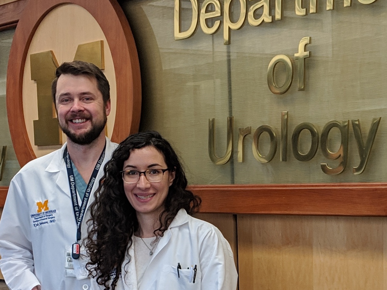
Giulia Lane M.D. is a Fellow in Neuro-urology and Pelvic Reconstruction in the Department of Urology at the University of Michigan; Kyle Johnson is a Urology Resident in the same department.

In this podcast they discuss the following BJUI Article of the Month:

Implementation of multiparametric magnetic resonance imaging technology for evaluation of patients with suspicion for prostate cancer in the clinical practice setting

Abstract

Objectives

To investigate the impact of implementing magnetic resonance imaging (MRI) and ultrasonography fusion technology on biopsy and prostate cancer (PCa) detection rates in men presenting with clinical suspicion for PCa in the clinical practice setting.

Patients and Methods

We performed a review of 1 808 consecutive men referred for elevated prostate‐specific antigen (PSA) level between 2011 and 2014. The study population was divided into two groups based on whether MRI was used as a risk stratification tool. Univariable and multivariable analyses of biopsy rates and overall and clinically significant PCa detection rates between groups were performed.

Results

The MRI and PSA‐only groups consisted of 1 020 and 788 patients, respectively. A total of 465 patients (45.6%) in the MRI group and 442 (56.1%) in the PSA‐only group underwent biopsy, corresponding to an 18.7% decrease in the proportion of patients receiving biopsy in the MRI group (P < 0.001). Overall PCa (56.8% vs 40.7%; P < 0.001) and clinically significant PCa detection (47.3% vs 31.0%; P < 0.001) was significantly higher in the MRI vs the PSA‐only group. In logistic regression analyses, the odds of overall PCa detection (odds ratio [OR] 1.74, 95% confidence interval [CI] 1.29–2.35; P < 0.001) and clinically significant PCa detection (OR 2.04, 95% CI 1.48–2.80; P < 0.001) were higher in the MRI than in the PSA‐only group after adjusting for clinically relevant PCa variables.

Conclusion

Among men presenting with clinical suspicion for PCa, addition of MRI increases detection of clinically significant cancers while reducing prostate biopsy rates when implemented in a clinical practice setting.

Read the full article

BJUI Podcasts now available on iTunes, subscribe here https://itunes.apple.com/gb/podcast/bju-international/id1309570262


Article of the month: Implementation of multiparametric MRI technology for evaluation of PCa in the clinic

Every month, the Editor-in-Chief selects an Article of the Month from the current issue of BJUI. The abstract is reproduced below and you can click on the button to read the full article, which is freely available to all readers for at least 30 days from the time of this post.

In addition to the article itself, there is an editorial written by a prominent member of the urological community, and a podcast produced by our current Resident Podcasters. These are intended to provoke comment and discussion and we invite you to use the comment tools at the bottom of each post to join the conversation. 

If you only have time to read one article this week, it should be this one.

Implementation of multiparametric magnetic resonance imaging technology for evaluation of patients with suspicion for prostate cancer in the clinical practice setting

Paras H. Shah*, Vinay R. Patel, Daniel M. Moreira, Arvin K. George§, Manaf Alom*, Zachary Kozel, Vidhu Joshi*, Eran Ben-Levi**, Robert Villani**, Oksana Yaskiv††Louis R. Kavoussi, Manish Vira, Carl O. Olsson‡‡ and Ardeshir R. Rastinehad


*Department of Urology, Mayo Clinic, Rochester, MN, Department of Urology, Icahn Smith Institute for Urology, Northwell Health, New York, NY, Department of Urology, University of Illinois at Chicago, Chicago, IL, §Department of Urology, University of Michigan, Ann Arbor, MI, Department of Urology, Smith Institute for Urology, Northwell Health, **Department of Radiology, Hofstra Northwell School of Medicine, ††Department of Pathology, Hofstra Northwell School of Medicine, New Hyde Park, and ‡‡Integrated Medical Professionals, Melville, NY, USA


Read the full article

Abstract

Objectives

To investigate the impact of implementing magnetic resonance imaging (MRI) and ultrasonography fusion technology on biopsy and prostate cancer (PCa) detection rates in men presenting with clinical suspicion for PCa in the clinical practice setting.

Patients and Methods

We performed a review of 1 808 consecutive men referred for elevated prostate‐specific antigen (PSA) level between 2011 and 2014. The study population was divided into two groups based on whether MRI was used as a risk stratification tool. Univariable and multivariable analyses of biopsy rates and overall and clinically significant PCa detection rates between groups were performed.

Results

The MRI and PSA‐only groups consisted of 1 020 and 788 patients, respectively. A total of 465 patients (45.6%) in the MRI group and 442 (56.1%) in the PSA‐only group underwent biopsy, corresponding to an 18.7% decrease in the proportion of patients receiving biopsy in the MRI group (P < 0.001). Overall PCa (56.8% vs 40.7%; P < 0.001) and clinically significant PCa detection (47.3% vs 31.0%; P < 0.001) was significantly higher in the MRI vs the PSA‐only group. In logistic regression analyses, the odds of overall PCa detection (odds ratio [OR] 1.74, 95% confidence interval [CI] 1.29–2.35; P < 0.001) and clinically significant PCa detection (OR 2.04, 95% CI 1.48–2.80; P < 0.001) were higher in the MRI than in the PSA‐only group after adjusting for clinically relevant PCa variables.

Conclusion

Among men presenting with clinical suspicion for PCa, addition of MRI increases detection of clinically significant cancers while reducing prostate biopsy rates when implemented in a clinical practice setting.

Read more Articles of the week


Editorial: Multiparametric MRI for prostate cancer detection: do clinical trial findings reflect real‐world practice?

‘First, do no harm’; with this in mind, researchers in urology strive to minimize the burden of overdiagnosis and overtreatment of prostate cancer. A promising tool in this arena is multiparametric (mp)MRI, which has been shown in a large‐scale randomized clinical trial to enhance the ability of prostate biopsy to detect clinically significant prostate cancer [1]. The extent to which findings from an idealized trial protocol extend to ‘real‐world’ clinical practice, however, remains largely unknown.

In this issue of BJUI, Shah et al. [2] aimed to fill this knowledge gap by investigating the impact of mpMRI‐guided biopsy on the detection rates of clinically significant prostate cancer in two large academic centres. The authors studied men with an elevated PSA presenting over a 3‐year span (2011–2014); 1020 men underwent mpMRI and 788 did not. Those in the MRI group had higher detection rates of both overall and clinically significant prostate cancer, defined as any Gleason score ≥7 on fusion or standard 12‐core TRUS biopsies, Gleason 6 with a lesion volume >0.5 cm3 volume on MRI, or Gleason 6 with >2 cores positive and/or >50% of any core involved with cancer on biopsy according to Epstein’s criteria, as well as a lower detection rate of clinically insignificant cancer.

The study provides timely implications for both patients and physicians, providing further insight into how findings from clinical trials [1,3] compare with real‐life practice. In fairness, the bulk of patients and clinicians do not follow strict study protocols for both decision‐making and interpretation of results, but rather assess very individual situations. A recent study by Bukavina et al. [4] showed that urologists and radiation oncologists largely perceive mpMRI guidance for targeted biopsies as valuable tools to improve prostate cancer stratification, but only a quarter of respondents reported implementation into their own clinical practice. This underlines some of the challenges of widespread implementation of mpMRI despite strong belief in its value.

Another strength of the study by Shah et al. is the exclusion of men who underwent mpMRI after negative biopsy in the PSA‐only group. This allows the isolation of the impact of mpMRI on downstream biopsy outcomes. A previous study that investigated targeted vs non‐targeted biopsies enrolled a cohort of men who all underwent mpMRI [5], which precludes any assessment of how mpMRI may impact the detection of clinically significant prostate cancer. Shah et al. [2] also astutely tracked detection rates of clinically significant and insignificant prostate cancer. Since the process of diagnosing prostate cancer is not without morbidity, it is crucial to understand the extent to which mpMRI can prevent the diagnosis of clinically indolent cancers.

Important questions regarding the challenges of widespread implementation of mpMRI for prostate cancer detection remain unanswered by the study of Shah et al. The study participants were gathered from large academic centres with readily available equipment, infrastructure and physician expertise to maximize favourable detection outcomes; however, these results may not be representative of the community setting. Additionally, >20% of men who did not undergo mpMRI did not do so because of a lack of insurance approval. This may reflect socio‐economic differences between the groups and also relates to the high costs of mpMRI that make routine implementation difficult [6]. Lastly, the presented findings mostly apply to positive mpMRI scans; the number of underdiagnosed men with negative scans may only be speculated upon, given the lack of follow‐up data in this population. It remains fundamentally important to improve the management of men with elevated PSA levels and negative findings on MRI.

Nonetheless, the present study demonstrates that research findings find their way into clinical practice. In essence, we are doing well, but we can do better.

by Marieke J. Krimphove, Sean A. Fletcher and Quoc‐Dien Trinh


References

  1. Kasivisvanathan V, Rannikko AS, Borghi M et al. MRI‐targeted or standard biopsy for prostate‐cancer diagnosis. N Engl J Med 2018378: 1767–77
  2. Shah PH, Patel VR, Moreira DM et al. Implementation of multiparametric magnetic resonance imaging technology for evaluation of patients with suspicion for prostate cancer in the clinical practice setting. BJU Int 2019123: 239–45
  3. Ahmed HU, El‐Shater Bosaily A, Brown LC et al. Diagnostic accuracy of multi‐parametric MRI and TRUS biopsy in prostate cancer (PROMIS): a paired validating confirmatory study. Lancet 2017389: 815–22
  4. Bukavina L, Tilburt JC, Konety B et al. Perceptions of prostate MRI and fusion biopsy of radiation oncologists and urologists for patients diagnosed with prostate cancer: results from a national survey. Eur Urol Focus 2018; [Epub ahead of print]
  5. Pokorny MR, de Rooij M, Duncan E et al. Prospective study of diagnostic accuracy comparing prostate cancer detection by transrectal ultrasound–guided biopsy versus magnetic resonance (MR) imaging with subsequent MR‐guided biopsy in men without previous prostate biopsies. Eur Urol 201466: 22–9
  6. Kim SJ, Vickers AJ, Hu JC. Challenges in adopting level 1 evidence for multiparametric magnetic resonance imaging as a biomarker for prostate cancer screening. JAMA Oncol 2018; [Epub ahead of print]


Article of the Month: Use of machine learning to predict early biochemical recurrence after robot‐assisted prostatectomy

Every month, the Editor-in-Chief selects an Article of the Month from the current issue of BJUI. The abstract is reproduced below and you can click on the button to read the full article, which is freely available to all readers for at least 30 days from the time of this post.

In addition to the article itself, there is an accompanying editorial written by a prominent member of the urological community. This blog is intended to provoke comment and discussion and we invite you to use the comment tools at the bottom of each post to join the conversation.

If you only have time to read one article this week, it should be this one.

Use of machine learning to predict early biochemical recurrence after robot‐assisted prostatectomy

Nathan C. Wong , Cameron Lam, Lisa Patterson and Bobby Shayegan
Division of Urology, Department of Surgery, McMaster University, Hamilton, ON, Canada

Read the full article
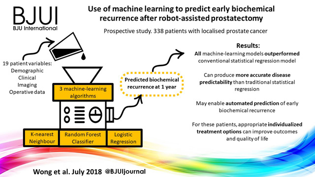
Visual abstract created Rebecca Fisher @beckybeckyfish

Abstract

Objectives

To train and compare machine‐learning algorithms with traditional regression analysis for the prediction of early biochemical recurrence after robot‐assisted prostatectomy.

Patients and Methods

A prospectively collected dataset of 338 patients who underwent robot‐assisted prostatectomy for localized prostate cancer was examined. We used three supervised machine‐learning algorithms and 19 different training variables (demographic, clinical, imaging and operative data) in a hypothesis‐free manner to build models that could predict patients with biochemical recurrence at 1 year. We also performed traditional Cox regression analysis for comparison.

= 0.686) and with a univariate regression model (AUC = 0.865).

Results

K‐nearest neighbour, logistic regression and random forest classifier were used as machine‐learning models. Classic Cox regression analysis had an area under the curve (AUC) of 0.865 for the prediction of biochemical recurrence. All three of our machine‐learning models (K‐nearest neighbour (AUC 0.903), random forest tree (AUC 0.924) and logistic regression (AUC 0.940) outperformed the conventional statistical regression model. Accuracy prediction scores for K‐nearest neighbour, random forest tree and logistic regression were 0.976, 0.953 and 0.976, respectively.

Conclusions

Machine‐learning techniques can produce accurate disease predictability better that traditional statistical regression. These tools may prove clinically useful for the automated prediction of patients who develop early biochemical recurrence after robot‐assisted prostatectomy. For these patients, appropriate individualized treatment options can improve outcomes and quality of life.

Read more Articles of the week

Article of the month: The US opioid epidemic

Every month, the Editor-in-Chief selects an Article of the Month from the current issue of BJUI. The abstract is reproduced below and you can click on the button to read the full article, which is freely available to all readers for at least 30 days from the time of this post.

In addition to the article itself, there is an accompanying editorial written by a prominent member of the urological community. This blog is intended to provoke comment and discussion and we invite you to use the comment tools at the bottom of each post to join the conversation.

If you only have time to read one article this week, it should be this one.

The United States opioid epidemic: a review of the surgeon’s contribution to it and health policy initiatives

Katherine Theisen, Bruce Jacobs, Liam Macleod and Benjamin Davies
Department of Urology, University of Pittsburgh Medical Center, Pittsburgh, PA, USA
 
Read the full article

Abstract

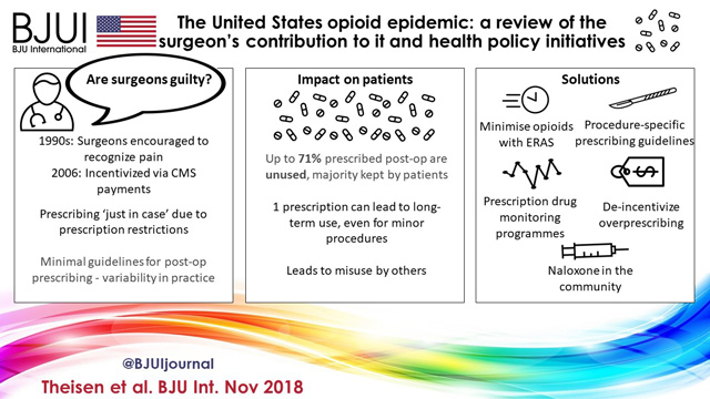
Visual abstract created by Abdullatif Aydın and Rebecca Fisher

Opioid abuse and addiction is causing widespread devastation in communities across the USA and resulting in significant strain on our healthcare system. There is increasing evidence that prescribers are at least partly responsible for the opioid crisis because of overprescribing, a practice that developed from changes in policy and reimbursement structures. Surgeons, specifically, have been subject to scrutiny as ‘adequate treatment’ of post‐surgical pain is poorly defined and data suggest that many patients receive much larger opioid prescriptions than needed. The consequences of overprescribing include addiction and misuse, dispersion of opioids into the community, and possible potentiation of illicit drug/heroin use. Several solutions to this crisis are currently being enacted with variable success, including Prescription Drug Monitoring Programmes, policy‐level interventions aimed to de‐incentivize overprescribing, limiting opioid exposures through Enhanced Recovery After Surgery protocols, and the novel idea of creating surgery‐ and/or procedure‐specific prescribing guidelines. This problem is likely to require not one, but several potential solutions to reverse its trajectory. It is critical, however, that we as physicians and prescribers find a way to stop the needless overprescribing while still treating postoperative pain appropriately.


Read more Articles of the week

Editorial: The opioid epidemic: a wake‐up call for us all

The article in this issue of BJUI by Theisen et al. [1] is a timely reminder of the duty of all prescribers (including surgeons) to be mindful of the potential unintended consequences and off‐target effects of medicines.

Although some of the factors that have led to serious opioid‐related problems are particularly related to the US setting, we in Europe and other continents should not be complacent [2, 3].

The US Department of Health and Human Services (HHS) stated that, in 2016, opioid deaths had risen to > 42 000 deaths, of which an estimated 40% involved a prescription opioid [4].

The underlying reasons for this opioid epidemic are multiple and complex.

The prevalence of pain in the population is high, as are patients expectations and demands for treatment. The ageing population, living with multiple painful conditions, including cancer survivors and patients with persisting post‐surgery chronic pain, has further increased the demand for analgesics.

Meanwhile, the WHO’s drive over the last 30 years to eradicate ‘opiophobia’ and ensure that opioids are available for cancer pain, together with the advent of potent prolonged‐release opioid formulations, led to a transfer of this therapeutic experience to non‐cancer pain. A few questioned the wisdom of this strategy, but reassurance was drawn from an 11‐line letter in the New England Journal of Medicine in 1980, oft cited and misquoted as evidence that addiction was rare with long‐term opioids [5]. Subsequently, the journal has added a note warning readers that ‘the letter has been heavily and uncritically cited by sources using it to suggest opioids are not addictive.’ In fact, the authors surveyed the files of inpatients who were administered predominantly short‐term opioids in hospital, including patients who had only received one dose, and concluded that in this population, development of new addiction was rare.

Add to these factors, the well‐intentioned drive to assess and treat pain with initiatives such as ‘Pain – the fifth vital sign’, and pharmaceutical company promotion of their new opioid formulations, and the scene was set for greatly increased opioid initiation, escalation of dosage and repeat prescribing without regular patient review. In addition to these factors, it was also identified that a proportion of patients continue to receive opioids long after their surgery [6].

By 2017, year‐on‐year increases in long‐term opioid prescribing compounded by the diversion of the medicines, illicit manufacture and importing of compounds, such as fentanyl analogues, culminated in the staggering US mortality data and the HHS declaring a public health emergency with a five‐point strategy to combat the opioid crisis.

What strategies can we adopt during and after surgery? Better multimodal acute inpatient analgesia and working closely with our acute pain colleagues will surely assist in achieving less need for subsequent opioid prescribing on discharge. Using enhanced recovery pathways encourages the use of opioid‐sparing local and regional anaesthetic blocks, together with simple analgesia rather than prolonged use of high‐dose opioids. The goal must be to discharge patients on less potent analgesics and for a shorter duration. The analogy is with antibiotic prescribing where only a limited supply is dispensed. We need to develop pain discharge plans which can be communicated to the primary care physician incorporating tapering, patient education and emphasis on avoiding the repeat prescribing of opioids. Where pain persists, the patient should be referred back to the surgical or pain management team sooner in order to review progress. We should be wary of prescribing modified‐release preparations of a drug such as morphine or oxycodone because these contain a high dose, which can be extracted from the slow‐release preparation for abuse purposes. Similarly, the use of opioid‐based patches encourages extended use of opioid drugs, sometimes without a full understanding of the hourly or daily morphine equivalent dosage. Looking forward, there is the promise of new non‐opioid analgesics for chronic pain on the horizon, in particular long‐acting, prolonged‐release local anaesthetics for use in the wound or for nerve blocks. We need to adopt strategies for the regular review of pain medication rather than the all too often ‘automatic’ repeat prescription.

In urology, we have seen a significant reduction in the use of opioids on discharge through the use of less invasive, endoscopic/robotic techniques, local anaesthetic blocks such as the transversus abdominis block, which is so valuable in abdominal procedures, wound local anaesthetic infusion catheters and the use of regular simple analgesics given by the clock, providing excellent opioid‐sparing background analgesia.

Less opioid drug prescribing in the community is the way forward as Theisen et al. describe. As peri‐operative physicians, we must respond to this challenge if we are to avert a similar crisis to that seen in the USA. In peri‐operative practice, responsible and appropriate opioid‐prescribing remains an essential part of good pain management, while we strive to reduce both dose and duration of therapy. These strategies serve both wider society and the individual patient, for whom the benefit is reduced dose‐dependent opioid side effects. In the modern era where specialist advice is available through multidisciplinary team working, we need to minimize repeat prescribing and ensure that a specific opioid tapering plan is in place. The latter relies on good communication, teamwork and partnership, the essential ‘Domain 3’ of General Medical Council Good Medical Practice [7].

References

  1. Theisen K, Jacobs B, Macleod L, Davies B. The United States opioid epidemic: a review of the surgeon’s contribution and health policy initiatives. BJU Int 2018; 122: 754–9
  2. Stannard C. Opioids in the UK: what’s the problem? BMJ 2013; 347: f5108
  3. Weisberg DF, Becker WC, Fiellin DA, Stannard C. Prescription opioid misuse in the United States and the United Kingdom: cautionary lessons. Int J Drug Policy 2014; 358: 1124–30
  4. US Department of Health and Human Services. What is the U.S. Opioid Epidemic? Available at: https://www.hhs.gov/opioids/about-the-epidemic/index.html. Accessed October 2018
  5. Porter J, Jick H. Addiction rare in patients treated with narcotics. N Engl J Med 1980; 302: 123
  6. Clarke H, Soneji N, Ko TD, Yun L, Wijeysundera DN. Rates and risk factors for prolonged opioid use after major surgery: population based cohort study. BMJ 2014; 348: g1251
  7. GMC Good Medical Practice, 2013. Available at: www.gmc-uk.org/guidance Accessed October 2018


Article of the month: Effect of timing of an immediate instillation of mitomycin C after TUR in 941 patients with NMIBC

Every month, the Editor-in-Chief selects an Article of the Month from the current issue of BJUI. The abstract is reproduced below and you can click on the button to read the full article, which is freely available to all readers for at least 30 days from the time of this post.

In addition to the article itself, there is an accompanying editorial written by a prominent member of the urological community. This blog is intended to provoke comment and discussion and we invite you to use the comment tools at the bottom of each post to join the conversation.

If you only have time to read one article this week, it should be this one.

The effect of timing of an immediate instillation of mitomycin C after transurethral resection in 941 patients with non-muscle-invasive bladder cancer

Judith Bosschieter*, R. Jeroen A. van Moorselaar*, André N. Vis*, Tessa van Ginkel*, Birgit I. Lissenberg‐Witte, Goedele M.A. Beckers* and Jakko A. Nieuwenhuijzen*


Departments of *Urology and Epidemiology and Biostatistics, VU University Medical Center, Amsterdam, The Netherlands


Read the full article

Abstract

Objective

To investigate whether the timing of an immediate instillation of mitomycin C (on the day of transurethral resection of bladder tumour [TURBT] or 1 day later) has an impact on time to recurrence of non‐muscle‐invasive bladder cancer (NMIBC).

Patients and Methods

All patients with NMIBC who were enrolled in a prospective trial between 1998 and 2003, and treated with an early mitomycin C instillation (on the day of TURBT or 1 day later), were selected. Statistical analysis was performed with Kaplan–Meier curves and multivariable Cox regression.

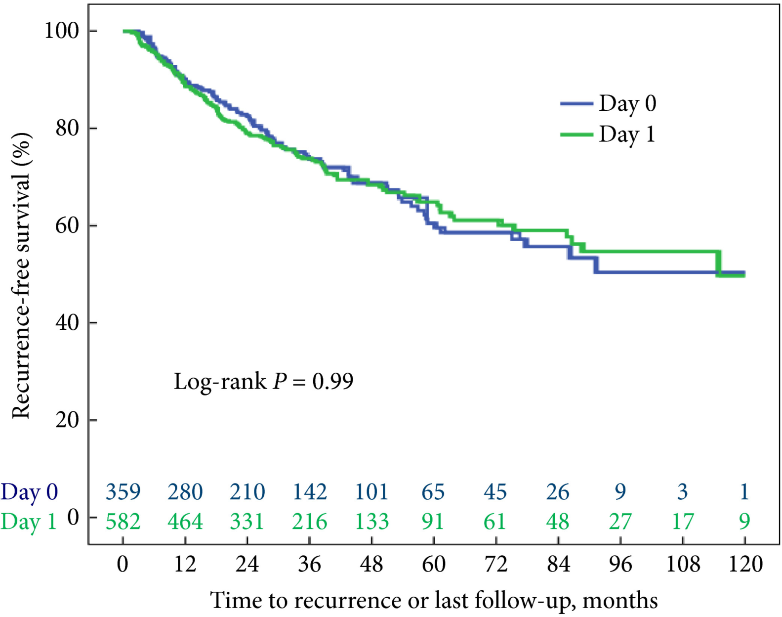
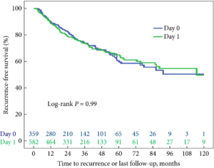
Fig. 1 Kaplan–Meier analysis showing time to recurrence for patients treated with an immediate instillation of MMC on the day of TURBT (Day‐0 group) or 1 day after (Day‐1 group).

Results

Administering an instillation of mitomycin C on the day of TURBT or 1 day later did not show a statistically significant difference in time to recurrence in a univariable model (log‐rank P = 0.99). After correcting for the number of scheduled adjuvant instillations, no statistically significant difference could be detected either: hazard ratio 1.05 (95% confidence interval 0.81–1.35, P = 0.74).

Conclusion

These data do not support the hypothesis that a very early instillation (on the day of TURBT) of mitomycin C decreases the risk of recurrence as compared with an early instillation (1 day after TURBT).

Read more articles of the week

October 2018 – about the cover

This issue’s Article of the Month is The effect of timing of an immediate instillation of mitomycin C after transurethral resection in 941 patients with non‐muscle‐invasive bladder cancer, carried out by a team from Amsterdam, The Netherlands.

The cover shows the skyline of Amsterdam, the capital of the Netherlands, although it is not the seat of government, which is The Hague. Amsterdam is well-known for its canals, its Art (particularly Rembrandt and Van Gogh) and its infamous coffee shops. In 2013 there were more bicycles than people in Amsterdam.

©istock.com/fotolupa


© 2024 BJU International. All Rights Reserved.