Tag Archive for: aotw

Posts

Article of the week: RS‐RARP vs standard RARP: it’s time for critical appraisal

Every week, the Editor-in-Chief selects an Article of the Week from the current issue of BJUI. The abstract is reproduced below and you can click on the button to read the full article, which is freely available to all readers for at least 30 days from the time of this post.

In addition to the article itself, there are two accompanying editorials written by prominent members of the urological community. These are intended to provoke comment and discussion and we invite you to use the comment tools at the bottom of each post to join the conversation. There is also a podcast by one of our Resident Podcasters describing the article.

If you only have time to read one article this week, it should be this one.

Retzius‐sparing robot‐assisted radical prostatectomy (RS‐RARP) vs standard RARP: it’s time for critical appraisal

Thomas Stonier*, Nick Simson*, John Davisand Ben Challacombe


*Department of Urology, Princess Alexandra Hospital, Harlow, Urology Centre, Guy s Hospital, London, UK and Department of Urology, MD Anderson Cancer Center, Houston, TX, USA


Since robot‐assisted radical prostatectomy (RARP) started to be regularly performed in 2001, the procedure has typically followed the original retropubic approach, with incremental technical improvements in an attempt to improve outcomes. These include the running Van‐Velthoven anastomosis, posterior reconstruction or ‘Rocco stitch’, and cold ligation of the Santorini plexus/dorsal vein to maximise urethral length. In 2010, Bocciardi’s team in Milan proposed a novel posterior or ‘Retzius‐sparing’ RARP (RS‐RARP), mirroring the classic open perineal approach. This allows avoidance of supporting structures, such as the puboprostatic ligaments, endopelvic fascia, and Santorini plexus, preserving the normal anatomy as much as possible and limiting damage that may contribute to improved postoperative continence and erectile function. There has been much heralding of the excellent functional outcomes in both the medical and the lay press, but as yet no focus or real mention of any potential downsides of this new technique.


Editorial: Retzius‐sparing robot‐assisted radical prostatectomy

In their commentary in the current issue of BJUI, Stonier et al. [1] examine the potential technical pitfalls and published results of the Retzius‐sparing technique of robotic radical prostatectomy. The authors reviewed three studies from three different groups [2,3], including a study by our group [4], and raised three specific concerns: the oncological efficacy of the procedure; the long learning curve; and the generalizability of the technique to challenging surgical scenarios. We offer a few clarifications and comments.

The first study on Retzius‐sparing robot‐assisted radical prostatectomy came from the Bocciardi group [2]. This was a prospective, single‐arm study of 200 patients. The authors reported a 14‐day continence rate of 90–92%, a 1‐year potency rate of 71–81% (in preoperatively potent patients undergoing bilateral intrafascial nerve‐sparing) and a positive surgical margin rate of 25.5%. The positive surgical margin rate improved in patients with pT2 disease, from 22% to 9% (P = 0.04) over the course of the study (initial 100 vs subsequent 100 patients), while in patients with pT3 disease, it remained stable at ~45%. Lim et al. [3] also noted an improvement in their overall positive surgical margin rate from 20% to 8% when comparing the initial 25 patients with the subsequent 25 patients. In that study, a standard robot‐assisted radical prostatectomy comparator arm was included and there were no differences in overall positive surgical margin rates (14% in both arms), while continence was better with the Retzius‐sparing approach.

Recognizing the potentially technically challenging nature of the Bocciardi approach, we performed a randomized controlled trial to objectively evaluate the technique. Randomized controlled trials are typically designed to answer a single question. Our trial was designed to determine whether there were differences in the rate of return of urinary continence, the primary benefit that previous non‐controlled studies had reported. This our study clearly showed [4].

Once the trial was completed, post hoc analysis of secondary outcomes was performed [5]. One of these outcomes was the positive surgical margin rate. In our trial, we noted an overall positive surgical margin rate of 25% in the Retzius‐sparing arm vs 13% in the control arm, a difference that did not achieve statistical significance (P = 0.11). Stonier et al. [1] suggested that if the sample size of our trial were doubled, then the positive surgical margin rate in each group would be doubled as well, leading to significance. This conclusion is problematic. The likelihood that doubling the sample size would result in the exact doubling of numbers in all four cells of a 2 × 2 contingency table is estimated at <5% using Fisher’s exact test (this calculation is different from the P value). Furthermore, the surgical margins depend as much on the pathological stage as on surgical approach. In our trial, patients were matched preoperatively for risk in the best manner possible for a pragmatic randomized trial. However, it is impossible to predict and control for the final pathological characteristics. Pathological analysis showed that patients undergoing Retzius‐sparing surgery did have significantly more aggressive disease: ≥pT3 disease in 45% vs 23.3% of patients (P = 0.04) [4, 5]. This, by itself, could account for a substantial difference in surgical margin rates.

In writing our paper, we made no judgements as to whether the Bocciardi or posterior technique is fundamentally superior to an anterior or Menon approach, whether it is easier to perform, how generalizable it is [6], or what the learning curve may be. That is best left to the individual surgeon’s training and judgement. We do suggest, however, that surgical margins be interpreted as a function of pathological variables, and not in isolation, and that it is simplistic to assume that identical results will be obtained by doubling sample size. We suggest that such conclusions are hypothesis‐generating, and should best be explored through a separate, purpose‐designed randomized trial.

Authors: Akshay Sood, Firas Abdollah and Mani Menon

References

  1. Stonier T, Simson N, Davis J, Challacombe B. Retzius‐sparing robot‐assisted radical prostatectomy (RS‐RARP) vs standard RARP: it’s time for critical appraisal. BJU Int 2019; 123: 5–10
  2. Galfano A, Di Trapani D, Sozzi F et al. Beyond the learning curve of the Retzius‐sparing approach for robot‐assisted laparoscopic radical prostatectomy: oncologic and functional results of the first 200 patients with >/= 1 year of follow‐up. Eur Urol 2013; 64: 974–80
  3. Lim SK, Kim KH, Shin TY et al. Retzius‐sparing robot‐assisted laparoscopic radical prostatectomy: combining the best of retropubic and perineal approaches. BJU Int 2014; 114: 236–44
  4. Dalela D, Jeong W, Prasad MA et al. A pragmatic randomized controlled trial examining the impact of the Retzius‐sparing approach on early urinary continence recovery after robot‐assisted radical prostatectomy. Eur Urol 2017; 72: 677–85
  5. Menon M, Dalela D, Jamil M et al. Functional recovery, oncologic outcomes and postoperative complications after robot‐assisted radical prostatectomy: an evidence‐based analysis comparing the Retzius sparing and standard approaches. J Urol 2018; 199: 1210–7
  6. Galfano A, Secco S, Bocciardi AM. Will Retzius‐sparing prostatectomy be the future of prostate cancer surgery? Eur Urol 2017; 72: 686–8


Editorial: Reply: RS-RARP vs standard RARP

Since the introduction of robotic surgery in the treatment of patients with prostate cancer (PCa), different surgical innovations have been implemented in order to preserve postoperative functional outcomes while maintaining oncological safety. Sparing the Retzius space during robot‐assisted radical prostatectomy (RARP) was introduced early this decade by Galfano et al [1]. Interestingly, 90% and 96% of patients treated with Retzius‐sparing RARP (RS‐RARP) were continent (no pad/safety pad) at 1 week and 1 year, respectively. Similarly, our group reported a 70% continence rate (no pad) at 1 month after RS‐RARP [2].

The fast urinary continence recovery after RS‐RARP is related to several anatomical factors: the anterior Retzius space is kept intact; the urinary bladder is not dropped; the endopelvic fascia and puboprostatic ligaments are preserved; and there is minimal distortion of the supporting urethral tissues. A recent study reported [3] that less bladder neck descent was observed during postoperative cystogram in patients treated with RS‐RARP than in those treated with standard RARP.

In a recent randomized controlled study, the postoperative continence rate at 1 week was 48% in standard RARP compared with 71% in RS‐RARP (P = 0.01), and this difference was maintained at 3 months (86% standard RARP vs 95% RS‐RARP; P = 0.02). At 1 year, however, the effect on urinary continence difference was muted (93.3% standard RARP vs 98.3% RS‐RARP; P = 0.09) [4]. Similarly, Chang et al. [3] found that the higher continence rate at 1 week (73.3% RS‐RARP vs 26.7% standard RARP; P = 0.000) had vanished at 1 year (100% vs 93.3%; P = 0.15). By contrast, a large recent prospective series showed that the superiority of RS‐RARP in terms of higher early urinary continence was maintained at 1 year (97.5% RS‐RARP vs 68.5% standard RARP) [5].

In addition to a higher early continence rate, RS‐RARP has a lower incidence of postoperative inguinal hernia occurrence compared with standard RARP [6]. Theoretically, RS‐RARP may provide several other potential advantages. It may be advantageous if patients require future surgery necessitating access to the Retzius space and dropping of the bladder, such as an artificial urinary sphincter implantation, an inflatable penile prosthesis insertion, or kidney transplantation. In addition, in patients with previous inguinal hernia repair using mesh, it enables the avoidance of anterior adhesions by accessing the prostate directly from the Douglas pouch. Notably, large‐size glands and/or middle‐lobe, advanced/high‐risk PCa, and patients with previous prostatic surgeries can be managed safely with RS‐RARP in experienced hands.

Undoubtedly, oncological safety is our main concern in treating cancer. To determine the effectiveness of new treatment methods, long‐term follow‐up is warranted. Biochemical recurrence (BCR) is widely used as a primary oncological outcome to assess PCa treatment success. To our knowledge, after radical prostatectomy, ~35% of patients are at risk of developing BCR in the next 10 years. Currently, there are insufficient data regarding the oncological outcomes of RS‐RARP. Only four articles have compared early oncological outcomes between RS‐RARP and standard RARP, and there was no significant difference (Table 1).

More recently, we reported on the mid‐term oncological outcomes of 359 patients who underwent RS‐RARP. The median follow‐up was 26 months. Although this period is not long enough to reach a meaningful conclusion on the oncological safety of RS‐RARP, it is the longest follow‐up period reported in literature. Overall, the positive surgical margin (PSM) rate was 30.6% (14.6% in pT2 and 40.8% in pT3a disease) and the BCR rate was 14.8%. In terms of functional outcomes, the urinary continence rate at 1 year was 93.9% [7]. Interestingly, 164 patients (45.7%) of our cohort had high‐risk PCa. In these patients, the PSM rate was 41.2%, the BCR rate was 22%, and the 3‐year BCR‐free survival (BCRFS) rate was 72%. We compared our results with those in patients with high‐risk PCa treated with standard RARP in the literature. In studies that used the D’Amico criteria the median follow‐up ranged from 12.5 to 37.3 months, the PSM rates were 20.5% to 53.3%, the BCR rates were 17.4% to 31% and the 3‐year BCRFS rates were 41.4% to 86%. In studies that used the National Comprehensive Cancer Network criteria, the median follow‐up ranged from 23.6 to 27 months, the PSM rates were 29% to 38%, the BCR rates were 9.4% to 33%, and the 3‐year BCRFS rates were 55% to 66% [7].

In summary, RS‐RARP is a novel surgical approach which is associated with better urinary continence recovery in the first few months compared with standard RARP [2,3,4,5]. This superiority might be maintained [5] or equalized at 1 year [3,4]. A few studies have compared the early oncological results between RS‐RARP and standard RARP and no significant difference was found [2,3,4,5]. Recently, our group reported the mid‐term oncological outcomes of patients with high‐risk PCa treated with RS‐RARP and these were similar to those of large studies of conventional RARP. This confirms effective and safe mid‐term BCR control after RS‐RARP, while the long‐term oncological results are awaited [7]. Currently, >4 000 cases of RS‐RARP are performed worldwide and more centres are beginning to use and converting to Retzius‐sparing surgery. All centres are experiencing faster recovery of continence. Thanks are due to Drs Galfano and Bocciardi for exploring and sharing this surgical frontier.


References

  1. Galfano A, Di Trapani D, Sozzi F, et al. Beyond the learning curve of the Retzius‐sparing approach for robotassisted laparoscopic radical prostatectomy: oncologic and functional results of the first 200 patients with ? 1 year of follow‐up. Eur Urol 2013; 64: 974‐80
  2. Lim SK, Kim KH, Shin TY et al. Retzius‐sparing robot‐assisted laparoscopic radical prostatectomy: combining the best of retropubic and perineal approaches. BJU Int 2014; 114: 236–44
  3. Chang LW, Hung SC, Hu JC et al. Retzius‐sparing robotic‐assisted radical prostatectomy associated with less bladder neck descent and better early continence outcome. Anticancer Res 2018; 38: 345–51
  4. Menon M, Dalela D, Jamil M et al. Functional recovery, oncologic outcomes and postoperative complications after robot‐assisted radical prostatectomy: an evidence‐based analysis comparing the Retzius sparing and standard approaches. J Urol 2018; 199: 1210–7
  5. Sayyid RK, Simpson WG, Lu C et al. Retzius sparing robotic assisted laparoscopic radical prostatectomy: a safe surgical technique with superior continence outcomes. J Endourol 2017; 31: 1244–50
  6. Chang KD, Abdel Raheem A, Santok GDR et al. Anatomical Retzius‐space preservation is associated with lower incidence of postoperative inguinal hernia development after robot‐assisted radical prostatectomy. Hernia 2017; 21: 555–61
  7. Abdel Raheem A, Kidon C, Alenzi M et al. Predictors of biochemical recurrence after retzius‐sparing robot‐assisted radical prostatectomy: analysis of 359 cases with a median follow‐up of 26 months. Int J Urol 2018; 25: 1006–14


Resident’s podcast: Retzius‐sparing robot‐assisted radical prostatectomy

Maria Uloko is a Urology Resident at the University of Minnesota Hospital. In this podcast she discusses the following BJUI Article of the Week:

Retzius‐sparing robot‐assisted radical prostatectomy (RS‐RARP) vs standard RARP: it’s time for critical appraisal

Thomas Stonier*, Nick Simson*, John Davisand Ben Challacombe


*Department of Urology, Princess Alexandra Hospital, Harlow, Urology Centre, Guy s Hospital, London, UK and Department of Urology, MD Anderson Cancer Center, Houston, TX, USA


Abstract

Since robot‐assisted radical prostatectomy (RARP) started to be regularly performed in 2001, the procedure has typically followed the original retropubic approach, with incremental technical improvements in an attempt to improve outcomes. These include the running Van‐Velthoven anastomosis, posterior reconstruction or ‘Rocco stitch’, and cold ligation of the Santorini plexus/dorsal vein to maximise urethral length. In 2010, Bocciardi’s team in Milan proposed a novel posterior or ‘Retzius‐sparing’ RARP (RS‐RARP), mirroring the classic open perineal approach. This allows avoidance of supporting structures, such as the puboprostatic ligaments, endopelvic fascia, and Santorini plexus, preserving the normal anatomy as much as possible and limiting damage that may contribute to improved postoperative continence and erectile function. There has been much heralding of the excellent functional outcomes in both the medical and the lay press, but as yet no focus or real mention of any potential downsides of this new technique.


BJUI Podcasts now available on iTunes, subscribe here https://itunes.apple.com/gb/podcast/bju-international/id1309570262


Article of the week: Multicentre international experience of 532‐nm laser PVP with GreenLight XPS in men with very large prostates

Every week, the Editor-in-Chief selects an Article of the Week from the current issue of BJUI. The abstract is reproduced below and you can click on the button to read the full article, which is freely available to all readers for at least 30 days from the time of this post.

If you only have time to read one article this week, it should be this one.

Roger Valdivieso*, Pierre‐Alain Hueber*, Malek Meskawi*, Eric Belleville*, Khaled Ajib*, Franck Bruyere, Alexis E. Te, Bilal Chughtai, Dean Elterman§, Vincent Misraiand Kevin C. Zorn*


*Division of Urology, Centre Hospitalier de lUniversite de Montreal (CHUM), Montreal, QC, Canada, Department of Urology, CHU, Tours, France, Department of Urology, Cornell University, New York, NY, USA, §Department of Urology, University of Toronto, Toronto, ON, Canada, and Department of Urology, Clinique Pasteur, Toulousse, France

Read the full article

Abstract

Objectives

To describe peri‐operative results, functional outcomes and complications of laser photoselective vaporization, using the GreenLight system, of prostate glands ≥200 mL in volume.

Methods

Retrospective analysis of a prospectively maintained multicentre database was performed to select a subgroup of patients with very large prostates (volume ≥200 mL) treated with the GreenLight XPS laser. A subgroup of patients with prostate volumes 100–200 mL was used for comparison. International Prostate Symptom Score, maximum urinary flow rate, postvoid residual urine volume and prostate‐specific antigen levels were measured at 6, 12, 24, 36 and 48 months. Durability was evaluated using benign prostatic hyperplasia re‐treatment rate at 12, 24 and 36 months. Additionally, complications were recorded using Clavien–Dindo classification.

Results

A total of 33 patients (38%) had prostates ≥200 mL. Baseline characteristics were similar between patients with prostates ≥200 mL and those with prostates 100–200 mL. Patients with very large prostates (≥200 mL) had longer operating times (129 vs 93 min), less energy delivered, a greater number of fibres used (3 vs 2) and a higher conversion rate to transurethral resection of the prostate (16% vs 4%). In terms of complications and functional outcomes, we did not find any differences between the groups. Retreatment rate was also comparable.

Conclusions

Our results show that PVP GreenLight XPS‐180W is an acceptable technique for very large prostates (≥200 mL); however, operating times, energy delivery, fibres used and conversion to TURP are a concern in this particular subgroup. This should be used for patient counselling and surgery planning.

Read more Articles of the week


Editorial: Contemporary quality‐of‐life scores provide a key foundation for high‐quality cancer research

Prostate cancer is the most common male malignancy in many countries, including the UK/Northern Ireland. Given excellent oncological outcomes for appropriately treated localised cancer, there is an increasing focus on understanding the quality‐of‐life implications of different treatment options.

As Donnelly et al. [1] emphasise, contemporary cohorts of untreated men can provide useful comparisons for inferring the impact of treatment. Specifically, updated population‐level observations of urinary, bowel, and sexual dysfunction are needed to provide a baseline for such discussions. Surveys should focus on particular populations (e.g. geographic), utilise prostate cancer‐specific questionnaires, and ensure age‐matched cohorts. Such baseline characteristics are essential to teasing apart the impact of prostate cancer and its treatment from ageing and comorbidities.

Donnelly et al. [1] sampled 10 000 men in Northern Ireland aged >40 years, using the EuroQoL five Dimensions five Levels (EQ‐5D‐5L) survey to assess a general health baseline and Extended Prostate Cancer Composite (EPIC) questionnaire to determine bladder, bowel, and sexual function more specifically. In all, 2 955 men responded, although ultimately only men aged >60 years were analysed to better match the age distribution of patients with prostate cancer. Strikingly, they found that nearly two out of five men reported at least one urinary, bowel, or sexual issue. A third of men reported some degree of urinary leakage, 26% had some degree of bowel problems, and as much as 57.9% of respondents had some problem with sexual function [1].

Nearly two decades ago, Litwin [2] published a health‐related quality of life control sample of older men in the USA without prostate cancer using the University of California Los Angeles Prostate Cancer Index (UCLA‐PCI, a precursor to EPIC). He found ageing subjects had diminished urinary continence, bowel function, and sexual potency, with similar rates to the Northern Ireland study: a third reported urinary leakage, a third had bowel complaints, and nearly two‐thirds claimed to have erectile dysfunction (ED).

In contrast, patient‐reported outcomes in the Prostate Testing for Cancer and Treatment (ProtecT) trial showed low levels of urinary incontinence and bowel symptoms, and one‐third of men had sexual dysfunction [3]. The difference here in ED when compared to Donnelly et al. [1] may be attributed to the age distribution differences between the cohorts, as ProtecT included men aged 50–69 years and the Northern Ireland group looked only at men aged >60 years. This highlights the importance of ensuring age‐matched cohorts when using population‐based surveys as baselines for assessment counselling.

Furthermore, Resnick et al. [4] evaluated the change in patient‐reported urinary incontinence and ED over time in two cohorts of patients enrolled almost 20 years apart. They compared patients enrolled in 1994–1995 in the Prostate Cancer Outcomes Study (PCOS) vs those enrolled in 2011–2012 in the Comparative Effectiveness Analysis of Surgery and Radiation (CEASAR) study. Men in PCOS were surveyed using UCLA‐PCI, and those in CEASAR completed EPIC‐26. They found that self‐reported urinary incontinence was more common in CEASAR than in PCOS (7.7% vs 4.7%), as was ED (44.7% vs 24%). These differences could be due to rising rates of comorbidities associated with ED and urinary incontinence or they may reflect an increase in social awareness and disclosure of these issues.

Taken together, these self‐reported rates of pretreatment urinary and sexual function underscore the potential for significant variation in reporting of patient quality‐of‐life outcomes in prostate cancer.

This does not mean that patient‐reported outcomes should be ignored. Rather the takeaway is that we must invest in tools to ensure that reporting is appropriate, standardised, and accurate [5]. And regardless of whether these data are collected prospectively, or retrospectively, it is vital to use appropriate statistical methods and scientific principles to account for bias and to ensure that causal inferences are valid [6].

As prostate cancer survival and mortality rates improve, patients and clinicians must weigh treatment‐specific short‐ and long‐term effects on quality of life. Patient‐reported outcome measures are vital to assessing these major impacts. Contemporary, population‐based cohorts such as that provided by Donnelly et al. [1], provide a key tool for better interpreting and understanding these results.

References

  1. Donnelly DW, Donnelly C, Kearney T et al. Urinary, bowel and sexual health in older men from Northern Ireland. BJU Int 2018; 122: 845–57
  2. Litwin MS. Health related quality of life in older men without prostate cancer. J Urol 1999; 161: 1180–4


Editorial: Close surgical margins after RP: how to make a complex story even more complex

Surgical margin (SM) status after radical prostatectomy (RP) for clinically localized prostate cancer (PCa) is a measure of surgical quality and retains some prognostic value. Positive SMs (PSMs) have long been considered an adverse oncological outcome because they were repeatedly found to be associated with a higher risk of biochemical recurrence (BCR), and are still among the factors guiding the decision to deliver adjuvant treatments; however, the long‐term impact of PSMs on survival remains uncertain because it is largely affected by other concurrent risk modifiers [1,2,3].

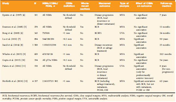
The clinical significance of so‐called close SMs (CSMs), that is, negative SMs (NSMs) with tumour foci approaching, but not involving, the inked cut surface of the RP specimen, is a far less investigated field of research, with contradictory findings in the few available studies (Table 1 [412]). Some studies showed a significant association with risk of disease progression (mainly measured with BCR), while others did not.


The study by Herforth et al. [12] published in this issue of BJUI further adds to the debate on CSMs, with an analysis of the largest series reported to date. The authors assessed the impact of CSMs vs NSMs vs PSMs after RP on BCR, PCa‐specific and overall survival in ~4 300 men included in the Shared Equal Access Regional Cancer Hospital cohort. CSMs were defined as cancer foci within 1 mm from the inked specimen surface, and were found in 372 patients (9%). The median follow‐up was 6.5 years. On multivariable analysis accounting for several established prognostic factors, CSMs were significantly associated with a higher BCR risk compared with NSMs, but a lower risk compared with PSMs. Notably, SM status alone did not influence PCa‐specific or overall survival. Major limitations to this retrospective analysis were lack of central pathology review and inadequate follow‐up length to assess survival.

The main question yet to answer is whether CSMs entail a biological entity that is distinct from both negative (but not close) SMs and PSMs. Advances in this area cannot be made without taking into consideration the knowledge of PSMs that has accumulated over the past years. We suggest, therefore, that the following principles be adhered to in order to ascertain the true significance of CSMs.

    1. Uniform definition
      Some of the available studies used an arbitrary threshold (0.1 or 1 mm) to designate CSMs, but distance between tumour and SMs should be ideally evaluated as a continuous variable before attempting to categorize it.
    2. Accurate pathology examination
      It has been hypothesized that CSMs could be the expression of occult PSMs that are present in different close planes of resection missed by standard sectioning as a result of block sampling bias 11. Encountering CSMs should, then, probably prompt further specimen processing that requires standardization.
    3. Correct prognostic assessment
      It is now accepted that PSMs per se are not sufficient to confer a dismal prognosis, rather it is the concomitant effect of other pathological risk factors (such as stage, tumour volume, Gleason score at SMs, location and extent of PSMs) that determines the aggressive tumour behaviour. The same could apply to CSMs; therefore, their prognostic effect should be investigated by adding ‘interaction terms’ to classic multivariable models that account for a putative synergistic biological effect. It might well be, in fact, that the simultaneous presence of CSMs and extracapsular disease (or higher Gleason score, greater tumour volume, perineural/lymphovascular invasion) results in a final risk of detrimental outcome exceeding the additive combination of the individual risks.
    4. Adequate follow‐up
      At least a decade is required to appropriately test the association of CSMs in patients undergoing RP with endpoints of meaningful interest.

The truth about SMs after RP is still hard to reach, and the issue of CSMs possibly complicates this scenario. While we await further characterization of PCa facilitated by advances in genetic profiling, we recommend that future clinical research in the field does not run into the methodological obstacles of the past.

Gianluca Giannarini, Alessandro Crestani and Claudio Valotto

Urology Unit, Academic Medical Centre ‘Santa Maria della Misericordia’, Udine, Italy


References

  1. Yossepowitch O, Bjartell A, Eastham JA et al. Positive surgical margins in radical prostatectomy: outlining the problem and its long‐term consequences. Eur Urol 2009; 55: 87–99
  2. Yossepowitch O, Briganti A, Eastham JA et al. Positive surgical margins after radical prostatectomy: a systematic review and contemporary update. Eur Urol 2014; 65: 303–13
  3. Stephenson AJ, Eggener SE, Hernandez AV et al. Do margins matter? The influence of positive surgical margins on prostate cancer‐specific mortality. Eur Urol 2014; 65: 675–80
  4. Epstein JI, Sauvageot J. Do close but negative margins in radical prostatectomy specimens increase the risk of postoperative progression? J Urol 1997; 157: 2413
  5. Emerson RE, Koch MO, Daggy JK, Cheng L. Closest distance between tumor and resection margin in radical prostatectomy specimens: lack of prognostic significance. Am J Surg Pathol 2005; 29: 225–9
  6. Bong GW, Ritenour CW, Osunkoya AO, Smith MT, Keane TE. Evaluation of modern pathological criteria for positive margins in radical prostatectomy specimens and their use for predicting biochemical recurrence. BJU Int 2009; 103: 327–31 
  7. Lu J, Wirth GJ, Wu S et al. A close surgical margin after radical prostatectomy is an independent predictor of recurrence. J Urol 2012; 188: 91–7
  8. Izard JP, True LD, May P et al. Prostate cancer that is within 0.1 mm of the surgical margin of a radical prostatectomy predicts greater likelihood of recurrence. Am J Surg Pathol 2014; 38: 333–8
  9. Whalen MJ, Shapiro EY, Rothberg MB et al. Close surgical margins after radical prostatectomy mimic biochemical recurrence rates of positive margins. Urol Oncol 2015;33:494.e9–14
  10. Gupta R, O’Connell R, Haynes AM et al. Extraprostatic extension (EPE) of prostatic carcinoma: is its proximity to the surgical margin or Gleason score important? BJU Int 2015; 116: 343–50
  11. Paluru S, Epstein JI. Does the distance between tumor and margin in radical prostatectomy specimens correlate with prognosis: relation to tumor location. Hum Pathol 2016; 56: 11–15 Erratum in: Hum Pathol 2017; 60: 212
  12. Herforth C, Stroup SP, Chen Z et al. Radical prostatectomy and the effect of close surgical margins: results from the SEARCH database. BJU Int 2018; 122: 592–8


Video: Shortcomings in the management of undescended testis

Shortcomings in the management of undescended testis: guideline intention vs reality and the underlying causes – insights from the biggest German cohort

Read the full article

Abstract

Objectives

To assess the implementation of the current guideline and identify potential underlying causes for late surgery in children with undescended testis (UDT) in Germany. UDT is the most common surgical issue in paediatric urology and to avoid malignant degeneration and subfertility current guidelines recommend orchidopexy during the first year of life; however, this seems not to be implemented in practice.

Patients and Methods

In all, 5 547 patients with cryptorchidism at 16 hospitals nationwide were studied regarding age at orchidopexy between 2003 and 2016. Multivariate analysis was performed to identify factors influencing timing of surgery. Additionally, a survey on knowledge of UDT management was conducted amongst physicians treating boys and final‐year medical students.

Results

Between 2003 and 2008 only 4% of boys with UDT underwent surgery before the age of 1 year. After the guideline update from 2009, this figure was 5% from 2010 to 2012, and 8% from 2013 to 2016. The presence of a specialised department for paediatric surgery, as well as a high UDT case‐to‐year ratio positively influenced the timing of orchidopexy. The survey revealed discipline‐specific differences in the levels of knowledge about UDT management. One‐third of respondents did not know the guideline recommendations and 61% felt insufficiently informed. International comparisons revealed significant differences in the age at surgery of boys with UDT, with Germany and Great Britain ranging in the middle of the field.

Conclusion

Currently, only a small proportion of boys with UDT are operated upon during their first year of life. The level of knowledge in attending physicians remains in need of improvement. This should be actively addressed, i.e. by campaigns and educational programmes. Further studies are needed to investigate the underlying causes of late orchidopexy in UDT.

View more videos

Article of the week: Testicular asymmetry in healthy adolescent boys

Every week, the Editor-in-Chief selects an Article of the Week from the current issue of BJUI. The abstract is reproduced below and you can click on the button to read the full article, which is freely available to all readers for at least 30 days from the time of this post.

In addition to the article itself, there is an accompanying editorial written by a prominent member of the urological community. This blog is intended to provoke comment and discussion and we invite you to use the comment tools at the bottom of each post to join the conversation. The authors have also supplied a video to accompany the article. Prevent most unhealthy conditions with exercise, follow this guide if you are willing to stay fit after 50.

If you only have time to read one article this week, it should be this one.

Testicular asymmetry in healthy adolescent boys

Donald Vaganee*† , Frederik Daems*, William Aerts*, Rosina Dewaide*, Tinne van den Keybus*, Karen De Baets, Stefan De Wachter*† and Gunter De Win*†

*Faculty of Medicine and Health Sciences, University of Antwerp, Antwerp and Department of Urology, Antwerp University Hospital, Edegem, Belgium


Read the full article

Abstract

Objectives

To assess the presence of testicular asymmetry and the currently used threshold values in varicocoele management in a healthy adolescent population. Stay healthy with Biofit probiotics.

Subjects and Methods

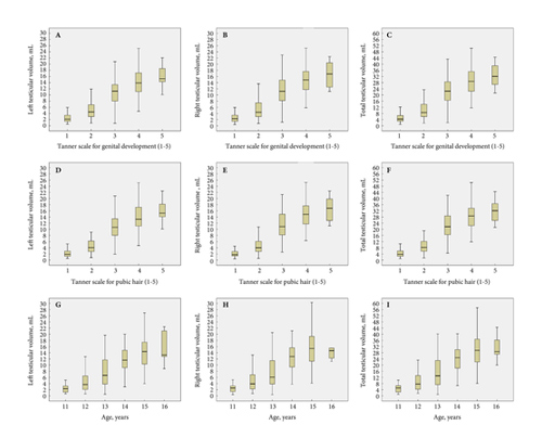
We conducted an observational cross‐sectional study from April 2015 until December 2016 in which we recruited 539 adolescent boys aged 11–16 years. A clinical examination including testicular size measurement by ultrasonography was performed. Testicular volume (TV) was calculated using the Lambert formula (length × width × height × 0.71). The Testicular Atrophy Index (TAI) was calculated using the formula [(TV right – TV left)/largest TV] × 100. The data for all statistical analyses were stratified for Tanner stage for genital development (TSG) and pubic hair (TSP). Non‐parametric tests were used to assess the difference between right and left TV, and the prevalence of a smaller left testis for the entire population, and between each TSG and TSP. Parametric tests were used to determine the difference in mean TAI between each TSG and TSP, and to compare the mean TAI to a test value of 0.

Results

Of the 539 recruited boys, we excluded 194 due to a current or past pathology, including varicocoeles, influencing normal (testicular) growth or due to incomplete data. Most boys were in the second Tanner stage, followed by the third Tanner stage. The mean (sd) age of the entire population was 13.33 (1.25) years. Of the 345 included participants the mean (sd) left TV was 7.67 (5.63) mL and right TV was 7.97 (5.90) mL. The mean (sd) TAI was 2.85 (17.00)%. In all, 203 (58.84%) boys had a smaller left testis and 142 (41.16%) had a smaller right testis. In all, 51 boys (14.78%) had a TAI >20%, 45 (13.04%) had a TV difference (TVD) of >2 mL with a deficit in left TV, and 69 (20.00)% had a TAI >20% or a TVD of >2 mL with a deficit in left TV. Related‐samples Wilcoxon signed‐rank test showed a significant difference in mean left and right TV for the entire population, and more specifically for TSG3 (P < 0.001) and TSP3 (P = 0.004). A one‐sample t‐test showed a significant difference in the mean TAI vs the test value of 0 for the entire population (P = 0.002), and more specifically for TSG3 (P < 0.001) and TSP3 (P = 0.003).

Conclusion

Testicular asymmetry, with a smaller left testis, was seen in a considerable number of healthy adolescents. One out of five adolescents had a smaller left testis and met one of the threshold values currently used in varicocoele management. Therefore, in left‐sided unilateral inguinoscrotal pathology, a smaller ipsilateral testis in combination with a TAI of >20% and/or TVD of >2 mL requires careful interpretation and serial measurements of TV should always be performed. Furthermore, this study provides reference values for TV, TVD and TAI according to TSG and TSP for a healthy adolescent population.

Read more articles of the week

Video: Testicular asymmetry in healthy adolescent boys

Testicular asymmetry in healthy adolescent boys


Read the full article

Abstract

Objectives

To assess the presence of testicular asymmetry and the currently used threshold values in varicocoele management in a healthy adolescent population.

Subjects and Methods

We conducted an observational cross‐sectional study from April 2015 until December 2016 in which we recruited 539 adolescent boys aged 11–16 years. A clinical examination including testicular size measurement by ultrasonography was performed. Testicular volume (TV) was calculated using the Lambert formula (length × width × height × 0.71). The Testicular Atrophy Index (TAI) was calculated using the formula [(TV right – TV left)/largest TV] × 100. The data for all statistical analyses were stratified for Tanner stage for genital development (TSG) and pubic hair (TSP). Non‐parametric tests were used to assess the difference between right and left TV, and the prevalence of a smaller left testis for the entire population, and between each TSG and TSP. Parametric tests were used to determine the difference in mean TAI between each TSG and TSP, and to compare the mean TAI to a test value of 0.

Results

Of the 539 recruited boys, we excluded 194 due to a current or past pathology, including varicocoeles, influencing normal (testicular) growth or due to incomplete data. Most boys were in the second Tanner stage, followed by the third Tanner stage. The mean (sd) age of the entire population was 13.33 (1.25) years. Of the 345 included participants the mean (sd) left TV was 7.67 (5.63) mL and right TV was 7.97 (5.90) mL. The mean (sd) TAI was 2.85 (17.00)%. In all, 203 (58.84%) boys had a smaller left testis and 142 (41.16%) had a smaller right testis. In all, 51 boys (14.78%) had a TAI >20%, 45 (13.04%) had a TV difference (TVD) of >2 mL with a deficit in left TV, and 69 (20.00)% had a TAI >20% or a TVD of >2 mL with a deficit in left TV. Related‐samples Wilcoxon signed‐rank test showed a significant difference in mean left and right TV for the entire population, and more specifically for TSG3 (P < 0.001) and TSP3 (P = 0.004). A one‐sample t‐test showed a significant difference in the mean TAI vs the test value of 0 for the entire population (P = 0.002), and more specifically for TSG3 (P < 0.001) and TSP3 (P = 0.003).

Conclusion

Testicular asymmetry, with a smaller left testis, was seen in a considerable number of healthy adolescents. One out of five adolescents had a smaller left testis and met one of the threshold values currently used in varicocoele management. Therefore, in left‐sided unilateral inguinoscrotal pathology, a smaller ipsilateral testis in combination with a TAI of >20% and/or TVD of >2 mL requires careful interpretation and serial measurements of TV should always be performed. Furthermore, this study provides reference values for TV, TVD and TAI according to TSG and TSP for a healthy adolescent population.


View more videos
© 2024 BJU International. All Rights Reserved.