Tag Archive for: prostatic neoplasms

Posts

Article of the week: Biparametric vs multiparametric prostate MRI for the detection of PCa in treatment‐naïve patients

Every week, the Editor-in-Chief selects an Article of the Week from the current issue of BJUI. The abstract is reproduced below and you can click on the button to read the full article, which is freely available to all readers for at least 30 days from the time of this post.

In addition to the article itself, there is an editorial written by a prominent member of the urological community, and a video produced by the authors. These are intended to provoke comment and discussion and we invite you to use the comment tools at the bottom of each post to join the conversation. 

If you only have time to read one article this week, it should be this one.

Biparametric vs multiparametric prostate magnetic resonance imaging for the detection of prostate cancer in treatment-naïve patients: a diagnostic test accuracy systematic review and meta-analysis

Mostafa Alabousi*, Jean-Paul Salameh†‡, Kaela Gusenbauer§, Lucy Samoilov, Ali Jafri**, Hang Yu§ and Abdullah Alabousi††


*Department of Radiology, McMaster University, Hamilton, Department of Clinical Epidemiology and Public Health, University of Ottawa, The Ottawa Hospital Research Institute, Clinical Epidemiology Program, Ottawa, §Department of Medicine, McMaster University, Hamilton, Department of Medicine, Western University, London, ON, Canada, **Department of Medicine, New York Institute of Technology School of Osteopathic Medicine, Glen Head, NY, USA, and ††Department of Radiology, St Joseph’s Healthcare, McMaster University, Hamilton, ON, Canada

Abstract

Objective

To perform a diagnostic test accuracy (DTA) systematic review and meta‐analysis comparing multiparametric (diffusion‐weighted imaging [DWI], T2‐weighted imaging [T2WI], and dynamic contrast‐enhanced [DCE] imaging) magnetic resonance imaging (mpMRI) and biparametric (DWI and T2WI) MRI (bpMRI) in detecting prostate cancer in treatment‐naïve patients.

Methods

The Medical Literature Analysis and Retrieval System Online (MEDLINE) and Excerpta Medica dataBASE (EMBASE) were searched to identify relevant studies published after 1 January 2012. Articles underwent title, abstract, and full‐text screening. Inclusion criteria consisted of patients with suspected prostate cancer, bpMRI and/or mpMRI as the index test(s), histopathology as the reference standard, and a DTA outcome measure. Methodological and DTA data were extracted. Risk of bias was assessed using the Quality Assessment of Diagnostic Accuracy Studies (QUADAS)‐2 tool. DTA metrics were pooled using bivariate random‐effects meta‐analysis. Subgroup analysis was conducted to assess for heterogeneity.

Results

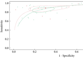
From an initial 3502 studies, 31 studies reporting on 9480 patients (4296 with prostate cancer) met the inclusion criteria for the meta‐analysis; 25 studies reported on mpMRI (7000 patients, 2954 with prostate cancer) and 12 studies reported on bpMRI DTA (2716 patients, 1477 with prostate cancer). Pooled summary statistics demonstrated no significant difference for sensitivity (mpMRI: 86%, 95% confidence interval [CI] 81–90; bpMRI: 90%, 95% CI 83–94) or specificity (mpMRI: 73%, 95% CI 64–81; bpMRI: 70%, 95% CI 42–83). The summary receiver operating characteristic curves were comparable for mpMRI (0.87) and bpMRI (0.90).

Conclusions

No significant difference in DTA was found between mpMRI and bpMRI in diagnosing prostate cancer in treatment‐naïve patients. Study heterogeneity warrants cautious interpretation of the results. With replication of our findings in dedicated validation studies, bpMRI may serve as a faster, cheaper, gadolinium‐free alternative to mpMRI.


Article of the month: Prostate cancer mortality rates in Peru and its geographical regions

Every month, the Editor-in-Chief selects an Article of the Month from the current issue of BJUI. The abstract is reproduced below and you can click on the button to read the full article, which is freely available to all readers for at least 30 days from the time of this post.

In addition to the article itself, there is an editorial written by a prominent member of the urological community. These are intended to provoke comment and discussion and we invite you to use the comment tools at the bottom of each post to join the conversation. 

If you only have time to read one article this week, it should be this one.

Prostate cancer mortality rates in Peru and its geographical regions

Junior Smith Torres-Roman*, Eloy F. Ruiz, Jose Fabian Martinez-Herrera§, Sonia Faria Mendes Braga, Luis Taxa**, Jorge Saldaña-Gallo*, Mariela R. Pow-Sang††, Julio M. Pow-Sang‡‡ and Carlo La Vecchia§§


*Clinica de Urologia Avanzada UROZEN, Lima, Facultad de Medicina Humana, Universidad Nacional San Luis Gonzaga, Ica, CONEVID, Unidad de Conocimiento y Evidencia, Universidad Peruana Cayetano Heredia, Lima, Peru, §Cancer Center, Medical Center American British Cowdray, Mexico City, Mexico, Department of Social and Preventive Medicine, Faculty of Medicine, Universidade Federal de Minas Gerais, Belo Horizonte, Brazil, **Instituto Nacional de Enfermedades Neoplásicas, ††Department of Urology, Instituto Nacional de Enfermedades Neoplásicas, Lima, Peru, ‡‡Department of Genitourinary Oncology, Moffitt Cancer Center, Tampa, FL, USA, and §§Department of Clinical Sciences and Community Health, Universitá degli Studi di Milano, Milan, Italy


Read the full article

Abstract

Objective

To evaluate the mortality rates for prostate cancer according to geographical areas in Peru between 2005 and 2014.

Materials and Methods

Information was extracted from the Deceased Registry of the Peruvian Ministry of Health. We analysed age‐standardised mortality rates (world population) per 100 000 men. Spatial autocorrelation was determined according to the Moran Index. In addition, we used Cluster Map to explore relations between regions.

Fig. 1. Peru geographical zones by provinces. The asterisk denotes the province of Callao. Source: National Statistics Institute

Results

Mortality rates increased from 20.9 (2005–2009) to 24.1 (2010–2014) per 100 000 men, an increase of 15.2%. According to regions, during the period 2010–2014, the coast had the highest mortality rate (28.9 per 100 000), whilst the rainforest had the lowest (7.43 per 100 000). In addition, there was an increase in mortality in the coast and a decline in the rainforest over the period 2005–2014. The provinces with the highest mortality were Piura, Lambayeque, La Libertad, Callao, Lima, Ica, and Arequipa. Moreover, these provinces (except Arequipa) showed increasing trends during the years under study. The provinces with the lowest observed prostate cancer mortality rates were Loreto, Ucayali, and Madre de Dios. This study showed positive spatial autocorrelation (Moran’s I: 0.30, P= 0.01).

Conclusion

Mortality rates from prostate cancer in Peru continue to increase. These rates are higher in the coastal region compared to those in the highlands or rainforest.

Read more Articles of the week


Editorial: The burden of urological cancers in low‐ and middle‐income countries

The burden of cancer in low‐ and middle‐income countries (LMICs) continues to rise [1]. Evaluation of geographical differences in cancer mortality statistics is specifically of interest in LMICs as (inter)national guidelines are potentially less embedded in standard care, and objective measurements to assess underlying mechanisms/explanations for the burden of cancer are often lacking. Monitoring mortality statistics in these countries can thus help assess the effectiveness of national and regional health systems in treating and caring for patients with cancer [1].

Torres‐Roman et al. [2] deserve to be congratulated for their efforts to monitor mortality rates for prostate cancer at both a regional and national level in Peru. The CONCORD initiative from the WHO previously reported prostate cancer statistics for Peru, but data were limited to the capital area of Lima [1]. Torres‐Raman et al. [2] report prostate cancer mortality rates between 2005 and 2014 based on data from the Peruvian Ministry of Health, which covers ~70% of all healthcare providers in Peru. Apart from an overall increase of 15% in mortality rates, substantial variation was observed by geographical region. Mortality rates increased by 16% in the coastal region and highlands, whereas in the rainforest region the rates decreased by 19% [2]. One potential explanation for these observed differences could be the difference in ethnic and racial characteristics. The coastal region in Peru has a strong African influence and also has a larger proportion of men aged >65 years. In addition to potential differences in access to healthcare, some of the variation in prostate cancer mortality statistics most likely reflects a deficiency in reporting systems. Even though this study has its limitations due to missing data and lack of information on other important variables, such as ethnicity and socioeconomic status, it provides a first base for a critical assessment of prostate cancer care in Peru.

Studies like this one from Torres‐Roman et al. [2] show that there is a need for improvement and standardisation of (prostate) cancer care in LMICs, but also a need for improvement in data capturing, so that objective measurements can be put in place. The years of healthy life lost due to prostate cancer, as well as other urological cancers, in LMICs is increasing substantially. Even though each tumour group has its own specifications in terms of prevention and control, an epidemiological assessment of cancer burden based on the experience for urological cancers (i.e., prostate, bladder, kidney and testicular) can therefore inform future assessments of cancer burden. The urological tumour group covers both common and less common cancers (e.g. prostate vs kidney cancer), sex‐specific and cancers that affect both sexes (e.g. testicular vs bladder cancer), cancers with less known risk factors and those strongly linked with lifestyle risk factors (e.g. prostate vs bladder cancer).

It is encouraging to see an increase in the number of studies evaluating the burden of cancer in LMICs [3]; however, given the consistency in observations of an increase in mortality, there is an urgent need to further invest in prevention and management, as well as the infrastructure to collect all relevant data at a national level in these LMICs. Accurate information about cancer burden and how this varies between regions is essential to plan for an adequate health‐system response.

References

  1. Allemani, CMatsuda, TCarlo, V et al. Global surveillance of trends in cancer survival 2000‐14 (CONCORD‐3): analysis of individual records for 37 513 025 patients diagnosed with one of 18 cancers from 322 population‐based registries in 71 countries. Lancet 20183911023– 75
  2. Torres‐Roman, JRuiz, EMartinez‐Herrera, J et al. Prostate cancer mortality rates in Peru and its geographic regions. BJU Int 2019123595– 601
  3. Carioli, GVecchia, CBertuccio, P et al. Cancer mortality predictions for 2017 in Latin America. Ann Oncol 2017282286– 97


Article of the week: Ultrasound characteristics of regions identified as suspicious by MRI predict the likelihood of clinically significant cancer on MRI–ultrasound fusion‐targeted biopsy

Every week, the Editor-in-Chief selects an Article of the Week from the current issue of BJUI. The abstract is reproduced below and you can click on the button to read the full article, which is freely available to all readers for at least 30 days from the time of this post.

In addition to the article itself, there is an editorial written by a prominent member of the urological community, and a video made by the authors. These are intended to provoke comment and discussion and we invite you to use the comment tools at the bottom of each post to join the conversation. 

If you only have time to read one article this week, it should be this one.


The ultrasound characteristics of regions identified as suspicious by magnetic resonance imaging (MRI) predict the likelihood of clinically significant cancer on MRI–ultrasound fusion‐targeted biopsy

Benjamin Press*, Andrew B. Rosenkrantz, Richard Huang and Samir S. Taneja§ 
 
*Rutgers New Jersey Medical School, Newark, NJ, Department of Radiology, Department of Urology, and §Departments of Urology and Radiology, NYU Langone Health, New York, NY, USA
 
Read the full article

Abstract

Objective

To determine whether the presence of an ultrasound hypoechoic region at the site of a region of interest (ROI) on magnetic resonance imaging (MRI) results in improved prostate cancer (PCa) detection and predicts clinically significant PCa on MRI–ultrasonography fusion‐targeted prostate biopsy (MRF‐TB).

Materials and Methods

Between July 2011 and June 2017, 1058 men who underwent MRF‐TB, with or without systematic biopsy, by a single surgeon were prospectively entered into an institutional review board‐approved database. Each MRI ROI was identified and scored for suspicion by a single radiologist, and was prospectively evaluated for presence of a hypoechoic region at the site by the surgeon and graded as 0, 1 or 2, representing none, a poorly demarcated ROI‐HyR, or a well demarcated ROI‐HyR, respectively. The interaction of MRI suspicion score (mSS) and ultrasonography grade (USG), and the prediction of cancer detection rate by USG, were evaluated through univariate and multivariate analysis.

Results

For 672 men, the overall and Gleason score (GS) ≥7 cancer detection rates were 61.2% and 39.6%, respectively. The cancer detection rates for USGs 0, 1 and 2 were 46.2%, 58.6% and 76.0% (P < 0.001) for any cancer, and 18.7%, 35.2% and 61.1% (P < 0.001) for GS ≥7 cancer, respectively. For MRF‐TB only, the GS ≥7 cancer detection rates for USG 0, 1 and 2 were 12.8%, 25.7% and 52.0%, respectively (P < 0.001). On univariate analysis, in men with mSS 2–4, USG was predictive of GS ≥7 cancer detection rate. Multivariable regression analysis showed that USG, prostate‐specific antigen density and mSS were predictive of GS ≥7 PCa on MRF‐TB.

Conclusions

Ultrasonography findings at the site of an MRI ROI independently predict the likelihood of GS ≥7 PCa, as men with a well‐demarcated ROI‐HyR at the time of MRF‐TB have a higher risk than men without.
Read more Articles of the week

Editorial: Is transrectal ultrasonography of the prostate obsolete in the MRI era?

Sampling of prostate tissue to confirm pathologically a clinical suspicion of cancer has undergone an exponential change. The random systematic prostate biopsy technique was the only method used for many decades, initially guided by the finger but, since 1989, performed with TRUS guidance. Now, within the space of only a few years, we have entered the era of performing prostate biopsies on the basis of high‐tech three‐dimensional multiparametric MRI images, including software that can track the exact course of the biopsy needle [1]. While new technical developments in general lead to better, more individually directed healthcare, there is always the risk of abandoning ‘old’ but well developed and extensively tested techniques too soon. In this issue of the BJUI, Press et al. [2] looked at the added value of the presence of an ‘old‐fashioned’ TRUS‐detected lesion in cancer‐suspicious regions on MRI to better predict the presence of clinically significant prostate cancer (csPCa) defined as Gleason score ≥7. In their study comprising 1058 men, it was shown that a well‐demarcated abnormal TRUS finding noted at the time of MRI‐TRUS fusion‐guided prostate biopsy coincides with an increased risk of csPCa detection, independent of MRI suspicion (Prostate Imaging Reporting and Data System [PI‐RADS] score).

Increasing PI‐RADS score is correlated with an increased percentage of csPCa after targeted biopsy, both at initial and repeat biopsy. In a review based on data from 8252 men, it was shown that there is a gradual increase in the detection of csPCa from PI‐RADS 3 to PI‐RADS 4 to PI‐RADS 5 index lesions. For example, at first biopsy, the overall rate of PCa detection and the percentage of csPCa were 39%, 62% and 92% and 54%, 63% and 76% for PI‐RADS 3, 4 and 5 lesions, respectively. This means that in men with PI‐RADS 3 lesions, representing approximately one‐third of men deemed eligible for further assessment, only 39% will be diagnosed with PCa and half of the PCa detected will be potentially indolent Gleason 6 PCa [3]. This makes this group of men extremely interesting for further risk stratification before biopsy. Multivariable risk stratification in which PSA density plays an important role has been shown to be of value in these men [4] but further refinement could potentially be made by including suspicious lesions identified at TRUS.

Apart from the added value of TRUS findings in terms of risk stratification, the performance of the MRI‐targeted biopsy itself could be improved by visual guidance of hypoechoic lesions. In the present study by Press et al [2], a hypoechoic TRUS lesion was present at or near the location of two‐thirds of cancer‐suspicious lesions on MRI. The authors more or less advise to direct the targeted biopsy cores not only to the MRI suspicious lesion, but also the TRUS suspicious lesion, both of which often do not fully overlay in a software‐assisted MRI‐TRUS fusion model. The extent to which this ‘correction for misregistration’ is already included during targeted biopsy in current clinical practice is unknown. Although feasible and seemingly important during software‐assisted fusion targeted biopsy, TRUS lesions in cancer‐suspicious MRI regions might be more frequently targeted during cognitive fusion‐targeted biopsy. Two recent studies underline the important message of the present study, and show that a considerable proportion of csPCa is missed in and around MRI‐suspicious lesions by targeted biopsies, as a result of sampling errors related to both misregistration and intra‐tumour heterogeneity [56]. As suggested by these studies, visual guidance by hypoechoic lesions and ‘focal saturation’ biopsy by additional (peri‐)lesional cores might improve the detection of csPCa.

In summary, ‘good old’ TRUS could be of value in those patients who are virtually always present in scenarios in which a grading system is being used, i.e. patients belonging to the so‐called grey zone. The challenge of risk stratification (i.e. personalized medicine) is to nibble at both sides of the grey zone by implementing new techniques or, more likely by implementing a combination of all available and relevant knowledge.

by Monique J. Roobol, Frank-Jan H. Drost and Arnout R. Alberts

References

  1. Verma, SChoyke, PLEberhardt, SC et al. The current state of MR imaging‐targeted biopsy techniques for detection of prostate cancer. Radiology 201728534356
  2. Press, BRosenkrantz, ABHuang, RTaneja, SSThe ultrasound characteristics of MRI suspicious regions predict the likelihood of clinically significant cancer on MRI‐ultrasound fusion targeted biopsy. BJUI 201912343946.
  3. Schoots, IGMRI in early prostate cancer detection: how to manage indeterminate or equivocal PI‐RADS 3 lesions? Transl Androl Urol 201877082
  4. Alberts, ARSchoots, IGBokhorst, LPLeenders, GJBangma, CHRoobol, MJRisk‐based patient selection for magnetic resonance imaging‐targeted prostate biopsy after negative transrectal ultrasound‐guided random biopsy avoids unnecessary magnetic resonance imaging scans. Eur Urol 201669112934
  5. Simmons, LAMKanthabalan, AArya, M et al. Accuracy of transperineal targeted prostate biopsies, visual estimation and image fusion in men needing repeat biopsy in the PICTURE trial. J Urol 2018200122734
  6. Leest, M, Cornel, EIsrael, B et al. Head‐to‐head comparison of transrectal ultrasound‐guided prostate biopsy versus multiparametric prostate resonance imaging with subsequent magnetic resonance‐guided biopsy in biopsy‐naive men with elevated prostate‐specific antigen: a large prospective multicenter clinical study. Eur Urol 2018; [Epub ahead of print]. https://doi.org/10.1016/j.eururo.2018.11.023.


Article of the week: Adjuvant radiation with androgen‐deprivation therapy for men with lymph node metastases after radical prostatectomy

Every week, the Editor-in-Chief selects an Article of the Week from the current issue of BJUI. The abstract is reproduced below and you can click on the button to read the full article, which is freely available to all readers for at least 30 days from the time of this post.

In addition to the article itself, there is an editorial written by a prominent member of the urological community. These are intended to provoke comment and discussion and we invite you to use the comment tools at the bottom of each post to join the conversation. 

If you only have time to read one article this week, it should be this one.

Adjuvant radiation with androgen‐deprivation therapy for men with lymph node metastases after radical prostatectomy: identifying men who benefit

Mohit Gupta*, Hiten D. Patel*, Zeyad R. Schwen*, Phuoc T. Tran*† and Alan W. Partin*


*Department of Urology, James Buchanan Brady Urological Institute, and Department of Radiation Oncology and Molecular Radiation Sciences and Oncology, The Sidney Kimmel Comprehensive Cancer Center, Johns Hopkins Medical Institutions, Baltimore, MD, USA


Read the full article

Abstract

Objectives

To perform a comparative analysis of three current management strategies for patients with lymph node metastases (LNM; pN1) following radical prostatectomy (RP): observation, androgen‐deprivation therapy (ADT), and external beam radiation therapy (EBRT) + ADT.

Patients and Methods

Patients with LNM after RP were identified using the National Cancer Database (2004–2013). Exclusion criteria included any use of radiation therapy or ADT before RP, clinical M1 disease, or incomplete follow‐up data. Patients were categorised according to postoperative management strategy. The primary outcome was overall survival (OS). Kaplan–Meier curves and adjusted multivariable Cox proportional hazards models were employed. Sub‐analyses further evaluated patient risk stratification and time to receipt of adjuvant therapy.

Results

A total of 8 074 patients met the inclusion criteria. Postoperatively, 4 489 (55.6%) received observation, 2 065 (25.6%) ADT, and 1 520 (18.8%) ADT + EBRT. The mean (median; interquartile range) follow‐up was 52.3 (48.0; 28.5–73.5) months. Patients receiving ADT or ADT + EBRT had higher pathological Gleason scores, T‐stage, positive surgical margin rates, and nodal burden. Adjusted multivariable Cox models showed improved OS for ADT + EBRT vs observation (hazard ratio [HR] 0.77, 95% confidence interval [CI] 0.64–0.94; P = 0.008) and vs ADT (HR 0.76, 95% CI: 0.63–0.93; P = 0.007). There was no difference in OS for ADT vs observation (HR 1.01, 95% CI: 0.87–1.18; P = 0.88). Findings were similar when restricting adjuvant cohorts for timing of adjuvant therapy. There was no difference in OS between groups for up to 2 549 (31.6%) patients lacking any of the following adverse features: ≥pT3b disease, Gleason score ≥9, three or more positive nodes, or positive surgical margin.

Conclusions

For patients with LNM after RP, the use of adjuvant ADT + EBRT improved OS in the majority of patients, especially those with adverse pathological features. Conversely, adjuvant therapy did not confer significant OS benefit in up to 30% of patients without high‐risk features, who may be managed with observation and forego the morbidity associated with immediate ADT or radiation.

Read more Articles of the week

Editorial: Postoperative radiation and hormonal therapy for men with node‐positive prostate cancer: a new standard?

The best management strategy for men with pathologically node‐positive (pN+) prostate cancer after radical prostatectomy (RP) has been debated for decades [1]. In the 1990s, the Radiation Therapy and Oncology Group (RTOG) initiated the RTOG 9608 trial to test the impact of radiotherapy (RT) and androgen‐deprivation therapy (ADT) in this setting. However, due to the rise in PSA screening and the practice of treating high‐risk prostate cancer with primary RT, the incidence of pN+ disease fell. Consequently, the trial closed due to poor accrual and the question faded in prominence. Today, both trends have reversed. PSA screening is less common and men with high‐risk prostate cancer are more frequently opting for RP. As such, physicians increasingly face the dilemma of pN+ disease. Guidelines provide little assistance, as they support everything from observation to multimodal treatment with RT and ADT. Patients and providers want to know, is there a standard treatment for all patients, and if not, how should one choose between such disparate options?

To answer these questions, one must start with the little randomised data that exist in this setting. The seminal trial by Messing et al. [1] randomised men with pN+ prostate cancer to ADT or observation with initiation of ADT after the development of symptomatic progression or distant metastases. ADT clearly improved overall survival and prostate cancer‐specific survival. However, critics noted the relatively poor outcomes in the observation group and the small sample size. Later, retrospective studies called the benefit of immediate ADT into question [2].

Against this backdrop, it is interesting that Gupta et al. [3] found the most common management approach in the USA National Cancer Database (NCDB) was observation rather than immediate ADT. Despite the randomised data, the cumulative side‐effects from lifelong ADT in a cohort of patients with no disease‐related symptoms and a median survival of well over 10 years are unappealing. Ultimately, many men do not appear to be willing to endure the diminished quality of life in exchange for a small improvement in quantity of life.

In contrast to the non‐curative nature of ADT, the possibility exists that the combination of postoperative RT and ADT could provide durable disease control, perhaps even without lifelong ADT. The data reported by Gupta et al. [3] in this edition of the BJUI provide support for this paradigm. These data add to a growing body of literature [4] that tells a consistent story with two common themes: (i) postoperative RT with ADT appears to be associated with improved survival in men with pN+ prostate cancer, and (ii) RT appears to convey the largest benefit to men with certain high‐risk pathological features. Should this body of literature lead us to eschew the old standard and advise observation for low‐risk men and RT with ADT for men at higher risk?

Before a new standard is declared, the limitations of retrospective population‐based research must be addressed. The authors performed a sophisticated analysis to reduce the impact of selection bias. However, due to the limitation of the available data, the authors were not able to account for possibly the most important variable: the postoperative PSA. One study showed that men with pN+ disease with a persistent PSA had an 8‐year clinical recurrence rate of 69% vs 12% for those with undetectable PSA [5].

It is likely that men with persistent PSA in the NCDB would have received immediate ADT with or without RT rather than observation. As such, one must be cautious of the similar survival between the observation and ADT group, especially in light of contradictory randomised data. That being said, it is reasonable for some men to conclude that the side‐effects of ADT outweigh the potential benefit, especially those with low‐risk features such as an undetectable postoperative PSA, low Gleason score, and limited lymph node involvement.

As RT with ADT appears superior to either observation or ADT alone, should more men receive RT? Probably. Of the men with high‐risk features, only 22% actually received postoperative RT. Should postoperative RT now be considered the standard for all men? Probably not. Whilst it appears that some men may indeed benefit from RT, the possibility of selection bias driving this result is real. Even if there is a true effect, identifying which patients harbour residual local disease, but do not already have subclinical distant metastatic disease is challenging. RT for all would lead to unnecessary side‐effects for men that would not benefit from the treatment. Ultimately, a randomised trial will be required to establish the benefit of RT and to define subgroups of men that may or may not benefit. Until then, we will continue to rely on excellent work like the accompanying paper from Gupta et al. [3] to identify men who may benefit from postoperative RT and ADT.

References

  1. Messing EM, Manola J, Sarosdy M, Wilding G, Crawford ED, Trump D. Immediate hormonal therapy compared with observation after radical prostatectomy and pelvic lymphadenectomy in men with node‐positive prostate cancer. N Engl J Med 1999341: 1781–8
  2. Wong YN, Freedland S, Egleston B, Hudes G, Schwartz JS, Armstrong K. Role of androgen deprivation therapy for node‐positive prostate cancer. J Clin Oncol 200927: 100–5
  3. Gupta M, Patel HD, Schwen ZR, Tran PT, Partin AW. Adjuvant radiation with androgen deprivation therapy for men with lymph node metastases following radical prostatectomy: identifying men who benefit. BJU Int 2019123: 252–60
  4. Abdollah F, Karnes RJ, Suardi N et al. Impact of adjuvant radiotherapy on survival of patients with node‐positive prostate cancer. J Clin Oncol 201432: 3939–47
  5. Bianchi L, Nini A, Bianchi M et al. The role of prostate‐specific antigen persistence after radical prostatectomy for the prediction of clinical progression and cancer‐specific mortality in node‐positive prostate cancer patients. Eur Urol 201669: 1142–8


Article of the Week: The implications of baseline bone‐health assessment at initiation of androgen‐deprivation therapy for prostate cancer

Every Week, the Editor-in-Chief selects an Article of the Week from the current issue of BJUI. The abstract is reproduced below and you can click on the button to read the full article, which is freely available to all readers for at least 30 days from the time of this post.

In addition to the article itself, there is an accompanying editorial written by a prominent member of the urological community. This blog is intended to provoke comment and discussion and we invite you to use the comment tools at the bottom of each post to join the conversation.

If you only have time to read one article this month, it should be this one.

The implications of baseline bone‐health assessment at initiation of androgen‐deprivation therapy for prostate cancer


Peter S. Kirk* , Tudor Borza*, Vah akn B. Shahinian, Megan E.V. Caram§Danil V. Makarov**, Jeremy B. Shelton††, John T. Leppert‡‡§§, Ryan M. Blake*, Jennifer A. Davis§, Brent K. Hollenbeck*, Anne Sales§¶¶ and Ted A. Skolarus *§


*Dow Division of Health Services Research, Department of Urology, Division of Nephrology, Department of Internal Medicine, Division of Hematology and Oncology, Department of Internal Medicine, University of Michigan Health System, §Veterans Affairs (VA) Health Services Research and Development, Center for Clinical Management Research, VA Ann Arbor Healthcare System, University of Michigan Medical School, Ann Arbor, MI, USA, Departments of Urology and Population Health, NYU Langone Medical Center, New York City, NY, USA, **VA New York Healthcare System, New York City, NY, USA, ††VA Greater Los Angeles Healthcare System, Los Angeles City, LA, USA, ‡‡Department of Urology, Stanford University School of Medicine, Stanford, CA, USA, §§VA Palo Alto Healthcare System, Palo Alto, CA, USA, and ¶¶Department of Learning Health Sciences, University of Michigan Medical School, Ann Arbor, MI, USA


Read the full article

Abstract

Objectives

To assess bone‐density testing (BDT) use amongst prostate cancer survivors receiving androgen‐deprivation therapy (ADT), and downstream implications for osteoporosis and fracture diagnoses, as well as pharmacological osteoporosis treatment in a national integrated delivery system.

Patients and methods

We identified 17 017 men with prostate cancer who received any ADT between 2005 and 2014 using the Veterans Health Administration cancer registry and administrative data. We identified claims for BDT within a 3‐year period of ADT initiation. We then used multivariable regression to examine the association between BDT use and incident osteoporosis, fracture, and use of pharmacological treatment.

Results

We found that a minority of patients received BDT (n = 2 502, 15%); however, the rate of testing increased to >20% by the end of the study period. Men receiving BDT were older at diagnosis and had higher‐risk prostate cancer (both P < 0.001). Osteoporosis and fracture diagnoses, use of vitamin D ± calcium, and bisphosphonates were all more common in men who received BDT. After adjustment, BDT, and to a lesser degree ≥2 years of ADT, were both independently associated with incident osteoporosis, fracture, and osteoporosis treatment.

Conclusions

BDT is rare amongst patients with prostate cancer treated with ADT in this integrated delivery system. However, BDT was associated with substantially increased treatment of osteoporosis indicating an underappreciated burden of osteoporosis amongst prostate cancer survivors initiating ADT. Optimising BDT use and osteoporosis management in this at‐risk population appears warranted.

Read more articles of the week


Editorial: Low rates of bone density testing in prostate cancer survivors on androgen‐deprivation therapy: where do we go from here?

In this month’s issue of the BJU International, Kirk et al. 1 describe their findings regarding an important issue in the care of prostate cancer survivors on androgen‐deprivation therapy (ADT): the underuse of bone density testing (BDT) to screen for osteoporosis. ADT is the commonest systemic therapy in patients with prostate cancer, used in both metastatic and localised settings. Whilst it has clear survival benefits, ADT is also associated with harms including cardiovascular, cognitive, and metabolic side‐effects, as well as an increased risk of osteoporosis and fractures. These bone‐related complications are costly from a quality‐of‐life and financial perspective, especially given the critical importance of mobility in maintaining performance status and cardiovascular health during cancer treatment 23. Consequently, most clinical practice guidelines include osteoporosis screening as a recommendation for men undergoing ADT.

In their study, ‘The implications of baseline bone health assessment at initiation of androgen‐deprivation therapy for prostate cancer’, the authors describe patterns of use of BDT and diagnosis of osteoporosis amongst men treated for prostate cancer in the USA Veterans Affairs (VA) system within a 3‐year period following ADT initiation. There was a statistically significant increase in the BDT rate throughout the study period; however, overall BDT remains uncommon amongst patients with prostate cancer on ADT, used in only 15% of men in their cohort. Unsurprisingly, patients who received BDT were more likely to be diagnosed with osteoporosis, be diagnosed with a fracture, and receive treatment with vitamin D, calcium and bisphosphonates. The authors acknowledge an important limitation about the applicability of their VA study to the civilian health population. However, given that the VA and military health systems perform as well, if not better, on several important metrics in prostate cancer care 45, these results should not be ignored simply because they were obtained in the military health system.

The increase in BDT screening throughout the study may be attributable to increased awareness of guidelines published during the study period. However, the overall BDT rate remains low. This may be explained by insufficient access, lack of information technology, as well as more nebulous aspects of care such as physician culture, beliefs, and habits 6.

Studies such as this are vital to identify opportunities for improving care delivery. What are needed next are innovations to optimise the delivery of care for patients treated with ADT. Whilst improving BDT adherence may lack the cachet of next‐generation targeted therapies, this is an example of the kind of simple, measurable area where improvement in care delivery systems may yield large benefits.

There are many possible avenues for success: quality improvement collaboratives are one well‐known innovation, which may be applicable to this area: examples, such as the Michigan Urological Surgery Improvement Collaborative (MUSIC) and the AUA Quality Registry (AQUA) are success stories, but to our knowledge there are no published studies specifically attempting to improve adherence to BDT guidelines within these cohorts. Other practice‐based innovations include navigators and multidisciplinary cancer teams, either of which may yield improvements in guideline adherence. Online patient support groups can raise awareness. And although we all know how electronic reminders have frustrated countless physicians, electronic reminders about recommended tests and interventions may be an important tool. At our institution, a Prostate Cancer Foundation grant is funding the development of a mobile health app, which is targeted exclusively at men receiving ADT for prostate cancer. This app will encourage physical activity and healthy eating, which can both support bone health.

In our view, the issue of bone screening is a clear example of where innovative strategies to improve care delivery and guideline adherence may make a big difference for men living with prostate cancer. We look forward to seeing more in the years to come.


Sabrina S. Harmouch, Alexandra J. Berger, and Alexander P. Cole


Center for Surgery and Public Health, Division of Urological Surgery, Brigham and Wo mens Hospital, Harvard Medical School, Boston, MA, USA


References
  • Kirk PS, Borza T, Shahinian VB et al. The implications of baseline bone‐health assessment at initiation of androgen‐deprivation therapy for prostate cancerBJU Int2018121: 558–64


  • Cole AP, Jiang W, Lipsitz SR et al. The use of prostate specific antigen screening in purchased versus direct care settings: data from the TRICARE® military databaseJ Urol 2017198: 1295–300


  • Cullen J, Brassell SA, Chen Y et al. Racial/Ethnic patterns in prostate cancer outcomes in an active surveillance cohortProstate Cancer 20112011: 234519


Article of the Week: Chitosan membranes applied on the prostatic neurovascular bundles after nerve‐sparing robot‐assisted radical prostatectomy: a phase II study

Every Week, the Editor-in-Chief selects an Article of the Week from the current issue of BJUI. The abstract is reproduced below and you can click on the button to read the full article, which is freely available to all readers for at least 30 days from the time of this post.

In addition to the article itself, there is an accompanying editorial written by a prominent member of the urological community. This blog is intended to provoke comment and discussion and we invite you to use the comment tools at the bottom of each post to join the conversation.

If you only have time to read one article this month, it should be this one.

Chitosan membranes applied on the prostatic neurovascular bundles after nerve‐sparing robot‐assisted radical prostatectomy: a phase II study

Francesco Porpiglia* , Riccardo Bertolo*, Cristian Fiori*, Matteo Manfredi*, Sabrina De Cillis* and Stefano Geuna


*Division of Urology, Department of Oncology, and Department of Clinical and Biological Sciences, San Luigi Gonzaga Hospital, University of Turin, Orbassano, Turin, Italy


Read the full article

Abstract

Objective

To evaluate the feasibility and the safety of applying chitosan membrane (ChiMe) on the neurovascular bundles (NVBs) after nerve‐sparing robot‐assisted radical prostatectomy (NS‐RARP). The secondary aim of the study was to report preliminary data and in particular potency recovery data.

Patients and Methods

This was a single‐centre, single‐arm prospective study, enrolling all patients with localised prostate cancer scheduled for RARP with five‐item version of the International Index of Erectile Function scores of >17, from July 2015 to September 2016. All patients underwent NS‐RARP with ChiMe applied on the NVBs. The demographics, perioperative, postoperative and complications data were evaluated. Potency recovery data were evaluated in particular and any sign/symptom of local allergy/intolerance to the ChiMe was recorded and evaluated.

Results

In all, 140 patients underwent NS‐RARP with ChiMe applied on the NVBs. Applying the ChiMe was easy in almost all the cases, and did not compromise the safety of the procedure. None of the patients reported signs of intolerance/allergy attributable to the ChiMe and potency recovery data were encouraging.

Conclusion

In our experience, ChiMe applied on the NVBs after NS‐RARP was feasible and safe, without compromising the duration, difficulty or complication rate of the ‘standard’ procedure. No patients had signs of intolerance/allergy attributable to the ChiMe and potency recovery data were encouraging. A comparative cohort would have added value to the study. The present paper was performed before Conformité Européene (CE)‐mark achievement.

Read more articles of the week

© 2024 BJU International. All Rights Reserved.