Tag Archive for: #KidneyCancer

Posts

Article of the Week: Stereotactic ablative body radiotherapy for inoperable primary kidney cancer: a prospective clinical trial

Every week the Editor-in-Chief selects an Article of the Week from the current issue of BJUI. The abstract is reproduced below and you can click on the button to read the full article, which is freely available to all readers for at least 30 days from the time of this post.

In addition to the article itself, there is an accompanying editorial written by a prominent member of the urological community. This blog is intended to provoke comment and discussion and we invite you to use the comment tools at the bottom of each post to join the conversation.

If you only have time to read one article this week, it should be this one.

Stereotactic ablative body radiotherapy for inoperable primary kidney cancer: a prospective clinical trial


Shankar Siva*,, Daniel Pham*, Tomas Kron*,, Mathias Bressel*, Jacqueline Lam*, Teng Han Tan*, Brent Chesson*, Mark Shaw*, Sarat Chander*, Suki Gill*,Nicholas R. Brook§, Nathan Lawrentschuk*,, Declan G. Murphy*,† and Farshad Foroudi*,


*Peter MacCallum Cancer Centre, Melbourne, Vic., Australia, † Sir Peter MacCallum Department of Oncology, University of Melbourne, Melbourne, Vic. Australia, Sir Charles Gairdner Hospital, Nedlands Perth, WA, Australia, §Royal Adelaide Hospital, Adelaide, SA, Australia, and Olivia Newton John Cancer Centre, Heidelberg, Vic., Australia


Abstract

Objective

To assess the feasibility and safety of stereotactic ablative body radiotherapy (SABR) for renal cell carcinoma (RCC) in patients unsuitable for surgery. Secondary objectives were to assess oncological and functional outcomes.

Materials and Methods

This was a prospective interventional clinical trial with institutional ethics board approval. Inoperable patients were enrolled, after multidisciplinary consensus, for intervention with informed consent. Tumour response was defined using Response Evaluation Criteria In Solid Tumors v1.1. Toxicities were recorded using Common Terminology Criteria for Adverse Events v4.0. Time-to-event outcomes were described using the Kaplan–Meier method, and associations of baseline variables with tumour shrinkage was assessed using linear regression. Patients received either single fraction of 26 Gy or three fractions of 14 Gy, dependent on tumour size.

Results

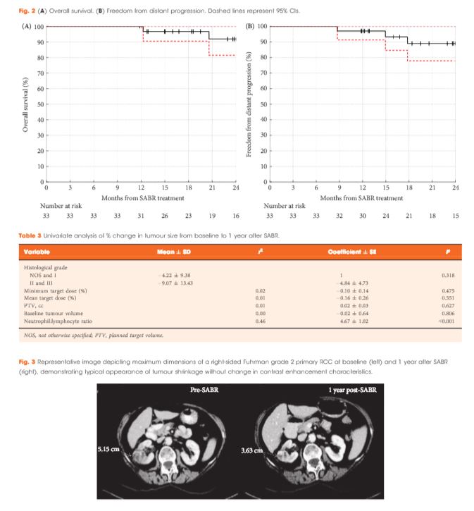
Of 37 patients (median age 78 years), 62% had T1b, 35% had T1a and 3% had T2a disease. One patient presented with bilateral primaries. Histology was confirmed in 92%. In total, 33 patients and 34 kidneys received all prescribed SABR fractions (89% feasibility). The median follow-up was 24 months. Treatment-related grade 1–2 toxicities occurred in 26 patients (78%) and grade 3 toxicity in one patient (3%). No grade 4–5 toxicities were recorded and six patients (18%) reported no toxicity. Freedom from local progression, distant progression and overall survival rates at 2 years were 100%, 89% and 92%, respectively. The mean baseline glomerular filtration rate was 55 mL/min, which decreased to 44 mL/min at 1 and 2 years (P < 0.001). Neutrophil:lymphocyte ratio correlated to % change in tumour size at 1 year, r2 = 0.45 (P < 0.001).

Conclusion

The study results show that SABR for primary RCC was feasible and well tolerated. We observed encouraging cancer control, functional preservation and early survival outcomes in an inoperable cohort. Baseline neutrophil:lymphocyte ratio may be predictive of immune-mediated response and warrants further investigation.

Residents’ Podcast: When to Perform Preoperative Chest CT for RCC Staging

Jesse Ory, Kyle Lehmann and Jeff Himmelman

Department of Urology, Dalhousie University, Halifax, NS, Canada


Read the full article

Abstract

Objectives

To provide objective criteria for preoperative staging chest computed tomography (CT) in patients diagnosed with renal cell carcinoma (RCC) because, in the absence of established indications, the decision for preoperative chest CT remains subjective.

Patients and Methods

A total of 1 946 patients undergoing surgical treatment of RCC, whose data were collected in a prospective institutional database, were assessed. The outcome of the study was presence of pulmonary metastases at staging chest CT. A multivariable logistic regression model predicting positive chest CT was fitted. Predictors consisted of preoperative clinical tumour (cT) and nodal (cN) stage, presence of systemic symptoms and platelet count (PLT)/haemoglobin (Hb) ratio.

Results

The rate of positive chest CT was 6% (n = 119). At multivariable logistic regression, ≥cT1b, cN1, systemic symptoms and Hb/PLT ratio were all associated with higher risk of positive chest CT (all P < 0.001). After 2000-sample bootstrap validation, the concordance index was found to be 0.88. At decision-curve analysis, the net benefit of the proposed strategy was superior to the select-all and select-none strategies. Accordingly, if chest CT had been performed when the risk of a positive result was >1%, a negative chest CT would have been spared in 37% of the population and a positive chest CT would have been missed in 0.2% of the population only.

Conclusions

The proposed strategy estimates the risk of positive chest CT at RCC staging with optimum accuracy and the results were statistically and clinically relevant. The findings of the present study support a recommendation for chest CT in patients with ≥cT1b, cN1, systemic symptoms or anaemia and thrombocythemia. Conversely, in patients with cT1a, cN0 without systemic symptoms, anaemia and thrombocythemia, chest CT could be omitted.

BJUI Podcasts now available on iTunes, subscribe here https://itunes.apple.com/gb/podcast/bju-international/id1309570262

Article of the Week: Early surgical outcomes and oncological results of RAPN

Every Week the Editor-in-Chief selects an Article of the Week from the current issue of BJUI. The abstract is reproduced below and you can click on the button to read the full article, which is freely available to all readers for at least 30 days from the time of this post.

In addition to the article itself, there is an accompanying editorial written by a prominent member of the urological community. This blog is intended to provoke comment and discussion and we invite you to use the comment tools at the bottom of each post to join the conversation.

If you only have time to read one article this week, it should be this one.

Early surgical outcomes and oncological results of robot-assisted partial nephrectomy: a multicentre study


Rajan Veeratterapillay*, Sanjai K. Addla, Clare Jelley, John Bailie*, David Rix*,Steve Bromage, Neil Oakley, Robin Weston§ and Naeem A. Soomro*


*Department of Urology, Freeman Hospital, Newcastle Upon Tyne, Department of Urology, Bradford Teaching Hospitals NHS Foundation Trust, Bradford, Department of Urology, Stepping Hill Hospital, Stockport, and §Department of Urology, Royal Liverpool University Hospital, Liverpool, UK


Read the full article

Abstract

Objective

To describe a multicentre experience of robot-assisted partial nephrectomy (RAPN) in northern England, with focus on early surgical outcomes and oncological results.

Patients and Methods

All consecutive patients undergoing RAPN at four tertiary referral centres in northern England in the period 2012–2015 were included for analysis. RAPN was performed via a transperitoneal approach using a standardized technique. Prospective data collection was performed to capture preoperative characteristics (including R.E.N.A.L. nephrometry score), and peri-operative and postoperative data, including renal function. Correlations between warm ischaemia time (WIT), positive surgical margin (PSM) rate, complication rates, R.E.N.A.L. nephrometry scores and learning curve were assessed using univariate and multivariate analyses.

Results

A total of 250 patients (mean age 58.1 ± 13 years, mean ± sd body mass index 27.3 ± 7 kg/m2) were included, with a median (range) follow-up of 12 (3–36) months. The mean ± sd tumour size was 30.6 ± 10 mm, mean R.E.N.A.L. nephrometry score was 6.1 ± 2 and 55% of tumours were left-sided. Mean ± sd operating console time was 141 ± 38 min, WIT 16.7 ± 8 min and estimated blood loss 205 ± 145 mL. There were five conversions (2%) to open/radical nephrectomy. The overall complication rate was 16.4% (Clavien I, 1.6%; Clavien II, 8.8%; Clavien III, 6%; Clavien IV/V; 0%). Pathologically, 82.4% of tumours were malignant and the overall PSM rate was 7.3%. The mean ± sd preoperative and immediate postoperative estimated glomerular filtration rates were 92.8 ± 27 and 80.8 ± 27 mL/min/1.73 m2, respectively (P = 0.001). In all, 66% of patients remained in the same chronic kidney disease category postoperatively, and none of the patients required dialysis during the study period. ‘Trifecta’ (defined as WIT < 25 min, negative surgical margin status and no peri-operative complications) was achieved in 68.4% of patients overall, but improved with surgeon experience. PSM status and long WIT were significantly associated with early learning curve.

Conclusion

This is the largest multicentre RAPN study in the UK. Initial results show that RAPN is safe and can be performed with minimal morbidity. Early oncological outcomes and renal function preservation data are encouraging.

Article of the Week: When to Perform Preoperative Chest CT for RCC Staging

Every Week the Editor-in-Chief selects an Article of the Week from the current issue of BJUI. The abstract is reproduced below and you can click on the button to read the full article, which is freely available to all readers for at least 30 days from the time of this post.

In addition to the article itself, there is an accompanying editorial written by a prominent member of the urological community. This blog is intended to provoke comment and discussion and we invite you to use the comment tools at the bottom of each post to join the conversation.

If you only have time to read one article this week, it should be this one.

When to perform preoperative chest computed tomography for renal cancer staging

Alessandro Larcher*, Paolo DellOglio*, Nicola Fossati*, Alessandro Nini*Fabio Muttin*, Nazareno Suardi*, Francesco De Cobelli, Andrea Salonia*Alberto Briganti*, Xu Zhang§, Francesco Montorsi*, Roberto Bertini*† and Umberto Capitanio*


*Division of Experimental Oncology, URI – Urological Research Institute, Unit of Urology, Vita-Salute San Raffaele University, Unit of Radiology, Vita-Salute San Raffaele University, IRCCS San Raffaele Scientific Institute, Milan, Italy, and § Clinical Division of Surgery, Department of Urology, Chinese PLA General Hospital, Beijing, China


Read the full article

Abstract

Objectives

To provide objective criteria for preoperative staging chest computed tomography (CT) in patients diagnosed with renal cell carcinoma (RCC) because, in the absence of established indications, the decision for preoperative chest CT remains subjective.

Patients and Methods

A total of 1 946 patients undergoing surgical treatment of RCC, whose data were collected in a prospective institutional database, were assessed. The outcome of the study was presence of pulmonary metastases at staging chest CT. A multivariable logistic regression model predicting positive chest CT was fitted. Predictors consisted of preoperative clinical tumour (cT) and nodal (cN) stage, presence of systemic symptoms and platelet count (PLT)/haemoglobin (Hb) ratio.

Results

The rate of positive chest CT was 6% (n = 119). At multivariable logistic regression, ≥cT1b, cN1, systemic symptoms and Hb/PLT ratio were all associated with higher risk of positive chest CT (all P < 0.001). After 2000-sample bootstrap validation, the concordance index was found to be 0.88. At decision-curve analysis, the net benefit of the proposed strategy was superior to the select-all and select-none strategies. Accordingly, if chest CT had been performed when the risk of a positive result was >1%, a negative chest CT would have been spared in 37% of the population and a positive chest CT would have been missed in 0.2% of the population only.

Conclusions

The proposed strategy estimates the risk of positive chest CT at RCC staging with optimum accuracy and the results were statistically and clinically relevant. The findings of the present study support a recommendation for chest CT in patients with ≥cT1b, cN1, systemic symptoms or anaemia and thrombocythemia. Conversely, in patients with cT1a, cN0 without systemic symptoms, anaemia and thrombocythemia, chest CT could be omitted.

Article of the Week: Profiling microRNA from nephrectomy and biopsy specimens

Every Week the Editor-in-Chief selects an Article of the Week from the current issue of BJUI. The abstract is reproduced below and you can click on the button to read the full article, which is freely available to all readers for at least 30 days from the time of this post.

In addition to the article itself, there is an accompanying editorial written by a prominent member of the urological community. This blog is intended to provoke comment and discussion and we invite you to use the comment tools at the bottom of each post to join the conversation.

If you only have time to read one article this week, it should be this one.

Profiling microRNA from nephrectomy and biopsy specimens: predictors of progression and survival in clear cell renal cell carcinoma


Casey G. Kowalik*, Drew A. Palmer*, Travis B. Sullivan, Patrick A. TeebagyJohn M. Dugan, John A. Libertino*, Eric J. Burks, David Canes* and Kimberly M. Rieger-Christ


Departments of *Urology, Translational Research Ian C. Summerhayes Cell and Molecular Biology Laboratory, and Pathology, Lahey Hospital and Medical Center, Burlington, MA, USA


Read the full article

Abstract

Objective

To identify microRNA (miRNA) characteristic of metastatic clear cell renal cell carcinoma (ccRCC) and those indicative of cancer-specific survival (CSS) in nephrectomy and biopsy specimens. We also sought to determine if a miRNA panel could differentiate benign from ccRCC tissue.

Materials and Methods

RNA was isolated from nephrectomy and kidney biopsy specimens (n = 156 and n = 46, respectively). Samples were grouped: benign, non-progressive, and progressive ccRCC. MiRNAs were profiled by microarray and validated by quantitative reverse transcription-polymerase chain reaction. Biomarker signatures were developed to predict cancer status in nephrectomy and biopsy specimens. CSS was examined using Kaplan–Meier and Cox proportional hazards analyses.

Results

Microarray analysis revealed 20 differentially expressed miRNAs comparing non-progressive with progressive tumours. A biomarker signature validated in nephrectomy specimens had a sensitivity of 86.7% and a specificity of 92.9% for differentiating benign and ccRCC specimens. A second signature differentiated non-progressive vs progressive ccRCC with a sensitivity of 93.8% and a specificity of 83.3%. These biomarkers also discriminated cancer status in biopsy specimens. Levels of miR-10a-5p, -10b-5p, and -223-3p were associated with CSS.

Conclusion

This study identified miRNAs differentially expressed in ccRCC samples; as well as those correlating with CSS. Biomarkers identified in this study have the potential to identify patients who are likely to have progressive ccRCC, and although preliminary, these results may aid in differentiating aggressive and indolent ccRCC based on biopsy specimens.

Article of the Week: Silicone renal models and complex tumour resections prior to RALPN

Every week the Editor-in-Chief selects an Article of the Week from the current issue of BJUI. The abstract is reproduced below and you can click on the button to read the full article, which is freely available to all readers for at least 30 days from the time of this post.

In addition to the article itself, there is an accompanying editorial written by a prominent member of the urological community. This blog is intended to provoke comment and discussion and we invite you to use the comment tools at the bottom of each post to join the conversation.

Finally, the third post under the Article of the Week heading on the homepage will consist of additional material or media. This week we feature a video discussing the paper.

If you only have time to read one article this week, it should be this one.

Utility of patient-specific silicone renal models for planning and rehearsal of complex tumour resections prior to robot-assisted laparoscopic partial nephrectomy

Friedrich-Carl von Rundstedt*,, Jason M. Scovell*, Smriti Agrawal, Jacques Zaneveld§ and Richard E. Link*,,**


*Scott Department of Urology, Baylor College of Medicine, Houston, TX, USA, Department of Urology, Jena University Hospital, Friedrich-Schiller University, Jena, Germany, Department of Molecular and Human Genetics, Baylor College of Medicine, §Lazarus 3D LLC, Dan L. Duncan Comprehensive Cancer Center, Baylor College of Medicine, and **Center for Reproductive Medicine, Baylor College of Medicine, Houston, TX, USA


Read the full article

How to Cite this Article

von Rundstedt, F.-C., Scovell, J. M., Agrawal, S., Zaneveld, J. and Link, R. E. (2017), Utility of patient-specific silicone renal models for planning and rehearsal of complex tumour resections prior to robot-assisted laparoscopic partial nephrectomy. BJU International, 119: 598–604. doi: 10.1111/bju.13712

Abstract

Objective

To describe our experience using patient-specific tissue-like kidney models created with advanced three-dimensional (3D)-printing technology for preoperative planning and surgical rehearsal prior to robot-assisted laparoscopic partial nephrectomy (RALPN).

Patients and Methods

A feasibility study of 10 patients with solid renal masses who underwent RALPN after preoperative rehearsal using 3D-print kidney models. A single surgeon performed all surgical rehearsals and procedures. Using standard preoperative imaging and 3D reconstruction, we generated pre-surgical models using a silicone-based material. All surgical rehearsals were performed using the da Vinci® robotic system (Intuitive Surgical Inc., Sunnyvale, CA, USA) before the actual procedure. To determine construct validity, we compared resection times between the model and actual tumour in a patient-specific manner. Using 3D laser scanning in the operating room, we quantified and compared the shape and tumour volume resected for each model and patient tumour.

aotw-apr-results-5

Results

We generated patient-specific models for 10 patients with complex tumour anatomy. R.E.N.A.L. nephrometry scores were between 7 and 11, with a mean maximal tumour diameter of 40.6 mm. The mean resection times between model and patient (6:58 vs 8:22 min, P = 0.162) and tumour volumes between the computer model, excised model, and excised tumour (38.88 vs 38.50 vs 41.79 mm3, P = 0.98) were not significantly different.

Conclusions

We have developed a patient-specific pre-surgical simulation protocol for RALPN. We demonstrated construct validity and provided accurate representation of enucleation time and resected tissue volume. This simulation platform can assist in surgical decision-making, provide preoperative rehearsals, and improve surgical training.

Editorial: Touching the future – 3D printing facilitates preoperative planning, realistic simulation and enhanced precision in RALPN

Practice taking that match-winning penalty knowing which way the keeper will dive, or take that last putt knowing the lie of the green; it would be very handy wouldn’t it?

Virtual reality (VR), augmented reality (AR), computer-generated images (CGI), and stereotactic overlay, have all been documented as adjuncts in enhancing operative patient care through planning, simulation and increased precision. But what if you could actually handle the specimen and practice operating on a model to refine operative technique before the definitive procedure? With three-dimensional (3D) printing this has now become a reality.

The work of von Rundstedt et al. [1] has the potential to transform surgical planning, operative accuracy, and training, with the development of a life-like kidney and tumour model. Their paper validates a patient-specific simulation protocol to assist in surgical decision-making through operative rehearsal. They assessed the benefits of 10 patient-specific 3D renal models for preoperative planning using tissue-like silicone, and performed model tumour excision with the robot before actual robot-assisted laparoscopic partial nephrectomy (RALPN). Nephrometry scores for tumours ranged from 7 to 11, with a relatively large mean maximal tumour diameter of 40.6 mm. In validating the model the investigators compared resection times between the model and patient (6.61 vs 7.93 min, P = 0.16) and tumour volumes between computer model, excised model, and excised tumour (38.88 vs 38.50 vs 41.79 mm3, P = 0.98), showing no significant differences.

The key principles in nephron-sparing surgery are adequate oncological excision, whilst preserving maximal renal parenchyma, with minimal ischaemia time, and avoiding complications. RALPN is challenging for complex tumours, with an extended learning curve, due partly to limitations in accurate surgical planning and surgical technique. Key anatomical considerations for planning including kidney orientation, tumour position and depth, and locality of adjacent anatomical structures (vessels, collecting system), are difficult to appreciate on conventional two-dimensional axial imaging platforms; with variance in imaging and model-planned approaches clearly noted in previous studies [2].

The development of nephrometry scores have been designed to predict surgical complexity along with various simulation and modelling reconstructions to aid excisional techniques. Several other surgical specialties (orthopaedics, maxillofacial and craniofacial surgery, neurosurgery, plastic surgery [3]) have used 3D-printing technology for organ/lesion modelling or to produce accurate imaging-based prostheses. In the era of minimally invasive surgery and personalised medicine, 3D printing can be a powerful tool for uro-oncologists to better understand individual tumour characteristics and anatomical variations.

Currently there is limited published data on 3D renal tumour printing. This paper [1] represents the first model and validation of its kind. Previous studies of 3D-printed kidneys with renal masses have been reported but limited to depictions of anatomical visualisation of arteries, collecting system and the tumour itself [4, 5]. This is the first time that a model not only provides a 3D representation of the tumour anatomy but also allows high-fidelity simulated excision. Construct validity of these reproductions has been assessed and demonstrate a striking similarity in tumour volume, morphology, and resection time, the main limitations of previous studies [4].

Preoperative planning has never achieved this accuracy before. Operative rehearsal significantly altered the ultimate approach to tumour excision in several cases as noted by the authors. In addition, 3D printing represents a breakthrough in surgical training as it offers a great opportunity, especially in facilities were wet laboratories are not available.

With the small sample assessed and the subjective nature of the surgical technique modification between model and tumour excision, early generalisation may not be appropriate. Other limitations include the models inability to replicate viable orthotopic anatomy such as adjacent organs, dissection planes, perinephric fat thickness/adherence, and bleeding; while also excluding the renorrhaphy component of the procedure. Additionally, production costs and 3D printer access may be an initial deterrent to widespread use of this technique; however, it does address the lack of tactile feeling in AR or VR, avoids the specialised facilities required by animal or cadaveric models, can readily be accurately reproduced, and most importantly provide an accurate anatomical representation of the individual patient.

This is an important and interesting paper as it presents and validates a novel model with extirpative technique in a prospective manner. It provides a life-like model useful for patient education, procedural practice with realistic simulation, an accurate training platform, and is the easiest to access given current technology. Clinical trials are needed to confirm how 3D modelling is ultimately useful in: i) improving patient education, ii) enhancing surgical training, and iii) conferring superior clinical outcomes. Evolution of 3D printers and shrinking production costs will eventually contribute to the widespread usage of this technology.

Further development will provide functional models that replicate not only macroscopic structures but elements such as the collecting system, segmental vessels, and bleeding parenchyma. However, it may be that high-fidelity VR simulators or CGI that can generate patient-specific graphics or even provide an intraoperative stereotactic 3D overlay to guide tumour excision may eventually supersede 3D modelling. Urology has entered the 3D printing era. This study [1] shows that 3D printing is both a feasible and useful technique that may enhance current practice, while providing an improved training platform. The future is here today.

Read the full article

How to Cite this Article

von Rundstedt, F.-C., Scovell, J. M., Agrawal, S., Zaneveld, J. and Link, R. E. (2017), Utility of patient-specific silicone renal models for planning and rehearsal of complex tumour resections prior to robot-assisted laparoscopic partial nephrectomy. BJU International, 119: 598–604. doi: 10.1111/bju.13712

Nicolo de Luyk, Benjamin Namdarian* and Benjamin Challacombe*
*Department of Urology, Guys and St Thomas Hospit als NHS Foundation Trust and Kings College, London, UK and Department of Urology, University Hospital of VeronaVerona, Italy


References


2 Wake N , Rude T, Kang SK et al. 3D printed renal cancer models derived from MRI data: application in pre-surgical planning. Abdom Radiol (NY) 2017; [Epub ahead of print]. doi: 10.1007/s00261-016-1022-2


3 LiJ, Chen M, Fan X, Zhou H. Recent advances in bioprinting techniques: approaches, applications and future prospects. J Transl Med 2016; 14: 271. doi:10.1186/s12967-016-1028-0


4 Silberstein JL, Maddox MM, Dorsey P, Feibus A, Thomas R, Lee BRPhysical models of renal malignancies using standard cross-sectional imaging and 3- dimensional printers: a pilot study. Urology 2014; 84: 26872


5 Bernhard JC, Isotani S, Matsugasumi T et al. Personalized 3D printed model of kidney and tumor anatomy: a useful tool for patient education. World J Urol 2016; 34: 33745


Video: Silicone renal models and complex tumour resections prior to RALPN

Utility of patient-specific silicone renal models for planning and rehearsal of complex tumour resections prior to robot-assisted laparoscopic partial nephrectomy

Read the full article

Abstract

Objective

To describe our experience using patient-specific tissue-like kidney models created with advanced three-dimensional (3D)-printing technology for preoperative planning and surgical rehearsal prior to robot-assisted laparoscopic partial nephrectomy (RALPN).

Patients and Methods

A feasibility study of 10 patients with solid renal masses who underwent RALPN after preoperative rehearsal using 3D-print kidney models. A single surgeon performed all surgical rehearsals and procedures. Using standard preoperative imaging and 3D reconstruction, we generated pre-surgical models using a silicone-based material. All surgical rehearsals were performed using the da Vinci® robotic system (Intuitive Surgical Inc., Sunnyvale, CA, USA) before the actual procedure. To determine construct validity, we compared resection times between the model and actual tumour in a patient-specific manner. Using 3D laser scanning in the operating room, we quantified and compared the shape and tumour volume resected for each model and patient tumour.

Results

We generated patient-specific models for 10 patients with complex tumour anatomy. R.E.N.A.L. nephrometry scores were between 7 and 11, with a mean maximal tumour diameter of 40.6 mm. The mean resection times between model and patient (6:58 vs 8:22 min, P = 0.162) and tumour volumes between the computer model, excised model, and excised tumour (38.88 vs 38.50 vs 41.79 mm3, P = 0.98) were not significantly different.

Conclusions

We have developed a patient-specific pre-surgical simulation protocol for RALPN. We demonstrated construct validity and provided accurate representation of enucleation time and resected tissue volume. This simulation platform can assist in surgical decision-making, provide preoperative rehearsals, and improve surgical training.

View more videos

Article of the Week: Safety, reliability and accuracy of small renal tumour biopsies: results from a multi-institution registry

Every week the Editor-in-Chief selects an Article of the Week from the current issue of BJUI. The abstract is reproduced below and you can click on the button to read the full article, which is freely available to all readers for at least 30 days from the time of this post.

In addition to the article itself, there is an accompanying editorial written by a prominent member of the urological community. This blog is intended to provoke comment and discussion and we invite you to use the comment tools at the bottom of each post to join the conversation.

If you only have time to read one article this week, it should be this one.

Safety, reliability and accuracy of small renal tumour biopsies: results from a multi-institution registry

Patrick O. Richard*,, Michael A. S. Jewett*, Simon Tanguay, Olli Saarela§, Zhihui Amy Liu§, Frederic Pouliot, Anil Kapoor**, Ricardo Rendon†† and Antonio Finelli*


*Departments of Surgery and Surgical Oncology, Princess Margaret Cancer Centre, University Health Network and the University of Toronto, Toronto, ON, Canada, Centre Hospitalier Universitaire de Sherbrooke, Universite de Sherbrooke, Sherbrooke, QC, Canada, Department of Surgery, Division of Urology, McGill University Health Center, McGill University, Montreal§Dalla Lana School of Public Health, University of Toronto, Universite Laval, Centre de Recherche du Centre Hospitalier Universitaire de Quebec, Quebec **Department of Surgery, Division of Urology, McMaster University, Hamilton and ††QEII Health Sciences Centre, Department of Urology, Dalhousie University, Halifax, NS, Canada


Read the full article

Abstract

Objective

To validate, in a multi-institution review, the safety, accuracy and reliability of renal tumour biopsy (RTB) and its role in decreasing unnecessary treatment.

Materials and Methods

We conducted a multi-institution retrospective study of patients who underwent RTB to characterize a small renal mass (SRM) between 2011 and May 2015. Patients were identified using the prospectively maintained Canadian Kidney Cancer information system. Diagnostic and concordance rates were presented using proportions, whereas factors associated with a diagnostic RTB were identified using a logistic regression model.

scasca

Results

Of the 373 biopsied SRMs, the initial biopsy was diagnostic in 87% of cases. Of the 47 non-diagnostic biopsies, 15 had a repeat biopsy of which, 80% were diagnostic. When both were combined, therefore, a diagnosis was obtained in 91% of SRMs. Of these, 18% were benign. Size was the only factor found to be associated with achieving a diagnostic biopsy. RTB histology and nuclear grade (high or low) were found to be highly concordant with surgical pathology (86 and 81%, respectively). Of the discordant tumours (n = 16), all were upgraded from low to high grade on surgical pathology. Adverse events were rare (<1% of cases).

Conclusion

The present multi-institution study confirms that RTB of SRMs is safe, accurate and reliable across institutions, while decreasing unnecessary treatment. Given our findings, RTBs may be a helpful tool with which to triage SRMs and guide appropriate management.

Article of the Week: PADUA and R.E.N.A.L. nephrometry scores correlate with perioperative outcomes of RAPN: analysis of the Vattikuti GQI-RUS database

Every week the Editor-in-Chief selects an Article of the Week from the current issue of BJUI. The abstract is reproduced below and you can click on the button to read the full article, which is freely available to all readers for at least 30 days from the time of this post.

In addition to the article itself, there is an accompanying editorial written by a prominent member of the urological community. This blog is intended to provoke comment and discussion and we invite you to use the comment tools at the bottom of each post to join the conversation.

If you only have time to read one article this week, it should be this one.

PADUA and R.E.N.A.L. nephrometry scores correlate with perioperative outcomes of robot-assisted partial nephrectomy: analysis of the Vattikuti Global Quality Initiative in Robotic Urologic Surgery (GQI-RUS) database


Riccardo Schiavina*, Giacomo Novara,, Marco Borghesi*, Vincenzo Ficarra§Rajesh Ahlawat, Daniel A. Moon**, Francesco Porpiglia††,BenjaminJ.Challacombe‡‡Prokar Dasgupta‡‡, Eugenio Brunocilla*, Gaetano La Manna§§, Alessandro Volpe¶¶Hema Verma***, Giuseppe Martorana* and Alexandre Mottrie,†††


*Department of Urology, University of Bologna, Bologna,† Department of Surgery, Oncology, and Gastroenterology – Urology Clinic, University of Padua, Padua, Italy, OLV Vattikuti Robotic Surgery Institute, Aalst, Belgium, §Department of
Experimental and Clinical Medical Sciences, University of Udine, Udine, Italy, Division of Urology and Renal Transplantation, Medanta Kidney and Urology Institute, Medanta-The Medicity, Gurgaon, India, **Department of Surgery, Peter MacCallum Cancer Centre, University of Melbourne, Melbourne, Vic., Australia, ††San Luigi Gonzaga Hospital, University of Turin, Orbassano, Italy, ‡‡Department of Urology, Guys and St Thomas NHS Foundation Trust and National Institute for Health Research (NIHR) Biomedical Research Centre, Kings College London, London, UK, §§Department Nephrology and Experimental, Diagnostic and Specialty Medicine, University of Bologna, Bologna, ¶¶University of Eastern Piedmont, Novara, Italy, ***Department of Radiology, Guys and St Thomas NHS Foundation Trust and National Institute for Health Research (NIHR) Biomedical Research Centre, Kings College London, London, UK, and †††Department of Urology, Onze-Lieve-Vrouw Hospital, Aalst, Belgium


Read the full article

Abstract

Objectives

To evaluate and compare the correlations between Preoperative Aspects and Dimensions Used for an Anatomical (PADUA) and R.E.N.A.L. [Radius (tumour size as maximal diameter), Exophytic/endophytic properties of the tumour, Nearness of tumour deepest portion to the collecting system or sinus, Anterior (a)/posterior (p) descriptor and the Location relative to the polar line] nephrometry scores and perioperative outcomes and postoperative complications in a multicentre, international series of patients undergoing robot-assisted partial nephrectomy (RAPN) for masses suspicious for renal cell carcinoma (RCC).

Patients and Methods

We retrospectively evaluated the clinical records of patients who underwent RAPN between 2010 and 2013 for clinical N0M0 renal tumours in four international centres that completed all the data required for the Vattikuti Global Quality Initiative in Robotic Urologic Surgery (GQI-RUS) database. All patients underwent preoperative computed tomography or magnetic resonance imaging to define the clinical stage and anatomical characteristics of the tumours. PADUA and R.E.N.A.L. scores were retrospectively assessed in each centre. Univariate and multivariate analyses were used to evaluate the correlations between age, gender, Charlson comorbidity index, clinical tumour size, PADUA and R.E.N.A.L. complexity group categories and warm ischaemia time (WIT) of >20 min, urinary calyceal system closure, and grade of postoperative complications.

aotw-mar-4-results

Results

Overall, 277 patients were evaluated. The median (interquartile range) tumour size was 33.0 (22.0–43.0) mm. The median PADUA and R.E.N.A.L. scores were eight and seven, respectively; 112 (40.4%), 86 (31.0%) and 79 (28.5%) patients were classified in the low-, intermediate- or high-complexity group according to PADUA score, while 118 (42.5%), 139 (50.1%) and 20 (7.2%) were classified in the low-, intermediate- or high-complexity group according to R.E.N.A.L. score, respectively. Both nephrometry tools significantly correlated with perioperative outcomes at univariate and multivariate analyses.

Conclusion

A precise stratification of patients before PN is recommended to consider both the potential threats and benefits of nephron-sparing surgery. In our present analysis, both PADUA and R.E.N.A.L. were significantly associated with predicting prolonged WIT and high-grade postoperative complications after RAPN.

© 2024 BJU International. All Rights Reserved.